

 PDF
PDF ePub
ePub Citation
 Print
Print


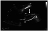
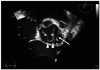
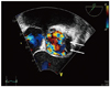
A 52-year-old man with sudden onset of dyspnea was transferred to our hospital. Transthoracic echocardiography showed an intimal flap ranged from sinus of Valsalva to sinotubular junction, with heterogenous hypoechoic materials within the flap (Fig. 1), and it caused severe aortic regurgitation. Computed tomography scan showed linear dissection flap and aneurysmal dilatation in the sinus of Valsalva. Transesophageal echocardiography (TEE) demonstrated a suspicious dissection flap in the left coronary cusp with destroyed aortic valve (Fig. 2), with no color Doppler signal within this flap (Fig. 3). We estimated the diagnosis as Type A aortic dissection requiring emergency operation. From the operative findings, however, the patient was diagnosed to have infective endocarditis involved the left coronary cusp of aortic valve and sinus of Valsalva which caused dissection. Therefore, we performed aortic valve replacement and sinus of Valsalva repair. The operation finished successfully. The pathologic findings of hypoechoic materials within the flap showed chronic inflammation with neutrophil infiltration. Even though the pathogen was not proved in the several times of blood culture, we treated the patient with 6 weeks of antibiotics and anticoagulation therapy.
A sinus of Valsalva aneurysm is a rare disorder. Although usually congenital, it may be associated with endocarditis, trauma, or aortic dissection.1)2) Once ruptured, sinus of Valsalva aneurysm may produce serious hemodynamic instability, such as acute heart failure or sudden death.3) When a sinus of Valsalva aneurysm is suspected, immediate diagnosis should be pursued with TEE. But, as in this case, it can be misdiagnosed despite performing a TEE.
 XML Download