

 PDF
PDF ePub
ePub Citation
 Print
Print


Introduction
Takayasu's arteritis (TA) is a chronic inflammatory disorder that results in stenosis of medium to large sized arteries. Aorta and its major branches are mainly involved with subclavian artery being most commonly affected. On the other hand, characteristics of pulmonary arterial involvement are not well established, although its incidence was reported to reach approximately 56% of TA patients.1)
We report here one case of right main pulmonary artery total
occlusion in a woman with TA, who had been diagnosed and treated with severe tricuspid regurgitation associated with idiopathic pulmonary arterial hypertension for several years elsewhere.
Case
A 52-year-old woman was referred to the outpatient clinic of rheumatology department with a 6-month history of dyspnea on exertion [New York Heart Association (NYHA) functional class II to III]. About 20 years ago, she visited a local clinic for non-healing toe wound and had a tentative diagnosis of TA. She had a previous history of vascular surgery for left carotid artery. Ten years later, she visited another local clinic with a complaint of dyspnea on exertion (NYHA functional class II). At that time, echocardiographic examination showed severe pulmonary hypertension with severe tricuspid regurgitation, based on which the attending physician prescribed diuretics and digoxin with a diagnosis of idiopathic pulmonary arterial hypertension. Despite the medications, however, she could not feel any improvement with regard to exertional dyspnea and she was advised to undergo surgery for severe tricuspid regurgitation. During preoperative work-ups, complete occlusion of right main pulmonary artery was incidentally detected. For further evaluation, she was referred to our hospital.
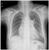
On physical examination, her blood pressure was 78/39 mmHg at right arm and 99/70 mmHg at left arm. Her chest radiography showed a prominent left hilar opacity and right atrial enlargement (Fig. 1). Her electrocardiogram showed right atrial enlargement and right ventricular hypertrophy. Laboratory parameters included increases in erythrocyte sedimentation rate (77 mm/hr) and C-reactive protein (2.14 mg/dL).
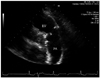
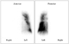
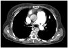
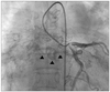
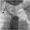
On echocardiography, right ventricle (RV) was severely dilated, its wall thickness was increased, and RV systolic function was decreased. Severe tricuspid regurgitation and severe pulmonary hypertension with maximal pressure gradient of 131 mmHg were noted, as well. On basal parasternal short axis view, the blood ejected from the RV to the main pulmonary artery appeared not to cross the right pulmonary artery. When we carefully traced the proximal right pulmonary artery, we could not clearly delineate its opening (Fig. 2). Lung perfusion scan showed the total absence of perfusion in the right lung with partial perfusion defects in the left lung (Fig. 3), but no pulmonary thromboembolism was clearly demonstrated on computed tomographic pulmonary angiography. The occlusion of right main pulmonary artery and left main pulmonary artery stenosis at the entrance were also clearly demonstrated on computed tomographic angiography (Fig. 4). According to the result of cardiac catheterization performed elsewhere, the mean right atrial pressure was severely elevated (18.4 mmHg), and catheter was impossible to go into the right pulmonary artery. Coronary angiography demonstrated a huge left circumflex coronary artery to right pulmonary artery fistula (Fig. 5). Arch aortography revealed total occlusion of the left subclavian and left common carotid artery in their proximities. Right subclavian artery showed near-total occlusion, as well (Supplemental Fig. 1). The diagnosis of TA was established based on the criteria of American college of Rheumatology for classification of TA; age at diagnosis (less than 40 years), difference in systolic blood pressure between arms (exceed 10 mmHg), arteriogram abnormality.
The patient was treated with corticosteroid, methotrexate, bosentan and symptomatic medications for right-sided heart failure. She is now being followed in the rheumatology clinic on a regular basis and her symptoms showed slight improvement with these medications.
Discussion
TA is a chronic inflammatory disease of aorta and its branches, and is acknowledged to be a disease of young Asian women.2) The etiology is unclear, but the hypothesis is generally accepted that cell-mediated autoimmunity triggered by unidentified factors such as infection and/or genetic factors plays a major role in the pathogenesis of TA.3) In this respect, suppression of inflammatory reaction before irreversible damage takes place is believed to be of clinical importance.
In 1908, Mikito Takayasu, Japanese ophthalmologist, first described a case about retinal arteriovenous fistula. At the same conference, 2 other cases with similar funduscopic findings were also reported, in whom pulse could not be palpable in one or both radial arteries. Several cases have been reported thereafter, and the term 'Takayasu's arteritis' was coined in 1939.2) A year later, pulmonary artery involvement of TA was first described in TA patients and it was reported that pathologic changes of affected pulmonary arteries were very similar to those observed in systemic vessels.4) Subsequently, prevalence of pulmonary artery involvement was estimated approximately 10% to 50% in TA patients, with a variety of pathological manifestations like stenosis, occlusion, or only vessel wall irregularity.5) Although abnormal pulmonary arteriograms or isolated pulmonary artery involvement was previously reported,5)6) there was no case reporting complete occlusion of one pulmonary artery, resulting in severe pulmonary hypertension.
The mainstay of TA treatment is glucocorticoid, but its relapse is common with glucocorticoid tapering. Another problem is that no study has proven survival benefit with its use. Cytotoxic agents did not show successful results in glucocorticoid-dependent or resistant patients, either. As an alternative treatment option, surgical correction of stenosed (not total occluded) vessels followed by glucocorticoid therapy was previously reported to improve outcome and decreased morbidity.7) Recently, biologic agents such as anti-tumor necrosis factor, interleukin-6 inhibitor are being under active investigation.8)
In this case, TA has not been previously treated at all because the patient's pulmonary hypertension was considered to be idiopathic in origin. After confirmation of TA diagnosis, corticosteroid treatment was immediately started, but she felt no symptomatic relief. Considering her long past medical history, her left pulmonary vasculature might be already damaged, more or less, on top of total occluded right pulmonary artery, which is the main reason why we prescribed bosentan to this patient. Of note, we found the presence of a fistula from the left circumflex coronary artery to the right pulmonary artery. Although we cannot provide definite explanation of the origin of the coronary-to-pulmonary fistula, this may be congenital in origin or may originate from de novo development along the natural course of TA. No matter what, this finding has never been reported before, as far as we know. With rigorous efforts to finding possible causes of her pulmonary hypertension at the initial stage, the outcome could have been changed.
Echocardiography is a useful noninvasive tool for predicting pulmonary artery systolic pressure using tricuspid regurgitation jet flow on the basis of modified Bernoulli equation [i.e., systolic pulmonary artery pressure = (maximal tricuspid regurgitation velocity)2 + right atrial pressure]. However, one important point that we should keep in mind is that, this simple equation is ultimately realized on the assumption that pulmonary arterial or valvular stenosis is not present. This assumption is very simple but is frequently missed when we perform echocardiographic examination in our daily clinical practice.9) In this case, more careful inspection of both proximal pulmonary arteries could have detected the occlusion of right pulmonary artery and could have been helpful for the appropriate treatment. Thus, comprehensive echocardiographic examination is of crucial value for assessing TA patients, especially in those with suspicious pulmonary hypertension.
 XML Download