

 PDF
PDF ePub
ePub Citation
 Print
Print


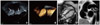
A 65-year-old woman was referred to the Department of Cardiovascular Surgery of our hospital for an operation with a presumptive diagnosis of right atrial myxoma. The mass was found incidentally on routine echocardiography during preoperative evaluation for total knee replacement. She had had hypertension and diabetes mellitus for 8 years. Her waist circumference was 111 cm and her body mass index was elevated at 37.8 kg/m2. She complained of mild exertional dyspnea. Electrocardiogram revealed normal sinus rhythm. Transthoracic echocardiography showed normal left ventricular dimensions and systolic function. However, it showed a poor echo window due to morbid obesity. Transesophageal echocardiography demonstrated a large right atrial mass attached to the interatrial septum with protrusion into the right atrium. The mass had a characteristic dumbbell-shaped appearance, separating into two parts (31 × 15 mm, 40 × 11 mm) due to sparing of the fossa ovalis (Fig. 1A and B). It was non-mobile and homogeneous echogenicity with a smooth surface. Fat tissue was also prominent around the aortic root. Cardiac magnetic resonance revealed a dumbbell shaped mass with no contrast enhancement involved on the interatrial septum from the coronary sinus to aortic root level. Subcutaneous, pericardial and epicardial fat tissue was revealed considerably (Fig. 1C). The mass showed high signal intensity on T1 weighted image (Fig. 1D) and dark signal on fat suppression image. Therefore, this obese woman did not undergo open heart surgery because her diagnosis on a multimodality imaging was extensive lipomatous hypertrophy of interatrial septum (LHIS).
LHIS is usually a benign condition caused by the excessive deposition of adipose tissue in the interatrial septum.1-3) It most often detected as an incidental finding on echocardiography in elderly obese patients. To avoid unnecessary surgical resection, we should consider this disease especially in elderly obese women.
 XML Download