Journal List > J Cardiovasc Ultrasound > v.20(3) > 1018231

Zhang and Yu: Clinical Implication of Mechanical Dyssynchrony in Heart Failure

Abstract

Mechanical dyssynchrony is a common phenomenon in patients with congestive heart failure, which usually identified by noninvasive cardiac imaging tools such as echocardiography. It demonstrates electromechanical delay in some regions of the failing heart which in turn contributes to further impairment of cardiac function. The diagnostic, therapeutic and prognostic values of mechanical dyssynchrony have been reported in a number of studies. Therefore, this review describes briefly the methods of measurement, but more importantly, explains the clinical implication of its assessment in heart failure related aspects including cardiac resynchronization therapy, functional mitral regurgitation, diastolic heart failure and mortality.

Introduction

Mechanical dyssynchrony describes the differences in the timing of contraction or relaxation between the left ventricular (LV) and right ventricular (RV) (interventricular dyssynchrony), or between different myocardial segments of the LV (intraventricular dyssynchrony). It is commonly observed in patients with congestive heart failure (CHF), which is caused by electromechanical delay in some regions of the failing heart and will result in further reduction of cardiac function. Its presence varies not only with the methodology of assessment, but also the characteristics of patients including the QRS duration, ejection fraction, loading condition, severity of coronary artery disease, and degree of LV hypertrophy or remodeling. Mechanical dyssynchrony has been suggested useful for exploration of disease mechanism, stratification of patient risk, selection of therapeutic modality, and prediction of prognosis in CHF population, in particular in those who are candidates of cardiac resynchronization therapy (CRT).

Assessment of Mechanical Dyssynchrony

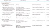
The analysis of mechanical dyssynchrony by echocardiography has been widely adopted due to its advantages of being easily available, non-invasive, radiation free and rapid technological development. The techniques range from conventional M-mode and Doppler echocardiography to more advanced tissue Doppler imaging (TDI), three-dimensional (3D) echocardiography and two-dimensional speckle tracking imaging, and most recently, 3D speckle tracking imaging. The parameters of systolic dyssynchrony include those signify the dispersion of the time to peak ventricular contraction, as represented by the standard deviation or maximal delay among a certain amount of LV segments, and the difference between the LV and RV. The majority of them were derived from CRT trials with cutoff values to define dyssynchrony and therefore suggested by the American Society of Echocardiography in an expert consensus statement (Table 1).1-10)
The assessment of diastolic dyssynchrony is similar to that of systolic dyssynchrony, in which the parameters calculate the dispersion of the time to ventricular relaxation, as referred to the standard deviation or maximal delay among a certain amount of LV segments.11-14) Of note, TDI velocity is almost the exclusive modality adopted for measurement, because of the discernable and consistent signal of early diastole (Table 1).

Dyssynchrony and CRT

The compelling evidence from multicenter clinical trials has established CRT as the most promising therapeutic modality in heart failure management over the past decade.15) CRT not only improves symptoms and cardiac function, but also reduces heart failure hospitalization and all-cause/cardiovascular mortality in patients with advanced CHF. However, it remains a major problem that non-responders of therapy are constantly observed in about one-third of patients receiving CRT, based on the current guidelines for patient selection in which the QRS duration ≥ 120 ms is regarded as the only marker of ventricular electromechanical delay.16)17) The presence of a prolonged QRS duration in CHF is associated with more advanced myocardial disease, more severe LV dysfunction, worse prognosis and higher all-cause mortality.18) However, a prolonged QRS duration may not necessarily be equivalent to significant mechanical dyssynchrony, even in the form of left bundle branch block (LBBB). Fung et al.19) observed that regional electrical conduction delay could be absent in CHF patients with LBBB. Using the 3D non-contact mapping electrograms, homogenous delay in LV the propagation of endocardial activation similar to that of normal subjects was found in some patients with typical LBBB, while typical delay in depolarization over the LV lateral or posterior wall was observed in others. In fact, the QRS duration on surface ECG is a rather inaccurate estimation of myocardial electrical activation which correlates poorly with the occurrence of systolic mechanical dyssynchrony.20-22)
The correction of cardiac electromechanical delay is suggested to be one of the major mechanisms for CRT benefits. Therefore, direct measurement of mechanical dyssynchrony by echocardiography has become clinically relevant in estimating the likelihood of response when ECG as a surrogate marker may fail. Over the last decade, a number of single-center studies have demonstrated that the lack of mechanical dyssynchrony assessed by noninvasive echocardiographic techniques is closely related to the lack of response in patients who received CRT. Nevertheless, those results were challenged by the Predictors of Response to CRT (PROSPECT) trial, the first multicenter trial in which no single echocardiographic measure of mechanical dyssynchrony could predict CRT responses with a good sensitivity and specificity.23) However, there were a number of major limitations in the design and execution of the PROSPECT trial, which challenged the value of conducting a highly flawed study that is unable to address the crucial question of role of echocardiography in predicting CRT response, and the implication of reducing the amount of unnecessary device implantation.24-27) The inadequacies in patient selection, study site training, echocardiographic standard for data acquisition and analysis, the lack of training and experience in dyssynchrony assessment by the three echocardiographic core-laboratories, and consideration of other contributing factors may explain the unexpected findings of the trial. They include extensive myocardial scar, absence of contractile reserve, severe mitral regurgitation, high pulmonary pressure, poor LV lead position and suboptimal device programming.16)28)29) Therefore, the PROSPECT trial shall not be regarded as the final conclusion about the inability of dyssynchrony assessment for predicting CRT response, but rather, dedicated training for knowledge and skill transfer cannot be overemphasized. Our recent study has confirmed the importance of systematic training to ensure the reproducibility of dyssynchrony analysis using TDI when comparing the reading between the "beginners" or the "graduates" of dyssynchrony training with the reference standard of the "experts".30)
A number of studies are conducted in the "post-PROSPECT era" to examine the ability of mechanical dyssynchrony in predicting CRT responses.31-46) Among them, a couple of studies were designed and conducted by the experienced centers in which shared protocol and standardized technique of dyssynchrony analysis could be ensured, as well as having a larger sample size with diversity of patients. Furthermore, hard endpoints over long-term follow up were selected, such as all-cause mortality and cardiovascular event. Mid-term LV reverse remodeling also frequently occurred as a primary endpoint in these trials, as its presence after CRT has been proved to correlate with improvement in clinical status and favorable long-term prognosis.47)48) More importantly, multivariate regression models were built up by including other factors at baseline such as age, gender, etiology of heart failure, severity of mitral regurgitation, presence of atrial fibrillation, and LV lead position with mechanical dyssynchrony, to demonstrate its independent or incremental predictive value in predicting CRT response.49-53)

Dyssynchrony and Functional Mitral Regurgitation

Functional mitral regurgitation (FMR) as a result of the dilation of LV cavity and/or alteration of LV chamber geometry is frequently observed in patients with CHF, in particular those with LV systolic dysfunction. In a large study including more than 2000 patients with symptomatic LV systolic dysfunction and ejection fraction < 40%, FMR of any grade assessed by angiography was present in about 60% of the patients.54) Another study investigated 1421 patients with LV ejection fraction ≤ 35%, using color Doppler echocardiography, there were moderate FMR in 30% of the patients and severe in 19%.55) Although mitral valve leaflets, papillary muscles and chordae tendineae appear normal in structure by surgical inspection or echocardiographic examination in FMR, the leaflets fail to coapt properly. FMR, as a complication of LV dilation and systolic dysfunction, can further aggravate LV volumetric overload and exacerbate left atrial (LA) pressure and volume overload, which will set up a vicious cycle of LV remodeling. Several studies revealed that the presence of FMR in heart failure was an independent predictor of worse survival.54)55) In addition, a dose-response relationship was observed that a 23% increased risk of death associated with the change from no FMR to mild FMR as well as the change from mild to moderate or severe FMR.54)
The basic mechanism of FMR is believed to be the mismatch between increased mitral leaflet tethering due to the outward displacement of papillary muscles and reduced closing force caused by LV systolic dysfunction.56) Furthermore, multiple factors are suggested to be involved in the pathogenesis of this force imbalance, including LV remodeling, leaflet tenting, annular dilation and dysfunction, as well as mechanical dyssynchrony. Consequently, these changes lead to the deformation of the mitral valve apparatus and reduction in the coaptation area of the leaflets. In echocardiographic studies, intraventricular mechanical dyssynchrony was found to be an important contributor to FMR. First of all, LV systolic dyssynchrony reduces the efficiency of contraction, resulting in decreased closing forces which worsened reduced leaflet coaptation and increased valve tenting. Secondly, uncoordinated contraction of the LV segments adjacent to the papillary muscles may increase mitral leaflet tethering and cause mal-alignment of the leaflet scallops leading to incomplete closure.57) Soyama et al.58) showed in 32 patients with dilated cardiomyopathy that the presence of FMR correlated with a significant delay in mechanical activity between the LV segments supporting the lateral and medial papillary muscle, as assessed by the difference in the time to peak systolic myocardial strain. Thirdly, LV mechanical dyssynchrony leads to changes in the mitral valve geometry and kinematics that may induce FMR. In animal models, a more widely opened mitral valve at end-diastole with delayed and dyssynchronous mitral valve closure was created by the RV apical pacing.59) The dyssynchronous contraction of the LV basal segments, attributable to the loss of mitral annular contraction, increase in systolic annular area and presence of mitral leaflet tethering, may worsen mitral regurgitation.60) Therefore, in a cross-sectional study which prospectively enrolled 136 CHF patients with LV ejection fraction < 50% and more than mild FMR, Liang et al.61) included variables of mitral valve deformation, LV global and regional remodeling, LV contractility, mitral annular size and function, and LV mechanical dyssynchrony for multivariate logistic regression analysis. As a result, mitral valve tenting area and LV global dyssynchrony, measured by the standard deviation of the time to peak systolic velocity among the 12 LV segments (Ts-SD) by TDI, were independent determinants of significant FMR.
Data from multicenter CRT trials revealed 13-50% reduction in FMR during 6- to 12-month follow up after the device therapy.56) Intriguingly, pre-pacing mechanical dyssynchrony was found to be one major determinant of FMR reduction after CRT.62)63) The improvement is suggested to be associated with decreased mechanical dyssynchrony,57)63) increased closing force,64) improved mitral valve deformation,62) and LV reverse remodeling.65) Mechanical dyssynchrony corrected by CRT would have direct impact on FMR and contribute to its improvement by interacting with several other aforementioned factors. Therefore, CRT would be a potential therapeutic option for selected CHF patients with significant FMR when valvular surgery as a current standard treatment carries high risk.61)66)

Dyssynchrony and Diastolic Heart Failure

Diastolic heart failure (DHF), or called heart failure with preserved ejection fraction, is a common condition among CHF population.67)68) In this condition, echocardiography with Doppler studies currently serves as a major diagnostic tool for the differentiation between DHF and systolic heart failure (SHF).69)70) Although it carries a significant risk of hospitalization and mortality similar to SHF, our knowledge of DHF is still limited with regard to its pathogenesis, diagnosis and evidence-based management. Hypertension, LV hypertrophy, diabetes and coronary artery disease have been recognized as main risk factors for developing clinically overt DHF, in which LV concentric remodeling, LV segmental wall motion abnormality, LV diastolic dysfunction and LA dilatation are commonly observed indices. Recently, the concept of LV mechanical dyssynchrony has also been extended to the investigation of patients with DHF as an additional factor involved in the pathogenesis. Our early publication demonstrated by TDI that isolated systolic, isolated diastolic, and combined dyssynchrony were observed in 25.0%, 21.7%, and 14.1% of DHF patients, though it was less prevalent than patients with SHF.13) The study by Wang et al.12) in their DHF population reported a similar prevalence of systolic dyssynchrony (33%) but a higher prevalence of diastolic dyssynchrony (58%). In patients with acute coronary syndrome accompanied by DHF, diastolic dyssynchrony was evident in 35% of patients and systolic dyssynchrony in 47%, while the prevalence of diastolic dyssynchrony was much higher than those without DHF.11) Interestingly, the presence of mechanical dyssynchrony also showed a dynamic change in hypertensive DHF patients, that the prevalence of systolic dyssynchrony increased dramatically during pharmacological stress test from 36% to 85% and diastolic dyssynchrony from 38% to 87%.71)
Although mechanical dyssynchrony is frequently observed in patients with DHF, a number of questions remain unanswered with regard to its contribution to the impairment of cardiac function and clinical manifestation of heart failure, in particular the differences from SHF. A wide QRS complex is very uncommon in DHF patients, therefore, the QRS duration is not a major determinant for the presence of systolic and diastolic dyssynchrony. Unlike patients with SHF, mechanical dyssynchrony in DHF may occur as a result of myocardial disease rather than electromechanical coupling delay. Coexistence but not cause-effect relationship of cardiac dysfunction and mechanical dyssynchrony was described in previous studies, while the correlation between the two facets of LV performance differed among studies.11-13)71) Therefore, apart from the severity of myocardial dysfunction, dyssynchronous LV relaxation and impairment of ventricular restoring forces may also interfere the LV filling and lead to a diastolic dyssynchrony,72) or vice versa. Interestingly, medical therapy for DHF, including diuretics, beta-blockers, calcium-channel blockers, angiotensin-converting enzyme inhibitors and/or angiotensin-receptor blockers, was associated with shortening of diastolic intraventricular delay, which in turn correlated with improvement of LV stiffness and reduction of filling pressure.12) However, it remains to define what extent LV dyssynchrony is involved in the pathophysiologic mechanism of DHF.

Dyssynchrony and Mortality in Heart Failure

The prognostic implication of mechanical dyssynchrony was initially reported by Bader et al.73) where 104 CHF patients with ejection fraction ≤ 45%, over half of them had wide QRS complexes, were examined by the use of pulsed TDI and followed up for one year. Although no mortality occurred at the end of follow up, 86 patients (83%) were admitted for decompensated CHF. As a result, intraventricular dyssynchrony was found to be most important independent predictor of heart failure hospitalization, and the other two independent predictors included LV ejection fraction and QRS width. In another early study of 106 CHF patients with LV ejection fraction < 35% and QRS duration ≤ 120 ms who were followed up for a mean of 17 ± 11 months, intraventricular dyssynchrony was measured by TDI as the Ts-SD from both basal and middle LV segments in apical 4- and 2-chamber views. A Ts-SD cutoff value of > 37 ms was associated with a significant increase in clinical event of including heart failure hospitalization or cardiac transplantation.74) The same group recently published their study on 167 CHF patients with a mean follow up of 33 months. Electrical dyssynchrony defined as the QRS duration ≥ 120 ms and mechanical delay as the septal-to-lateral wall delay ≥ 65 ms were investigated for their association with adverse events.75) In multivariate Cox regression analysis, the septal-to-lateral wall delay [hazard ratio (HR), 2.37; p = 0.002] showed a better predictive value than QRS duration (HR, 1.88; p = 0.028) for cardiac events. Moreover, patients with both electrical and mechanical dyssynchrony had a HR of 3.98 (p < 0.001) when compared with those with normal QRS duration and absence of mechanical dyssynchrony.75)
Recently, the impact of mechanical dyssynchrony on prognosis was explored in a subgroup of CHF patients who had ischemic cardiomyopathy.76)77) In the Valsartan in Acute Myocardial Infarction (VALIANT) echocardiography study, mechanical dyssynchrony was assessed in 381 patients with ventricular dysfunction or heart failure after myocardial infarction, who were followed up for a median period of 611 days.76) Consequently, LV dyssynchrony was independently associated with increased risk of death or heart failure hospitalization, while QRS width ≥ 120 ms which occurred in about 5% of patients failed to do so. Another study consisted of 215 patients with moderate systolic heart failure undergoing coronary artery bypass graft (CABG) surgery, in which mechanical dyssynchrony was calculated by TDI and myocardial viability by single photon emission computed tomography.77) Post-CABG dyssynchrony ≥ 72 ms and ≥ 5 viable segments were used to categorize patients into different groups. Patients without post-CABG dyssynchrony and with viable myocardium had the least clinical events compared to those with severe post-CABG dyssynchrony and nonviable myocardium (3% vs. 64%; p < 0.001). In addition, QRS duration did not predict cardiac events during the median follow up period of 359 days.
Importantly, QRS duration was not an independent prognosticator in CHF patients who did not exhibit wide QRS complexes. Therefore, all of these studies have suggested that assessment of mechanical dyssynchrony is helpful to provide important prognostic value on disease outcome on top of QRS duration.

Summary

Mechanical dyssynchrony is common in CHF patients, in particular in those with reduced ejection fraction and prolonged QRS complex. With cumulated knowledge in the advanced imaging techniques and expanded clinical applications of mechanical dyssynchrony, it appears that the assessment of mechanical dyssynchrony has a unique role in heart failure population. Not only being useful in CRT candidates, it can also be used to predict the development and progression of cardiac diseases, and as prognosticators. However, before the measurement of dyssynchrony is contemplated, it is imperative to receive systematic training in order to achieve high quality online image acquisition and knowledge of offline analysis. Furthermore, mechanical dyssynchrony varies with many conditions. Therefore, it is important to understand the right clinical context while applying knowledge of dyssynchrony: wide vs. narrow QRS complex, systolic vs. diastolic heart failure, resting vs. stress echocardiography, cause vs. effect, single vs. multiple contributors, and short- vs. long-term outcome.

Figures and Tables

Table 1
Major systolic and diastolic dyssynchrony indices with cutoff values reported
jcu-20-117-i001

2D: two-dimensional, 3D: three-dimensional, LV: left ventricular, RV: right ventricular, SD: standard deviation

References

1. Suffoletto MS, Dohi K, Cannesson M, Saba S, Gorcsan J 3rd. Novel speckle-tracking radial strain from routine black-and-white echocardiographic images to quantify dyssynchrony and predict response to cardiac resynchronization therapy. Circulation. 2006. 113:960–968.
crossref
2. Pitzalis MV, Iacoviello M, Romito R, Massari F, Rizzon B, Luzzi G, Guida P, Andriani A, Mastropasqua F, Rizzon P. Cardiac resynchronization therapy tailored by echocardiographic evaluation of ventricular asynchrony. J Am Coll Cardiol. 2002. 40:1615–1622.
crossref
3. Bax JJ, Bleeker GB, Marwick TH, Molhoek SG, Boersma E, Steendijk P, van der Wall EE, Schalij MJ. Left ventricular dyssynchrony predicts response and prognosis after cardiac resynchronization therapy. J Am Coll Cardiol. 2004. 44:1834–1840.
crossref
4. Gorcsan J 3rd, Kanzaki H, Bazaz R, Dohi K, Schwartzman D. Usefulness of echocardiographic tissue synchronization imaging to predict acute response to cardiac resynchronization therapy. Am J Cardiol. 2004. 93:1178–1181.
crossref
5. Yu CM, Gorcsan J 3rd, Bleeker GB, Zhang Q, Schalij MJ, Suffoletto MS, Fung JW, Schwartzman D, Chan YS, Tanabe M, Bax JJ. Usefulness of tissue Doppler velocity and strain dyssynchrony for predicting left ventricular reverse remodeling response after cardiac resynchronization therapy. Am J Cardiol. 2007. 100:1263–1270.
crossref
6. Yu CM, Fung JW, Zhang Q, Chan CK, Chan YS, Lin H, Kum LC, Kong SL, Zhang Y, Sanderson JE. Tissue Doppler imaging is superior to strain rate imaging and postsystolic shortening on the prediction of reverse remodeling in both ischemic and nonischemic heart failure after cardiac resynchronization therapy. Circulation. 2004. 110:66–73.
crossref
7. Yu CM, Fung WH, Lin H, Zhang Q, Sanderson JE, Lau CP. Predictors of left ventricular reverse remodeling after cardiac resynchronization therapy for heart failure secondary to idiopathic dilated or ischemic cardiomyopathy. Am J Cardiol. 2003. 91:684–688.
crossref
8. Notabartolo D, Merlino JD, Smith AL, DeLurgio DB, Vera FV, Easley KA, Martin RP, León AR. Usefulness of the peak velocity difference by tissue Doppler imaging technique as an effective predictor of response to cardiac resynchronization therapy. Am J Cardiol. 2004. 94:817–820.
crossref
9. Kapetanakis S, Bhan A, Murgatroyd F, Kearney MT, Gall N, Zhang Q, Yu CM, Monaghan MJ. Real-time 3D echo in patient selection for cardiac resynchronization therapy. JACC Cardiovasc Imaging. 2011. 4:16–26.
crossref
10. Cazeau S, Bordachar P, Jauvert G, Lazarus A, Alonso C, Vandrell MC, Mugica J, Ritter P. Echocardiographic modeling of cardiac dyssynchrony before and during multisite stimulation: a prospective study. Pacing Clin Electrophysiol. 2003. 26(1 Pt 2):137–143.
crossref
11. Lee AP, Zhang Q, Yip G, Fang F, Liang YJ, Xie JM, Lam YY, Yu CM. LV mechanical dyssynchrony in heart failure with preserved ejection fraction complicating acute coronary syndrome. JACC Cardiovasc Imaging. 2011. 4:348–357.
crossref
12. Wang J, Kurrelmeyer KM, Torre-Amione G, Nagueh SF. Systolic and diastolic dyssynchrony in patients with diastolic heart failure and the effect of medical therapy. J Am Coll Cardiol. 2007. 49:88–96.
crossref
13. Yu CM, Zhang Q, Yip GW, Lee PW, Kum LC, Lam YY, Fung JW. Diastolic and systolic asynchrony in patients with diastolic heart failure: a common but ignored condition. J Am Coll Cardiol. 2007. 49:97–105.
crossref
14. Shanks M, Bertini M, Delgado V, Ng AC, Nucifora G, van Bommel RJ, Borleffs CJ, Holman ER, van de Veire NR, Schalij MJ, Bax JJ. Effect of biventricular pacing on diastolic dyssynchrony. J Am Coll Cardiol. 2010. 56:1567–1575.
crossref
15. Yu CM, Hayes DL, Auricchio A. Cardiac resynchronization therapy. 2008. 2nd ed. Malden: Blackwell Futura.
16. Birnie DH, Tang AS. The problem of non-response to cardiac resynchronization therapy. Curr Opin Cardiol. 2006. 21:20–26.
crossref
17. Tang AS, Ellenbogen KA. A futuristic perspective on clinical studies of cardiac resynchronization therapy for heart failure patients. Curr Opin Cardiol. 2006. 21:78–82.
crossref
18. Kashani A, Barold SS. Significance of QRS complex duration in patients with heart failure. J Am Coll Cardiol. 2005. 46:2183–2192.
crossref
19. Fung JW, Yu CM, Yip G, Zhang Y, Chan H, Kum CC, Sanderson JE. Variable left ventricular activation pattern in patients with heart failure and left bundle branch block. Heart. 2004. 90:17–19.
crossref
20. Yu CM, Lin H, Zhang Q, Sanderson JE. High prevalence of left ventricular systolic and diastolic asynchrony in patients with congestive heart failure and normal QRS duration. Heart. 2003. 89:54–60.
crossref
21. Yu CM, Yang H, Lau CP, Wang Q, Wang S, Lam L, Sanderson JE. Regional left ventricle mechanical asynchrony in patients with heart disease and normal QRS duration: implication for biventricular pacing therapy. Pacing Clin Electrophysiol. 2003. 26(2 Pt 1):562–570.
crossref
22. Bleeker GB, Schalij MJ, Molhoek SG, Verwey HF, Holman ER, Boersma E, Steendijk P, Van Der Wall EE, Bax JJ. Relationship between QRS duration and left ventricular dyssynchrony in patients with end-stage heart failure. J Cardiovasc Electrophysiol. 2004. 15:544–549.
crossref
23. Chung ES, Leon AR, Tavazzi L, Sun JP, Nihoyannopoulos P, Merlino J, Abraham WT, Ghio S, Leclercq C, Bax JJ, Yu CM, Gorcsan J 3rd, St John Sutton M, De Sutter J, Murillo J. Results of the Predictors of Response to CRT (PROSPECT) trial. Circulation. 2008. 117:2608–2616.
crossref
24. Sanderson JE. Echocardiography for cardiac resynchronization therapy selection: fatally flawed or misjudged? J Am Coll Cardiol. 2009. 53:1960–1964.
25. Bax JJ, Gorcsan J 3rd. Echocardiography and noninvasive imaging in cardiac resynchronization therapy: results of the PROSPECT (Predictors of Response to Cardiac Resynchronization Therapy) study in perspective. J Am Coll Cardiol. 2009. 53:1933–1943.
crossref
26. Yu CM, Sanderson JE, Gorcsan J 3rd. Echocardiography, dyssynchrony, and the response to cardiac resynchronization therapy. Eur Heart J. 2010. 31:2326–2337.
crossref
27. Yu CM, Bax JJ, Gorcsan J 3rd. Critical appraisal of methods to assess mechanical dyssynchrony. Curr Opin Cardiol. 2009. 24:18–28.
crossref
28. Yu CM, Wing-Hong Fung J, Zhang Q, Sanderson JE. Understanding nonresponders of cardiac resynchronization therapy--current and future perspectives. J Cardiovasc Electrophysiol. 2005. 16:1117–1124.
crossref
29. Bax JJ, Abraham T, Barold SS, Breithardt OA, Fung JW, Garrigue S, Gorcsan J 3rd, Hayes DL, Kass DA, Knuuti J, Leclercq C, Linde C, Mark DB, Monaghan MJ, Nihoyannopoulos P, Schalij MJ, Stellbrink C, Yu CM. Cardiac resynchronization therapy: part 1--issues before device implantation. J Am Coll Cardiol. 2005. 46:2153–2167.
30. Zhang Q, Liang YJ, Zhang QH, Li RJ, Chua Y, Xie JM, Lee PW, Yu CM. Impact of a dedicated training program on the reproducibility of systolic dyssynchrony measures using tissue Doppler imaging. J Am Soc Echocardiogr. 2012. 25:210–217.
crossref
31. Lim P, Buakhamsri A, Popovic ZB, Greenberg NL, Patel D, Thomas JD, Grimm RA. Longitudinal strain delay index by speckle tracking imaging: a new marker of response to cardiac resynchronization therapy. Circulation. 2008. 118:1130–1137.
crossref
32. Cleland J, Freemantle N, Ghio S, Fruhwald F, Shankar A, Marijanowski M, Verboven Y, Tavazzi L. Predicting the long-term effects of cardiac resynchronization therapy on mortality from baseline variables and the early response a report from the CARE-HF (Cardiac Resynchronization in Heart Failure) Trial. J Am Coll Cardiol. 2008. 52:438–445.
crossref
33. Zhang Q, van Bommel RJ, Fung JW, Chan JY, Bleeker GB, Ypenburg C, Yip G, Liang YJ, Schalij MJ, Bax JJ, Yu CM. Tissue Doppler velocity is superior to strain imaging in predicting long-term cardiovascular events after cardiac resynchronisation therapy. Heart. 2009. 95:1085–1090.
crossref
34. Lafitte S, Reant P, Zaroui A, Donal E, Mignot A, Bougted H, Belghiti H, Bordachar P, Deplagne A, Chabaneix J, Franceschi F, Deharo JC, Dos Santos P, Clementy J, Roudaut R, Leclercq C, Habib G. Validation of an echocardiographic multiparametric strategy to increase responders patients after cardiac resynchronization: a multicentre study. Eur Heart J. 2009. 30:2880–2887.
crossref
35. Van Bommel RJ, Ypenburg C, Borleffs CJ, Delgado V, Marsan NA, Bertini M, Holman ER, Schalij MJ, Bax JJ. Value of tissue Doppler echocardiography in predicting response to cardiac resynchronization therapy in patients with heart failure. Am J Cardiol. 2010. 105:1153–1158.
crossref
36. Oyenuga O, Hara H, Tanaka H, Kim HN, Adelstein EC, Saba S, Gorcsan J 3rd. Usefulness of echocardiographic dyssynchrony in patients with borderline QRS duration to assist with selection for cardiac resynchronization therapy. JACC Cardiovasc Imaging. 2010. 3:132–140.
crossref
37. van Bommel RJ, Borleffs CJ, Ypenburg C, Marsan NA, Delgado V, Bertini M, van der Wall EE, Schalij MJ, Bax JJ. Morbidity and mortality in heart failure patients treated with cardiac resynchronization therapy: influence of pre-implantation characteristics on long-term outcome. Eur Heart J. 2010. 31:2783–2790.
crossref
38. Miyazaki C, Redfield MM, Powell BD, Lin GM, Herges RM, Hodge DO, Olson LJ, Hayes DL, Espinosa RE, Rea RF, Bruce CJ, Nelson SM, Miller FA, Oh JK. Dyssynchrony indices to predict response to cardiac resynchronization therapy: a comprehensive prospective single-center study. Circ Heart Fail. 2010. 3:565–573.
crossref
39. van Bommel RJ, Tanaka H, Delgado V, Bertini M, Borleffs CJ, Ajmone Marsan N, Holzmeister J, Ruschitzka F, Schalij MJ, Bax JJ, Gorcsan J 3rd. Association of intraventricular mechanical dyssynchrony with response to cardiac resynchronization therapy in heart failure patients with a narrow QRS complex. Eur Heart J. 2010. 31:3054–3062.
crossref
40. Gorcsan J 3rd, Oyenuga O, Habib PJ, Tanaka H, Adelstein EC, Hara H, McNamara DM, Saba S. Relationship of echocardiographic dyssynchrony to long-term survival after cardiac resynchronization therapy. Circulation. 2010. 122:1910–1918.
crossref
41. Tanaka H, Nesser HJ, Buck T, Oyenuga O, Jánosi RA, Winter S, Saba S, Gorcsan J 3rd. Dyssynchrony by speckle-tracking echocardiography and response to cardiac resynchronization therapy: results of the Speckle Tracking and Resynchronization (STAR) study. Eur Heart J. 2010. 31:1690–1700.
crossref
42. Delgado V, van Bommel RJ, Bertini M, Borleffs CJ, Marsan NA, Arnold CT, Nucifora G, van de Veire NR, Ypenburg C, Boersma E, Holman ER, Schalij MJ, Bax JJ. Relative merits of left ventricular dyssynchrony, left ventricular lead position, and myocardial scar to predict long-term survival of ischemic heart failure patients undergoing cardiac resynchronization therapy. Circulation. 2011. 123:70–78.
crossref
43. Inden Y, Ito R, Yoshida N, Kamiya H, Kitamura K, Kitamura T, Shimano M, Uchikawa T, Tsuji Y, Shibata R, Hirai M, Murohara T. Combined assessment of left ventricular dyssynchrony and contractility by speckled tracking strain imaging: a novel index for predicting responders to cardiac resynchronization therapy. Heart Rhythm. 2010. 7:655–661.
crossref
44. Jansen AH, Bracke F, van Dantzig JM, Peels KH, Post JC, van den Bosch HC, van Gelder B, Meijer A, Korsten HH, de Vries J, van Hemel NM. The influence of myocardial scar and dyssynchrony on reverse remodeling in cardiac resynchronization therapy. Eur J Echocardiogr. 2008. 9:483–488.
crossref
45. Parsai C, Bijnens B, Sutherland GR, Baltabaeva A, Claus P, Marciniak M, Paul V, Scheffer M, Donal E, Derumeaux G, Anderson L. Toward understanding response to cardiac resynchronization therapy: left ventricular dyssynchrony is only one of multiple mechanisms. Eur Heart J. 2009. 30:940–949.
crossref
46. Soliman OI, Geleijnse ML, Theuns DA, van Dalen BM, Vletter WB, Jordaens LJ, Metawei AK, Al-Amin AM, ten Cate FJ. Usefulness of left ventricular systolic dyssynchrony by real-time three-dimensional echocardiography to predict long-term response to cardiac resynchronization therapy. Am J Cardiol. 2009. 103:1586–1591.
crossref
47. Ypenburg C, van Bommel RJ, Borleffs CJ, Bleeker GB, Boersma E, Schalij MJ, Bax JJ. Long-term prognosis after cardiac resynchronization therapy is related to the extent of left ventricular reverse remodeling at mid-term follow-up. J Am Coll Cardiol. 2009. 53:483–490.
crossref
48. Yu CM, Bleeker GB, Fung JW, Schalij MJ, Zhang Q, van der Wall EE, Chan YS, Kong SL, Bax JJ. Left ventricular reverse remodeling but not clinical improvement predicts long-term survival after cardiac resynchronization therapy. Circulation. 2005. 112:1580–1586.
crossref
49. Gasparini M, Auricchio A, Regoli F, Fantoni C, Kawabata M, Galimberti P, Pini D, Ceriotti C, Gronda E, Klersy C, Fratini S, Klein HH. Four-year efficacy of cardiac resynchronization therapy on exercise tolerance and disease progression: the importance of performing atrioventricular junction ablation in patients with atrial fibrillation. J Am Coll Cardiol. 2006. 48:734–743.
crossref
50. Zhang Q, Yip GW, Chan YS, Fung JW, Chan W, Lam YY, Yu CM. Incremental prognostic value of combining left ventricular lead position and systolic dyssynchrony in predicting long-term survival after cardiac resynchronization therapy. Clin Sci (Lond). 2009. 117:397–404.
crossref
51. Zhang Q, Fung JW, Chan JY, Yip G, Lam YY, Liang YJ, Yu CM. Difference in long-term clinical outcome after cardiac resynchronisation therapy between ischaemic and non-ischaemic aetiologies of heart failure. Heart. 2009. 95:113–118.
crossref
52. Richardson M, Freemantle N, Calvert MJ, Cleland JG, Tavazzi L. CARE-HF Study Steering Committee and Investigators. Predictors and treatment response with cardiac resynchronization therapy in patients with heart failure characterized by dyssynchrony: a pre-defined analysis from the CARE-HF trial. Eur Heart J. 2007. 28:1827–1834.
crossref
53. Gradaus R, Stuckenborg V, Löher A, Köbe J, Reinke F, Gunia S, Vahlhaus C, Breithardt G, Bruch C. Diastolic filling pattern and left ventricular diameter predict response and prognosis after cardiac resynchronisation therapy. Heart. 2008. 94:1026–1031.
crossref
54. Trichon BH, Felker GM, Shaw LK, Cabell CH, O'Connor CM. Relation of frequency and severity of mitral regurgitation to survival among patients with left ventricular systolic dysfunction and heart failure. Am J Cardiol. 2003. 91:538–543.
crossref
55. Koelling TM, Aaronson KD, Cody RJ, Bach DS, Armstrong WF. Prognostic significance of mitral regurgitation and tricuspid regurgitation in patients with left ventricular systolic dysfunction. Am Heart J. 2002. 144:524–529.
crossref
56. Vinereanu D. Mitral regurgitation and cardiac resynchronization therapy. Echocardiography. 2008. 25:1155–1166.
crossref
57. Kanzaki H, Bazaz R, Schwartzman D, Dohi K, Sade LE, Gorcsan J 3rd. A mechanism for immediate reduction in mitral regurgitation after cardiac resynchronization therapy: insights from mechanical activation strain mapping. J Am Coll Cardiol. 2004. 44:1619–1625.
crossref
58. Soyama A, Kono T, Mishima T, Morita H, Ito T, Suwa M, Kitaura Y. Intraventricular dyssynchrony may play a role in the development of mitral regurgitation in dilated cardiomyopathy. J Card Fail. 2005. 11:631–637.
crossref
59. Langer F, Tibayan FA, Rodriguez F, Timek T, Zasio MK, Liang D, Daughters GT, Ingels NB, Miller DC. Altered mitral valve kinematics with atrioventricular and ventricular pacing. J Heart Valve Dis. 2005. 14:286–294. discussion 294.
60. Szymanski P, Klisiewicz A, Hoffman P. Asynchronous movement of mitral annulus: an additional mechanism of ischaemic mitral regurgitation. Clin Cardiol. 2007. 30:512–516.
crossref
61. Liang YJ, Zhang Q, Fang F, Lee AP, Liu M, Yan BP, Lam YY, Chan GC, Yu CM. Incremental value of global systolic dyssynchrony in determining the occurrence of functional mitral regurgitation in patients with left ventricular systolic dysfunction. Eur Heart J. 2012. [Epub ahead of print].
crossref
62. Ypenburg C, Lancellotti P, Tops LF, Bleeker GB, Holman ER, Piérard LA, Schalij MJ, Bax JJ. Acute effects of initiation and withdrawal of cardiac resynchronization therapy on papillary muscle dyssynchrony and mitral regurgitation. J Am Coll Cardiol. 2007. 50:2071–2077.
crossref
63. Ypenburg C, Lancellotti P, Tops LF, Boersma E, Bleeker GB, Holman ER, Thomas JD, Schalij MJ, Piérard LA, Bax JJ. Mechanism of improvement in mitral regurgitation after cardiac resynchronization therapy. Eur Heart J. 2008. 29:757–765.
crossref
64. Breithardt OA, Sinha AM, Schwammenthal E, Bidaoui N, Markus KU, Franke A, Stellbrink C. Acute effects of cardiac resynchronization therapy on functional mitral regurgitation in advanced systolic heart failure. J Am Coll Cardiol. 2003. 41:765–770.
crossref
65. St John Sutton MG, Plappert T, Abraham WT, Smith AL, DeLurgio DB, Leon AR, Loh E, Kocovic DZ, Fisher WG, Ellestad M, Messenger J, Kruger K, Hilpisch KE, Hill MR. Multicenter InSync Randomized Clinical Evaluation (MIRACLE) Study Group. Effect of cardiac resynchronization therapy on left ventricular size and function in chronic heart failure. Circulation. 2003. 107:1985–1990.
crossref
66. van Bommel RJ, Marsan NA, Delgado V, Borleffs CJ, van Rijnsoever EP, Schalij MJ, Bax JJ. Cardiac resynchronization therapy as a therapeutic option in patients with moderate-severe functional mitral regurgitation and high operative risk. Circulation. 2011. 124:912–919.
crossref
67. Dougherty AH, Naccarelli GV, Gray EL, Hicks CH, Goldstein RA. Congestive heart failure with normal systolic function. Am J Cardiol. 1984. 54:778–782.
crossref
68. Angeja BG, Grossman W. Evaluation and management of diastolic heart failure. Circulation. 2003. 107:659–663.
crossref
69. DeMaria AN, Wisenbaugh TW, Smith MD, Harrison MR, Berk MR. Doppler echocardiographic evaluation of diastolic dysfunction. Circulation. 1991. 84:3 Suppl. I288–I295.
70. Thomas JD, Weyman AE. Echocardiographic Doppler evaluation of left ventricular diastolic function. Physics and physiology. Circulation. 1991. 84:977–990.
crossref
71. Lee AP, Song JK, Yip GW, Zhang Q, Zhu TG, Li C, Chan A, Yu CM. Importance of dynamic dyssynchrony in the occurrence of hypertensive heart failure with normal ejection fraction. Eur Heart J. 2010. 31:2642–2649.
crossref
72. Kasner M, Westermann D, Schultheiss HP, Tschöpe C. Diastolic heart failure and LV dyssynchrony. Curr Pharm Biotechnol. 2012. [Epub ahead of print].
crossref
73. Bader H, Garrigue S, Lafitte S, Reuter S, Jaïs P, Haïssaguerre M, Bonnet J, Clementy J, Roudaut R. Intra-left ventricular electromechanical asynchrony. A new independent predictor of severe cardiac events in heart failure patients. J Am Coll Cardiol. 2004. 43:248–256.
crossref
74. Cho GY, Song JK, Park WJ, Han SW, Choi SH, Doo YC, Oh DJ, Lee Y. Mechanical dyssynchrony assessed by tissue Doppler imaging is a powerful predictor of mortality in congestive heart failure with normal QRS duration. J Am Coll Cardiol. 2005. 46:2237–2243.
crossref
75. Cho GY, Kim HK, Kim YJ, Choi DJ, Sohn DW, Oh BH, Park YB. Electrical and mechanical dyssynchrony for prediction of cardiac events in patients with systolic heart failure. Heart. 2010. 96:1029–1032.
crossref
76. Shin SH, Hung CL, Uno H, Hassanein AH, Verma A, Bourgoun M, Køber L, Ghali JK, Velazquez EJ, Califf RM, Pfeffer MA, Solomon SD. Valsartan in Acute Myocardial Infarction Trial (VALIANT) Investigators. Mechanical dyssynchrony after myocardial infarction in patients with left ventricular dysfunction, heart failure, or both. Circulation. 2010. 121:1096–1103.
crossref
77. Penicka M, Bartunek J, Lang O, Medilek K, Tousek P, Vanderheyden M, De Bruyne B, Maruskova M, Widimsky P. Severe left ventricular dyssynchrony is associated with poor prognosis in patients with moderate systolic heart failure undergoing coronary artery bypass grafting. J Am Coll Cardiol. 2007. 50:1315–1323.
crossref
TOOLS
Similar articles