Journal List > J Cardiovasc Ultrasound > v.20(2) > 1018266

Yoshida and Obase: Assessment of Mitral Valve Complex by Three-Dimensional Echocardiography: Therapeutic Strategy for Functional Mitral Regurgitation

Abstract

The mitral valve complex is consisted of annulus, leaflets, chordae tendineae, papillary muscle (PMs) and surrounding left ventricle. Functional mitral regurgitation (MR) results from left ventricular remodeling such as dilatation or distortion, which displaces the PMs and then tethers the mitral leaflets, restricting leaflet coaptation. Undersized annuloplasty, which has been widely accepted as a simple and effective procedure for functional MR, sometimes worsens the tethering of posterior leaflet and induces recurrent MR. In order to overcome such problems, several additional procedures to the simple annuloplasty have been produced. Three dimensional echocardiography plays an essential role to understand the geometry of mitral valve complex and contributes greatly to decision making of the surgical strategy in functional MR and its postoperative assessment.

Introduction

Conventional two-dimensional (2D) transthoracic echocardiography has been greatly contributing in the clinical diagnosis of mitral regurgitation (MR) and it is certainly the clinical standard for the evaluation of valve apparatus. Nevertheless, 2D has a disadvantage in the assessment of three-dimensional (3D) morphology. Three-dimensional approach allows us to visually understand the valve configuration and gives us additional information that has never been provided by 2D approach. Another important aspect about 3D approach is that the reconstructed 3D images can be quantitatively measured and analyzed in 3D spaces. Three-dimensional transesophageal echocardiography (TEE), which has been widely available recently, can have a significant impact in pre-, post- and intraoperative assessment of mitral valve apparatus. These information play a critical role in decision making of the surgical strategy in organic or functional MR.

Mitral Valve Complex

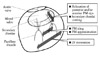
The whole mitral valve complex is consisted of annulus, leaflets, chordae tendineae, papillary muscles (PMs) and surrounding left ventricle (Fig. 1).1)2) With using multi-scanned 2D TEE images, well-trained echocardiographers can mentally reconstruct the 3D shape and position of the structures but it is not so easy to recognize or quantify the morphology and anatomical position of mitral valve complex. Some special software enable us to reconstruct the 3D image. The reconstructed 3D images can be quantitatively measured and analyzed in 3D spaces, which is impossible to do in 2D images. Three-dimensional evaluation of the mitral valve provides us important additional information in the clinical diagnosis, planning a surgical strategy or postoperative assessment.

Functional/Ischemic Mitral Regurgitation

Functional MR is defined as a regurgitation that is seen with structurally normal valve leaflets. Functional MR results from left ventricular remodeling such as dilatation or distortion, which displaces the PMs and then tethers the mitral leaflets, restricting leaflet coaptation.3)4) Ischemic MR is a complication of the chronic phase of myocardial infarction. It occurs commonly after myocardial infarction, with an estimated prevalence of 20% to 50%,5)6) and its presence has been shown to significantly worsen prognosis independent of ejection fraction.6)7) However, long-term effectiveness of the valve and ventricular repair surgery for the functional MR is still controversial.

Geometry in Functional/Ischemic MR

In a mitral valve with functional MR, geometric abnormalities of incomplete valve coaptation at systole are mainly characterized by leaflet tethering due to displacement of PMs and flattening of mitral annulus. These are caused by left ventricular remodeling, so that functional MR is called as ventricular disease rather than valvular disease. In patients with dilated cardiomyopathy, global ventricular remodeling occurs with resultant displacement of both PMs. In contrast, in patients with functional/ischemic MR due to inferior infarction, localized ventricular remodeling is more common with resultant posteromedial PM displacement.8) Real-time 3D echocardiography has proven that in patients with functional MR due to dilated cardiomyopathy, symmetrical PM displacement lead to progressive cordal tethering and leaflet tenting, resulting in predominantly central MR as a result of decreased leaflet coaptation.9-11) On the other hand, in patients with ischemic MR, left ventricular remodeling caused by abnormal wall motion results in uneven papillary displacement and asymmetric tethering associated with eccentric MR.9)12-14) Three dimensional TEE allows us to directly visualize the decreased leaflet coaptation and the differences of the coaptation depth in each segment.15)
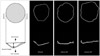
Mitral annulus shows a nonplanar saddle-shaped configuration in healthy individual.16) There are two peaks at the aortic insertion and posterior left ventricular wall and two low troughs closest to the apex located medially and laterally (Fig. 1).1)2)17) The saddle shape is most remarkable during mid-systole.18) The annulus acquires a more flattened shape at early-systole and end-diastole.19) The curvature contributes to the mechanism that mitral leaflets have an effective coaptation and to reduction the stresses imposed by left ventricular pressure during systole.20) Kwan et al.21) demonstrated the enlargement and less nonplanarity mainly in the anteroposterior direction during systole in patients with global left ventricular systolic dysfunction. These geometric changes were proportional to the global left ventricular systolic function. Our group has demonstrated dilatation and flattening of the mitral annulus in patients with ischemic MR22) and more deformation in anterior infarction compared to posterior infarction (Fig. 2 and 3).22)23)

Surgical Therapeutic Strategy for Functional MR

As previously mentioned, functional MR is a predictor of mortality in patient with left ventricular dysfunction. The survival of patients with functional MR, even with mild MR is significantly worse than in patients with the same degree of organic MR. It has been widely accepted that patients with moderate ischemic MR undergoing coronary artery bypass grafting should have concomitant mitral valve repair.24) However, the indication for surgical correction of mild to moderate functional MR is still a controversial issue. As a surgical procedure, mitral valve repair or mitral valve replacement is also controversial.25-29)

Need for Submitral Approach

Undersized annuloplasty has been widely accepted as a simple and at the same tine effective procedure.30)31) However it sometimes fails to ameliorate MR in patient with severely tethered mitral leaflet.32) A simple annuloplasty sometimes worsen the tethering of posterior leaflet and it induces recurrent MR33-36) and has a possibility to induce functional mitral stenosis.37-39) With these facts, additional procedures should be considered for patients with severely tethered valve or dilated left ventricle.

Submitral Procedure

Several submitral targeted surgical procedures for functional MR have been produced (Fig. 4).40-45) Relocation of PMs is effective for improving the mitral tethering; however, relocation of PM head for anterior leaflet alone has a possibility to remain the tethering of posterior leaflet. To overcome this problem, novel surgical procedure for functional MR named "papillary heads optimization" has been developed (Fig. 5). In this method, the PM head for posterior leaflet was connected to the PM head for anterior leaflet by using a polytetrafluoroethylene suture in each PM. After annuloplasty by a semi-rigid ring, its arms were re-suspended towards the mid-anterior mitral annulus and the sutures were passed through it and tied after the length adjustment. This method aims physiological relocation of both PM head for anterior leaflet and for posterior leaflet towards physiological position to avoid residual tethering of both leaflet after annuloplasty and prevention of further left ventricular remodeling by the sutures.

Visualizing the Subvalvular Structure

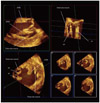
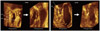
In the setting of submitral-targeted surgery such as papillary heads optimization, a detailed understanding of the submitral apparatus is essential for successful repair and for evaluating results of surgery. Adequate imaging is necessary for precise quantitative evaluation of the mitral valve structure. Three dimensional TEE has become widely available recently and it plays an essential role in understanding the status of the mitral valve apparatus. However, the conventional trans-left atrial view sometimes fails to visualize the subvalvular apparatus. On such occasion, transgastric approach should be employed.46) Transgastric view provides optimal image for quantitative analysis of the submitral apparatus (Fig. 6). A typical case which underwent papillary head optimization procedure is shown in Fig. 7. PM heads in each PM are clearly visualized. With the aid of quantitative software Real View (YD, Nara, Japan), these images allows accurate measurement of the pre and postoperative distance between PM head and mid-anterior annulus and the tenting volume, etc (Fig. 8). In this particular patient, the distance between posterior PM head for anterior leaflet and mid-anterior annulus were shorten from 26.5 mm to 23.5 mm during the surgery. On the other hand the distance between anterior PM head for anterior leaflet and mid-anterior annulus only changed from 24.1 mm to 24.0 mm. The distances between the mid anterior annulus and the both PM head for posterior leaflet were markedly reduced. Although MR was completely controlled in this patient, the colorized postoperative mitral leaflet by a Real View includes the red part in the lateral site, indicating slight residual tethering of leaflet in lateral site. Such information will assist further improvement of the quality of the surgery.

Summary

Three dimensional echocardiography plays an essential role to understand the geometry of mitral valve complex, including PM, PM head division, chordae tendineae, leaflets and annulus and contributes greatly to decision making of the surgical strategy in functional MR and its postoperative assessment.

Figures and Tables

Fig. 1
Mitral valve complex. The whole mitral valve complex geometry consisted of annulus, leaflets, chordae, papillary muscles and surrounding left ventricle. Mitral annulus shows a nonplanar saddle-shaped configuration in healthy individual. There are two peaks at the aortic insertion and posterior left ventricular wall and two low troughs closest to the apex located medially and laterally (Otto1) and Levine et al.2) with permission). LA: left atrium, LV: left ventricle.
jcu-20-69-g001
Fig. 2
Mitral annulus configuration. Three dimensional echocardiography allowed us to visualize the dilatation and flattening of the mitral annulus in patients with ischemic mitral regurgitation and more deformation in anterior infarction compared to posterior infarction (Watanabe et al.23) with permission). MI: myocardial infarction.
jcu-20-69-g002
Fig. 3
Visualizing the annular flattening and leaflet tenting. The reconstructed three-dimensional (3D) images by Real View allow us to understand the annular flattening or mitral leaflet tenting in patients with functional mitral regurgitation (MR) and it also can be quantitatively measured and analyzed in 3D spaces, which is impossible to do in two-dimensional images (Watanabe et al.22) with permission).
jcu-20-69-g003
Fig. 4
Surgical procedures for subvalvular apparati or left ventricle. Undersized annuloplasty sometimes fails to ameliorate mitral regurgitation (MR) in patient with severely tethered mitral leaflet and worsen the tethering of posterior leaflet, resulting recurrent MR. In addition, undersized annuloplasty has a possibility to induce functional mitral stenosis. Therefore, additive procedures should be considered for patients with severely tethered valve. PM: papillary muscle.
jcu-20-69-g004
Fig. 5
Papillary heads optimization method. In Papillary heads optimization method, the papillary muscle (PM) head for posterior leaflet was connected to the PM head for anterior leaflet by using a PTFE suture in each PM. After annuloplasty by a semi-rigid ring, its arms were re-suspended towards the mid-anterior mitral annulus and the sutures were passed through it and tied after the length adjustment. PTFE: polytetrafluoroethylene.
jcu-20-69-g005
Fig. 6
Visualizing the submitral structure by transgastric approach. Visualizing submitral structure at mid systole from a three-dimensional dataset acquired by transgastric approach using QLAB (Philips Medical Systems, Andver, MA, USA), with coronal section (commissure-commissure direction) view (upper left), sagittal section (anterior-posterior leaflet direction) view of posterior papillary muscle (PPM) (upper right) and short axis view (lower left and right). APM: anterior papillary muscle, pp: PPM head for posterior leaflet, pa: PPM head for anterior leaflet.
jcu-20-69-g006
Fig. 7
Pre- and postoperative observation of papillary heads optimization method. Postoperative connected papillary muscle head for anterior leaflet and for posterior leaflet of each papillary muscle by papillary heads optimization procedure is clearly visualized by three-dimensional transesophageal echocardiography with transgastric approach. PPM: posterior papillary muscle, APM: anterior papillary muscle, pp: PPM head for posterior leaflet, pa: PPM head for anterior leaflet, aa: APM head for anterior leaflet, ap: APM head for posterior leaflet.
jcu-20-69-g007
Fig. 8
Quantitative analysis of submitral structure. With the aid of quantitative software, acquired three-dimensional volume dataset allows accurate measurement of the pre and postoperative distance between papillary muscle head and mid-anterior annulus and the tenting volume, etc. Vo: mid-anterior annulus point, pp: posterior papillary muscle (PPM) head for posterior leaflet, pa: PPM head for anterior leaflet, aa: anterior papillary muscle (APM) head for anterior leaflet, ap: APM head for posterior leaflet, PM: papillary muscle.
jcu-20-69-g008

References

1. Otto CM. Textbook of clinical echocardiography. 2009. 4th ed. Philadelphia: Saunders/Elsevier.
2. Levine RA, Weyman AE, Handschumacher MD. Three-dimensional echocardiography: techniques and applications. Am J Cardiol. 1992. 69:121H–130H. discussion 131H-4H.
crossref
3. He S, Fontaine AA, Schwammenthal E, Yoganathan AP, Levine RA. Integrated mechanism for functional mitral regurgitation: leaflet restriction versus coapting force: in vitro studies. Circulation. 1997. 96:1826–1834.
crossref
4. Yiu SF, Enriquez-Sarano M, Tribouilloy C, Seward JB, Tajik AJ. Determinants of the degree of functional mitral regurgitation in patients with systolic left ventricular dysfunction: a quantitative clinical study. Circulation. 2000. 102:1400–1406.
crossref
5. Bursi F, Enriquez-Sarano M, Nkomo VT, Jacobsen SJ, Weston SA, Meverden RA, Roger VL. Heart failure and death after myocardial infarction in the community: the emerging role of mitral regurgitation. Circulation. 2005. 111:295–301.
crossref
6. Grigioni F, Enriquez-Sarano M, Zehr KJ, Bailey KR, Tajik AJ. Ischemic mitral regurgitation: long-term outcome and prognostic implications with quantitative Doppler assessment. Circulation. 2001. 103:1759–1764.
crossref
7. Bursi F, Barbieri A, Grigioni F, Reggianini L, Zanasi V, Leuzzi C, Ricci C, Piovaccari G, Branzi A, Modena MG. Prognostic implications of functional mitral regurgitation according to the severity of the underlying chronic heart failure: a long-term outcome study. Eur J Heart Fail. 2010. 12:382–388.
crossref
8. Kumanohoso T, Otsuji Y, Yoshifuku S, Matsukida K, Koriyama C, Kisanuki A, Minagoe S, Levine RA, Tei C. Mechanism of higher incidence of ischemic mitral regurgitation in patients with inferior myocardial infarction: quantitative analysis of left ventricular and mitral valve geometry in 103 patients with prior myocardial infarction. J Thorac Cardiovasc Surg. 2003. 125:135–143.
crossref
9. Veronesi F, Corsi C, Sugeng L, Caiani EG, Weinert L, Mor-Avi V, Cerutti S, Lamberti C, Lang RM. Quantification of mitral apparatus dynamics in functional and ischemic mitral regurgitation using real-time 3-dimensional echocardiography. J Am Soc Echocardiogr. 2008. 21:347–354.
crossref
10. Tsukiji M, Watanabe N, Yamaura Y, Okahashi N, Obase K, Neishi Y, Toyota E, Kawamoto T, Okura H, Ogasawara Y, Yoshida K. Three-dimensional quantitation of mitral valve coaptation by a novel software system with transthoracic real-time three-dimensional echocardiography. J Am Soc Echocardiogr. 2008. 21:43–46.
crossref
11. Song JM, Fukuda S, Kihara T, Shin MS, Garcia MJ, Thomas JD, Shiota T. Value of mitral valve tenting volume determined by real-time three-dimensional echocardiography in patients with functional mitral regurgitation. Am J Cardiol. 2006. 98:1088–1093.
crossref
12. Watanabe N, Ogasawara Y, Yamaura Y, Yamamoto K, Wada N, Kawamoto T, Toyota E, Akasaka T, Yoshida K. Geometric differences of the mitral valve tenting between anterior and inferior myocardial infarction with significant ischemic mitral regurgitation: quantitation by novel software system with transthoracic real-time three-dimensional echocardiography. J Am Soc Echocardiogr. 2006. 19:71–75.
crossref
13. Daimon M, Saracino G, Gillinov AM, Koyama Y, Fukuda S, Kwan J, Song JM, Kongsaerepong V, Agler DA, Thomas JD, Shiota T. Local dysfunction and asymmetrical deformation of mitral annular geometry in ischemic mitral regurgitation: a novel computerized 3D echocardiographic analysis. Echocardiography. 2008. 25:414–423.
crossref
14. Kim K, Kaji S, An Y, Yoshitani H, Takeuchi M, Levine RA, Otsuji Y, Furukawa Y. Mechanism of asymmetric leaflet tethering in ischemic mitral regurgitation: 3D analysis with multislice CT. JACC Cardiovasc Imaging. 2012. 5:230–232.
crossref
15. Saito K, Okura H, Watanabe N, Obase K, Tamada T, Koyama T, Hayashida A, Neishi Y, Kawamoto T, Yoshida K. Influence of chronic tethering of the mitral valve on mitral leaflet size and coaptation in functional mitral regurgitation. JACC Cardiovasc Imaging. 2012. 5:337–345.
crossref
16. Ormiston JA, Shah PM, Tei C, Wong M. Size and motion of the mitral valve annulus in man. I. A two-dimensional echocardiographic method and findings in normal subjects. Circulation. 1981. 64:113–120.
crossref
17. Watanabe N, Ogasawara Y, Yamaura Y, Kawamoto T, Akasaka T, Yoshida K. Geometric deformity of the mitral annulus in patients with ischemic mitral regurgitation: a real-time three-dimensional echocardiographic study. J Heart Valve Dis. 2005. 14:447–452.
18. Levine RA, Handschumacher MD, Sanfilippo AJ, Hagege AA, Harrigan P, Marshall JE, Weyman AE. Three-dimensional echocardiographic reconstruction of the mitral valve, with implications for the diagnosis of mitral valve prolapse. Circulation. 1989. 80:589–598.
crossref
19. Kaplan SR, Bashein G, Sheehan FH, Legget ME, Munt B, Li XN, Sivarajan M, Bolson EL, Zeppa M, Arch MZ, Martin RW. Three-dimensional echocardiographic assessment of annular shape changes in the normal and regurgitant mitral valve. Am Heart J. 2000. 139:378–387.
crossref
20. Salgo IS, Gorman JH 3rd, Gorman RC, Jackson BM, Bowen FW, Plappert T, St John Sutton MG, Edmunds LH Jr. Effect of annular shape on leaflet curvature in reducing mitral leaflet stress. Circulation. 2002. 106:711–717.
crossref
21. Kwan J, Qin JX, Popović ZB, Agler DA, Thomas JD, Shiota T. Geometric changes of mitral annulus assessed by real-time 3-dimensional echocardiography: becoming enlarged and less nonplanar in the anteroposterior direction during systole in proportion to global left ventricular systolic function. J Am Soc Echocardiogr. 2004. 17:1179–1184.
crossref
22. Watanabe N, Ogasawara Y, Yamaura Y, Kawamoto T, Toyota E, Akasaka T, Yoshida K. Quantitation of mitral valve tenting in ischemic mitral regurgitation by transthoracic real-time three-dimensional echocardiography. J Am Coll Cardiol. 2005. 45:763–769.
crossref
23. Watanabe N, Ogasawara Y, Yamaura Y, Wada N, Kawamoto T, Toyota E, Akasaka T, Yoshida K. Mitral annulus flattens in ischemic mitral regurgitation: geometric differences between inferior and anterior myocardial infarction: a real-time 3-dimensional echocardiographic study. Circulation. 2005. 112:9 Suppl. I458–I462.
24. Vahanian A, Baumgartner H, Bax J, Butchart E, Dion R, Filippatos G, Flachskampf F, Hall R, Iung B, Kasprzak J, Nataf P, Tornos P, Torracca L, Wenink A;. Guidelines on the management of valvular heart disease: The Task Force on the Management of Valvular Heart Disease of the European Society of Cardiology. Eur Heart J. 2007. 28:230–268.
crossref
25. Rankin JS, Feneley MP, Hickey MS, Muhlbaier LH, Wechsler AS, Floyd RD, Reves JG, Skelton TN, Califf RM, Lowe JE, et al. A clinical comparison of mitral valve repair versus valve replacement in ischemic mitral regurgitation. J Thorac Cardiovasc Surg. 1988. 95:165–177.
crossref
26. Cohn LH, Rizzo RJ, Adams DH, Couper GS, Sullivan TE, Collins JJ Jr, Aranki SF. The effect of pathophysiology on the surgical treatment of ischemic mitral regurgitation: operative and late risks of repair versus replacement. Eur J Cardiothorac Surg. 1995. 9:568–574.
crossref
27. Al-Radi OO, Austin PC, Tu JV, David TE, Yau TM. Mitral repair versus replacement for ischemic mitral regurgitation. Ann Thorac Surg. 2005. 79:1260–1267.
crossref
28. Chan V, Ruel M, Mesana TG. Mitral valve replacement is a viable alternative to mitral valve repair for ischemic mitral regurgitation: a case-matched study. Ann Thorac Surg. 2011. 92:1358–1365.
crossref
29. Perrault LP, Moskowitz AJ, Kron IL, Acker MA, Miller MA, Horvath KA, Thourani VH, Argenziano M, D'Alessandro DA, Blackstone EH, Moy CS, Mathew JP, Hung J, Gardner TJ, Parides MK. Optimal surgical management of severe ischemic mitral regurgitation: to repair or to replace? J Thorac Cardiovasc Surg. 2012. 143:1396–1403.
crossref
30. Bolling SF, Pagani FD, Deeb GM, Bach DS. Intermediate-term outcome of mitral reconstruction in cardiomyopathy. J Thorac Cardiovasc Surg. 1998. 115:381–386.
crossref
31. Bolling SF, Deeb GM, Bach DS. Mitral valve reconstruction in elderly, ischemic patients. Chest. 1996. 109:35–40.
crossref
32. Calafiore AM, Gallina S, Di Mauro M, Gaeta F, Iacò AL, D'Alessandro S, Mazzei V, Di Giammarco G. Mitral valve procedure in dilated cardiomyopathy: repair or replacement? Ann Thorac Surg. 2001. 71:1146–1152.
crossref
33. Zhu F, Otsuji Y, Yotsumoto G, Yuasa T, Ueno T, Yu B, Koriyama C, Hamasaki S, Biro S, Kisanuki A, Minagoe S, Levine RA, Sakata R, Tei C. Mechanism of persistent ischemic mitral regurgitation after annuloplasty: importance of augmented posterior mitral leaflet tethering. Circulation. 2005. 112:9 Suppl. I396–I401.
34. Gelsomino S, Lorusso R, Caciolli S, Capecchi I, Rostagno C, Chioccioli M, De Cicco G, Billè G, Stefàno P, Gensini GF. Insights on left ventricular and valvular mechanisms of recurrent ischemic mitral regurgitation after restrictive annuloplasty and coronary artery bypass grafting. J Thorac Cardiovasc Surg. 2008. 136:507–518.
crossref
35. Troubil M, Marcian P, Gwozdziewicz M, Santavy P, Langova K, Nemec P, Lonsky V. Predictors of failure following restrictive annuloplasty for chronic ischemic mitral regurgitation. J Card Surg. 2012. 27:6–12.
crossref
36. Gelsomino S, van Garsse L, Lucà F, Lorusso R, Cheriex E, Rao CM, Caciolli S, Vizzardi E, Crudeli E, Stefàno P, Gensini GF, Maessen J. Impact of preoperative anterior leaflet tethering on the recurrence of ischemic mitral regurgitation and the lack of left ventricular reverse remodeling after restrictive annuloplasty. J Am Soc Echocardiogr. 2011. 24:1365–1375.
crossref
37. Magne J, Sénéchal M, Mathieu P, Dumesnil JG, Dagenais F, Pibarot P. Restrictive annuloplasty for ischemic mitral regurgitation may induce functional mitral stenosis. J Am Coll Cardiol. 2008. 51:1692–1701.
crossref
38. Kubota K, Otsuji Y, Ueno T, Koriyama C, Levine RA, Sakata R, Tei C. Functional mitral stenosis after surgical annuloplasty for ischemic mitral regurgitation: importance of subvalvular tethering in the mechanism and dynamic deterioration during exertion. J Thorac Cardiovasc Surg. 2010. 140:617–623.
crossref
39. Kainuma S, Taniguchi K, Daimon T, Sakaguchi T, Funatsu T, Kondoh H, Miyagawa S, Takeda K, Shudo Y, Masai T, Fujita S, Nishino M, Sawa Y. Osaka Cardiovascular Surgery Research (OSCAR) Group. Does stringent restrictive annuloplasty for functional mitral regurgitation cause functional mitral stenosis and pulmonary hypertension? Circulation. 2011. 124:11 Suppl. S97–S106.
crossref
40. Kron IL, Green GR, Cope JT. Surgical relocation of the posterior papillary muscle in chronic ischemic mitral regurgitation. Ann Thorac Surg. 2002. 74:600–601.
crossref
41. Hvass U, Tapia M, Baron F, Pouzet B, Shafy A. Papillary muscle sling: a new functional approach to mitral repair in patients with ischemic left ventricular dysfunction and functional mitral regurgitation. Ann Thorac Surg. 2003. 75:809–811.
crossref
42. Hvass U, Joudinaud T. The papillary muscle sling for ischemic mitral regurgitation. J Thorac Cardiovasc Surg. 2010. 139:418–423.
crossref
43. Matsui Y, Suto Y, Shimura S, Fukada Y, Naito Y, Yasuda K, Sasaki S. Impact of papillary muscles approximation on the adequacy of mitral coaptation in functional mitral regurgitation due to dilated cardiomyopathy. Ann Thorac Cardiovasc Surg. 2005. 11:164–171.
44. Masuyama S, Marui A, Shimamoto T, Komeda M. Chordal translocation: secondary chordal cutting in conjunction with artificial chordae for preserving valvular-ventricular interaction in the treatment of functional mitral regurgitation. J Heart Valve Dis. 2009. 18:142–146.
45. Fattouch K, Murana G, Castrovinci S, Mossuto C, Sampognaro R, Borruso MG, Bertolino EC, Caccamo G, Ruvolo G, Lancellotti P. Mitral valve annuloplasty and papillary muscle relocation oriented by 3-dimensional transesophageal echocardiography for severe functional mitral regurgitation. J Thorac Cardiovasc Surg. 2012. 143:4 Suppl. S38–S42.
crossref
46. Obase K, Komeda M, Okura H, Yoshida K. A new echocardiographic window to visualize the mitral valve complex during mitral valve repair for functional mitral regurgitation. J Thorac Cardiovasc Surg. 2012. 143:e42–e44.
crossref
TOOLS
Similar articles