

 PDF
PDF ePub
ePub Citation
 Print
Print


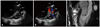
A 23-year-old basketball player was referred to the cardiology examination because of recurrent transient ischemic attacks (TIA). The patient was asymptomatic, with no history of cardiovascular risk factors or previous heart disease. Clinical and electrocardiography parameters were unremarkable. The echocardiography showed a sack-like aneurysm of the membranous ventricular septum (AMS), approximately 15 × 9 mm in size, protruding into the right ventricle (Fig. 1A). Color Doppler revealed blood flow directly from the left ventricular cavity into the AMS through ventricular septal defect (VSD), approximately 2-3 mm in diameter (Fig. 1B). There were no signs of right ventricular outflow tract obstruction, infection or thrombosis. A spontaneous echo-contrast in the AMS was detected, suggesting that AMS is the most likely source of emboli responsible for recurrent TIA.1) Cardiac magnetic resonance confirmed these echocardiography findings (Fig. 1C). Complete resection of AMS and closure of VSD were done by a pericardial patch. Two years after the surgical repair, no other neurological event has occurred.
AMS develops as a consequence of partial or complete spontaneous closure of a VSD, during various periods from the neonatal stage to 6 years of age.2) Large, hemodynamically significant, membranous VSD in infancy progresses to a functionally smaller defect with aneurysm formation later in the childhood.2) In most of the cases, formed aneurysms completely close VSD. Due to that reason, AMS is generally asymptomatic, and clinical examinations would not detect its presence.2) Therefore, AMS is the most frequently detected accidentally in adult patients during echocardiography, which is generally the only method needed for definite diagnosis. Computed tomography or cardiac magnetic resonance imaging can help delineate the extent of the AMS, its relationships to surrounding cardiac structures, and AMS thrombosis or inflammation. Clinical importance of AMS stems from potentially severe or fatal complications (e.g. tricuspid or aortic valve insufficiency, right ventricular outflow tract obstruction, rupture, thromboembolism, infectious endocarditis).3) Therefore, AMS should be operated soon after diagnosis, even in asymptomatic patients.4) The only recommended surgical option is complete AMS resection and VSD closure with a patch.4)
 XML Download