

 PDF
PDF ePub
ePub Citation
 Print
Print


Introduction
Persistent left superior vena cava (PLSVC) is one of the most common venous anormaly of the thorax. However, the incidence is in less than 0.5% in the general population but occurs in approximately 4% of patients with congenital heart disease.1) Sinus venosus atrial septal defect (SVD) accounts for 4-11% of all atrial septal defect (ASD) and partial anomalous pulmonary venous connection (PAPVC) is present in about 90% of patients with SVD. Anomalies of systemic venous connection are usually found incidentally in asymptomatic patients, but their identification is sometimes crucial especially when cardiac or abdominal aorta surgery is planned. We present a case of a complex anomaly of systemic and pulmonary venous return associated with SVD which was detected by computed tomography (CT) pulmonary angiography and echocardiography. Modern imaging techniques are not only useful for detecting congenital cardiac defects but also for detecting systemic and pulmonary venous connection anomalies. This case demonstrates the importance of knowledge of the anatomical basis of venous connection and identification of one lesion should prompt an appropriate investigation of other potential anomalies that may be less obvious.
Case
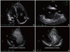
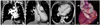
A 41-year-old man was admitted to the hospital because of palpitation and exertional dyspnea. On auscultation, fixed splitting of the second heart sound and a systolic murmur over the pulmonary valve area were heard. Electrocardiogram on admission showed paroxysmal supraventricular tachycardia. A chest X-ray demonstrated mild cardiac enlargement. Transthoracic echocardiography (TTE) showed enlargement of the right ventricle and an extremely dilated coronary sinus with normal function of both ventricles (Fig. 1A). On the second day of admission, we performed a CT pulmonary angiography. The study showed a PLSVC draining into the right atrium (Fig. 2A). CT also revealed an ASD with anomalous return of the right superior pulmonary vein to the right superior vena cava (SVC) (Fig. 2B and C). Transesophageal echocardiography (TEE) also demonstrated a SVD (Fig. 1B). Simultaneously, contrast echocardiography was performed. A right arm peripheral vein was injected with agitated saline, and normal opacification was noted in the right atrium, followed by opacification of the right ventricle (Fig. 1C). Next, the left arm peripheral vein was injected with agitated saline and abnormal opacification of the coronary sinus was first noted, followed by opacification of the right atrium and then right ventricle (Fig. 1D). On the third day of admission, coronary CT angiography was performed. Coronary CT angiography with 3-dimensional reconstruction demonstrated a more detailed structure of PLSVC drainage into the right atrium via a dilated coronary sinus (Fig. 2D). The patient underwent surgical correction with the double-patch technique. Five months later, follow-up transthoracic echocardiography showed near-normalization of the right ventricular and coronary sinus size.
Discussion
PLSVC occurs in approximately 0.3-0.5% of the general population and characteristically drains into the coronary sinus. During and after embryonic development of SVC, SVC develops on the right side from a portion of the right anterior cardinal vein. On the left side, part of the left anterior cardinal vein undergo normal regression to form the ligament of the left vena cava.1) PLSVC results from the persistence of the left anterior cardinal vein. Usually, PLSVC is asymptomatic and discovered incidentally during imaging study and pacemaker implantation or central catheterization but sometimes their elucidation is crucial especially during cardiovascular surgery.2) PLSVC should be considered whenever a dilated coronary sinus is identified at echocardiography and the diagnosis could be confirmed by saline contrast echocardiography.3) Other modern imaging modalities such as CT or magnetic resonance imaging (MRI) can be used to confirm the diagnosis. In our case, we could not consider the presence of PLSVC before performing CT pulmonary angiography just because of the focus on volume overload of right-sided heart chambers. We performed a contrast echocardiography based on the information obtained from CT pulmonary angiography.
In our case, PAPVC associated with SVD which is a rarer anomaly than PLSVC was also found. PAPVC is frequently associated with congenital heart disease such as an ASD. It is estimated that 10-15% of patients have an ASD and approximately 85% of PAPVC are associated with SVD.4)5) Usually, the diagnosis of PAPVC can be made by echocardiography, and cardiac catheterization along with angiography is often performed for confirmation of the diagnosis. Nowadays, CT, MRI and TEE with contrast examination are considered as sensitive methods for the detection of PAPVC.6) In our case, the diagnosis of PAPVC was missed on routine TTE, and a definitive diagnosis of PAPVC could be made on CT pulmonary angiography.
Some authors have previously reported a combined anomaly of systemic and pulmonary venous return associated with SVD.7)8) These authors especially emphasized the importance of new imaging modalities in diagnosing complex anomaly of systemic and pulmonary venous return associated with SVD. Also in our case, CT made a definitive diagnosis of the anomaly and provided more detailed structural information. However, this case report also illustrated that careful echocardiographic examination should be performed using several windows and even contrast for diagnosing the anomalies of systemic and pulmonary venous return combined with congenital heart disease. And consideration is required when the case has accompanying cardiac abnormalities besides PLSVC such as an ASD, when coronary sinus is extremely dilated and it is coexisting with enlargement of right ventricle.
 XML Download