Journal List > J Cardiovasc Ultrasound > v.20(1) > 1018256

Cacciapuoti, Scognamiglio, Paoli, Romano, and Cacciapuoti: Left Atrial Volume Index as Indicator of Left Ventricular Diastolic Dysfunction: Comparation between Left Atrial Volume Index and Tissue Myocardial Performance Index

Abstract

Background

To point out a possible correlation between left atrial volume index (LAVI) and left ventricular (LV) diastolic time interval to better define LV diastolic dysfunction, this study was performed.

Methods

In 62 hypertensive-hypertrophic patients without LV systolic dysfunction, LV volumes, myocardial mass index, ejection fraction% (EF%) and LAVI were measured by two-dimensional echocardiography. Instead, tissue Doppler echocardiography (TDE) was used to measure myocardial performance index (MPI) and its systo-diastolic time intervals, such as: iso-volumetric contraction time (IVCT); iso-volumetric relaxation time (IVRT); ejection time. LAVI, TDE-MPI and time intervals where also measured in 15 healthy controls, to obtain the reference values.

Results

Results shown a significant increase of LV volumes in hypertensives in comparison to the control group (p < 0.05). LV mass index also augmented (p < 0.001). Instead, EF% not significantly changed in hypertrophic patients in comparison with healthy controls. LAVI raised in hypertensives wih left ventricular hypertrophy, whereas IVCT resulted within the normal limits. On the contrary, IVRT significantly raised. Accordingly, MPI resulted higher in controls.

Conclusion

LAVI, MPI and its time intervals appear as reliable tools to non-invasively individualize LV diastolic dysfunction in systemic hypertension, in absence of mitral valve disease.

Introduction

The increased left ventricular diastolic filling pressure evolves in left ventricular (LV) diastolic dysfunction.1)2) This haemodynamic condition usually is demonstrated by the impairment of E/A mitral inflow ratio (E/A < 1) or by the change of normal pattern of pulmonary veins' flow. The combination of early inflow velocity curve and tissue Doppler imaging of the mitral annulus (E/E' ratio) better estimates this condition. But, in the absence of any mitral valve derangement, LV diastolic dysfunction directly affects Left Atrial Volume (LAV). This parameter can be easily measured by two-dimensional echocardiography and indexed to the body surface area (BSA) as left atrial volume index (LAVI).3)4) Therefore, LAVI also may be used as faithful indicator of LV diastolic dysfunction.5) On the other hand, LV function can be adequately evaluated by myocardial performance index (MPI).6)7) This (also called Tei index) can be measured either with conventional Doppler method or tissue Doppler echocardiography (TDE).8) This last method has the advantage to directly assess transmural myocardial velocities.9) In addition, TDE-MPI is more sensitive than the conventional Doppler MPI in to define LV function, especially in the presence of regional wall motion abnormality.9)
In this study, we evaluated the relationship between LAVI and diastolic LV function defined with TDE-MPI in a group of hypertensive patients with LV diastolic dysfunction and ejection fraction% (EF%) > 50%.

Methods

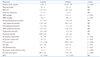
Since October 2009 to February 2011, 62 hypertensive patients (43 males and 29 females) aged from 45 to 61 years (mean age = 55 ± 6 years) and without any valvular heart diseases were examined. The leading epidemiological, metabolic and echocardiographic characteristics of controls and hypertensive patients (group II) were shown in Table 1. These were in sinus rhythm and have an echocardiographic finding of left ventricular hypertrophy (LVH).10) Coronary artery disease was excluded by coronary angiography in 24 of these, and by rest and effort myocardial SPECT in the remaining 38. Cumulative anti-hypertensive treatments given in patients of group II were shown in Table 2. In accordance with the recommendations for the evaluation of LV function by echocardiography,11) the patients were diagnosed as affected by LV diastolic dysfunction, with EF% > 50% (group II).12)
Fifteen (8 males and 7 females) healthy subjects (M and F; mean age = 54 ± 3 years) was also enrolled, as controls. In these, echocardiographic left ventricular function was defined too (group I).

Conventional and tissue Doppler echocardiography

Two groups were echocardiographically examined in the left-lateral position by using a iE33 machine (Philips, Amsterdam, the Netherlands). According to the biplane Simpson's rule, LV end-diastolic volume (LVEDV) and LV end-systolic volume (LVESV) in mL and EF% were defined.13) Inter ventricular septum (IVS) thickness was measured (in mm) during systole. LV wall thickness was also measured (in mm). Left ventricular mass and its indexed value was assessed by the method proposed by Devereux et al.10) Left atrial (LA) volume in systole was also measured just before the mitral valve opening, using the biplane Simpson's method, as a mean between the values recorded in apical four- and two-chamber approaches. Subsequently, LAV was indexed for BSA, such as LAVI in mL/m2.14) Finally, MPI was evaluated by using TDE method. Pulsed-wave TDE was performed by activating the tissue Doppler function. Sample volume was placed at the lateral annular mitral site in apical four chamber view, in order to record the following cardiac time intervals: iso-volumetric contraction time (IVCT) in ms; iso-volumetric relaxation time (IVRT) in ms; and ejection time (ET) in ms. Images were acquired with a variable frequency phased-array transducer. The filter settings were kept low, and gains were adjusted to the minimal optimal level to minimize noise and eliminate the signals produced by the transmitral flow. Three consecutive beats were measured and averaged for each parameter at a sweep speed of 100 mm/s. MPI was defined as the sum of IVCT and IVRT divided by ET.15) LAVI; MPI; IVCT; IVRT; and ET were defined in controls (group I) too, as reference values (Table 3).

Statistical analysis

Echocardiographic data are presented as a mean values ± SD. Statistical analyses were performed with SPSS statistical software (SPSS, Chicago, IL, USA). Differences between two groups were examined by an unpaired t test. A p value < 0.05 was considered significant. Finally, LAVI was compared to MPI, IVCT, IVRT and ET between controls (group I) and hypertensive patients (group II) by unpaired t-test.

Results

Mean values of LVEDV and LVESV were 95 ± 18 mL and 39 ± 17 mL respectively in controls (group I). These resulted 125 ± 15 mL (LVEDV), and 48 ± 11 mL (LVESV) in hypertensive patients (group II). Differences were significant (p < 0.05). IVS thickness was = 10 ± 0.4 mm in controls, it resulted 135 ± 0.5 mm in hypertensive-patients. Mean value of E/A waves ratio was 1.21 ± 0.38 in normal and 0.85 ± 0.27 in hypertensives (p < 0.01). Mitral deceleration time (DT) resulted = 135 ± 3.4 ms in healthy adults and 245 ± 31 in hypertensive patients (p < 0.01). On the other hand, LV walls' thickness was 9 ± 0.3 mm in controls (group I) and 17 ± 0.2 mm in hypertensives (group II). Differences were significant (p < 0.001). The mean value of LV mass index was 90 ± 21 g/m2 in control group. It increased to 178 ± 29 g/m2 in hypertensive group (p < 0.001). EF% resulted of 61 ± 0.8% in controls (group I) and of 57 ± 0.9% in hypertrophic patients (group II). Differences between two groups weren't significant (NS) (Table 1).
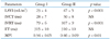
With reference to LAVI, a mean of 47 ± 5 mL/m2 was found in hypertensive-hypertrophic patients (group II). This value was significantly higher (p < 0.001) than that recorded in controls (group I) (23 ± 4 mL/m2). Normal values of TDE-MPI (0.34 ± 0.05) obtained in control-group significantly increased (p < 0.01) in patients with LV hypertrophy (0.46 ± 0.09). Particularly, IVCT resulted 28 ± 7 ms in healthy individuals, almost similar to that obtained in hypertensive patients (30 ± 8 ms), without significant differences (NS). On the contrary, IVRT was significantly (p < 0.001) prolonged (107 ± 9 ms) in hypertensives in comparison to healthy subjects (79 ± 6 ms). ET was within the limits both in normals (315 ± 10 ms) and in hypertensive patients (312 ± 10 ms) (NS) (Table 3).

Discussion

LAV may be calculated by three different methods: the biplane area lengh; the biplane modified Simpson's, and the prolate ellipse method.16) Significant differences among three diverse methods exist, even through all three shown highly satisfactory reproducibility. In this study, we used biplane Simpson's method indexed for BSA, to obtain LAVI mesaured in mL/m2. Mean value of LAVI reported by several AA is 22 ± 6 mL/m2.17-21) In our healthy controls, a mean value of 23 ± 4 mL/m2 was found. This was reported as reference value for our laboratory.
It is known that mechanical function of LA has described in three phases: reservoir; conduit, and contractile phase. The "reservoir" corresponds to the difference between maximal and minimum LA volumes occurring in the interval-just before the opening mitral valve and just before the aortic valve opening. "Conduit" is the early phase of ventricular diastole. The blood is passively transferred to left ventricle just after mitral valve opening. "Contractile" phase or "booster pump" is calculated as the difference between minimum and pre-atrial contraction. It serves to augment the stroke volume. The contribution of three phases of LA function changes according to the diastolic properties of LV. In normal conditions, the contribution of reservoir, conduit and contractile function of the LA to the LV filling is 40%, 35%, and 25% respectively. As LV relaxation worsens, the contribution of different LA phases gradually increases,22) in accordance with recent experiences performed in patients with LV diastolic dysfunction.23)24)
In the present study, we evaluated the relationship between LAVI and LV diastolic dysfunction due to LV hypertrophy. LV diastolic impairment was demonstrated by the increase of IVRT and TDE-MPI. Achieved results indicate that LAVI significantly raised in comparison to the contol values in hypertensives with LV hypertrophy. This faithfullly certainly reflects LV diastolic dyfunction consequent to LVH. The increase of IVRT and TDE-MPI (with normal values of IVCT) can be considered as an useful and reliable tool to identify LV diastolic LV dysfunction.25)26) Several research groups previously have shown that MPI and IVRT reflect LV diastolic dysfunction, independently of arterial pressure,27) heart failure28) or heart rate,29) in presence of preserved systolic function especially.30) A previous study has also demonstrated an association between LVH induced by systemic hypertension and left atrial dimension.31) Successively, Pritchett et al.32) evidenced that LAVI is a highly sensitive and specific tool for the detection of severe LV diastolic dysfunction (III degree of diastolic dysfunction). These AAs. = Authors also demonstrated that LAVI may better reflect the cumulative effect of increased LV filling pressures over time in comparison to the Doppler indexes, as E/A ratio, DT and E/E' ratio (that reflect increased LV filling pressures at one point in time). The incremental value of LAVI measurement is its prognostic implications towards cardiovascular death and/or adverse cardiovascular outcomes in hypertensive patients with LV diastolic dysfunction, as recently demonstrated by Leung et al.33)
In the present report, we firstly identified LV diastolic dysfunction using TDE-MPI. LAVI (in the absence of any mitral disease) appeared also expressive of LV diastolic dysfunction, further confirming the relationship between LAV and LV diastolic dysfunction. But, other studies performed in a wide range are requested to definitively demonstrate the relationship among LAVI, TDE-MPI and LV diastolic dysfunction.

Figures and Tables

Table 1
Epidemiological, metabolic, and echocardiographic characteristics of controls and enrolled patients
jcu-20-25-i001

Leading epidemiological, biochemical and echocardiographic characteristics of healthy controls and hypertensive-hypertrophic patients, with statistical significance. NS: not significant, BSA: body surface area, SBP: systolic blood pressure, DBP: diastolic blood pressure, LV: left ventricle, IVS: inter ventricular septum, EF%: ejection fraction%

Table 2
Cumulative anti-hypertensive drugs given in 62 hypertensives
jcu-20-25-i002
Table 3
LAVI, time intervals and MPI-TDE values in two groups
jcu-20-25-i003

Doppler results of LAVI, MPI and cardiac time intervals (IVCT, IVRT, ET) recorded in healthy controls and hypertensive patients. LAVI: left atrial volume index, IVCT: iso-volumetric contraction time, NS: not significant, IVRT: iso-volumetric relaxation time, ET: ejection time, MPI: myocardial performance index, TDE: tissue Doppler echocardiography

References

1. Appleton CP, Galloway JM, Gonzalez MS, Gaballa M, Basnight MA. Estimation of left ventricular filling pressures using two-dimensional and Doppler echocardiography in adult patients with cardiac disease. Additional value of analyzing left atrial size, left atrial ejection fraction and the difference in duration of pulmonary venous and mitral flow velocity at atrial contraction. J Am Coll Cardiol. 1993. 22:1972–1982.
crossref
2. Simek CL, Feldman MD, Haber HL, Wu CC, Jayaweera AR, Kaul S. Relationship between left ventricular wall thickness and left atrial size: comparison with other measures of diastolic function. J Am Soc Echocardiogr. 1995. 8:37–47.
crossref
3. Schabelman S, Schiller NB, Silverman NH, Ports TA. Left atrial volume estimation by two-dimensional echocardiography. Cathet Cardiovasc Diagn. 1981. 7:165–178.
crossref
4. Schiller NB, Botvinick EH. Noninvasive quantitation of the left heart by echocardiography and scintigraphy. Cardiovasc Clin. 1986. 17:45–93.
5. Moller JE, Hillis GS, Oh JK, Seward JB, Reeder GS, Wright RS, Park SW, Bailey KR, Pellikka PA. Left atrial volume: a powerful predictor of survival after acute myocardial infarction. Circulation. 2003. 107:2207–2212.
crossref
6. Tei C, Ling LH, Hodge DO, Bailey KR, Oh JK, Rodeheffer RJ, Tajik AJ, Seward JB. New index of combined systolic and diastolic myocardial performance: a simple and reproducible measure of cardiac function--a study in normals and dilated cardiomyopathy. J Cardiol. 1995. 26:357–366.
7. Tei C, Dujardin KS, Hodge DO, Kyle RA, Tajik AJ, Seward JB. Doppler index combining systolic and diastolic myocardial performance: clinical value in cardiac amyloidosis. J Am Coll Cardiol. 1996. 28:658–664.
crossref
8. Isaaz K. Tissue Doppler imaging for the assessment of left ventricular systolic and diastolic functions. Curr Opin Cardiol. 2002. 17:431–442.
crossref
9. Tekten T, Onbasili AO, Ceyhan C, Unal S, Discigil B. Value of measuring myocardial performance index by tissue Doppler echocardiography in normal and diseased heart. Jpn Heart J. 2003. 44:403–416.
crossref
10. Devereux RB, Alonso DR, Lutas EM, Gottlieb GJ, Campo E, Sachs I, Reichek N. Echocardiographic assessment of left ventricular hypertrophy: comparison to necropsy findings. Am J Cardiol. 1986. 57:450–458.
crossref
11. Nagueh SF, Appleton CP, Gillebert TC, Marino PN, Oh JK, Smiseth OA, Waggoner AD, Flachskampf FA, Pellikka PA, Evangelista A. Recommendations for the evaluation of left ventricular diastolic function by echocardiography. J Am Soc Echocardiogr. 2009. 22:107–133.
crossref
12. Lang RM, Bierig M, Devereux RB, Flachskampf FA, Foster E, Pellikka PA, Picard MH, Roman MJ, Seward J, Shanewise JS, Solomon SD, Spencer KT, Sutton MS, Stewart WJ. Chamber Quantification Writing Group. American Society of Echocardiography's Guidelines and Standards Committee. European Association of Echocardiography. Recommendations for chamber quantification: a report from the American Society of Echocardiography's Guidelines and Standards Committee and the Chamber Quantification Writing Group, developed in conjunction with the European Association of Echocardiography, a branch of the European Society of Cardiology. J Am Soc Echocardiogr. 2005. 18:1440–1463.
crossref
13. Schiller NB, Shah PM, Crawford M, DeMaria A, Devereux R, Feigenbaum H, Gutgesell H, Reichek N, Sahn D, Schnittger I, et al. Recommendations for quantitation of the left ventricle by two-dimensional echocardiography. American Society of Echocardiography Committee on Standards, Subcommittee on Quantitation of Two-Dimensional Echocardiograms. J Am Soc Echocardiogr. 1989. 2:358–367.
crossref
14. Abhayaratna WP, Seward JB, Appleton CP, Douglas PS, Oh JK, Tajik AJ, Tsang TS. Left atrial size: physiologic determinants and clinical applications. J Am Coll Cardiol. 2006. 47:2357–2363.
15. Galiuto L, Ignone G, DeMaria AN. Contraction and relaxation velocities of the normal left ventricle using pulsed-wave tissue Doppler echocardiography. Am J Cardiol. 1998. 81:609–614.
crossref
16. Jiamsripong P, Honda T, Reuss CS, Hurst RT, Chaliki HP, Grill DE, Schneck SL, Tyler R, Khandheria BK, Lester SJ. Three methods for evaluation of left atrial volume. Eur J Echocardiogr. 2008. 9:351–355.
crossref
17. Ujino K, Barnes ME, Cha SS, Langins AP, Bailey KR, Seward JB, Tsang TS. Two-dimensional echocardiographic methods for assessment of left atrial volume. Am J Cardiol. 2006. 98:1185–1188.
crossref
18. Pritchett AM, Jacobsen SJ, Mahoney DW, Rodeheffer RJ, Bailey KR, Redfield MM. Left atrial volume as an index of left atrial size: a population-based study. J Am Coll Cardiol. 2003. 41:1036–1043.
crossref
19. Tsang TS, Barnes ME, Gersh BJ, Bailey KR, Seward JB. Left atrial volume as a morphophysiologic expression of left ventricular diastolic dysfunction and relation to cardiovascular risk burden. Am J Cardiol. 2002. 90:1284–1289.
crossref
20. Vandenberg BF, Weiss RM, Kinzey J, Acker M, Stark CA, Stanford W, Burns TL, Marcus ML, Kerber RE. Comparison of left atrial volume by two-dimensional echocardiography and cine-computed tomography. Am J Cardiol. 1995. 75:754–757.
crossref
21. Patel VV, Ren JF, Jeffery ME, Plappert TJ, St John Sutton MG, Marchlinski FE. Comparison of left atrial volume assessed by magnetic endocardial catheter mapping versus transthoracic echocardiography. Am J Cardiol. 2003. 91:351–354.
crossref
22. Prioli A, Marino P, Lanzoni L, Zardini P. Increasing degrees of left ventricular filling impairment modulate left atrial function in humans. Am J Cardiol. 1998. 82:756–761.
crossref
23. Douglas PS. The left atrium: a biomarker of chronic diastolic dysfunction and cardiovascular disease risk. J Am Coll Cardiol. 2003. 42:1206–1207.
24. Teo SG, Yang H, Chai P, Yeo TC. Impact of left ventricular diastolic dysfunction on left atrial volume and function: a volumetric analysis. Eur J Echocardiogr. 2010. 11:38–43.
crossref
25. Leung DY, Boyd A, Ng AA, Chi C, Thomas L. Echocardiographic evaluation of left atrial size and function: current understanding, pathophysiologic correlates, and prognostic implications. Am Heart J. 2008. 156:1056–1064.
crossref
26. Waggoner AD, Bierig SM. Tissue Doppler imaging: a useful echocardiographic method for the cardiac sonographer to assess systolic and diastolic Tei C. New non-invasive index for combined systolic and diastolic ventricular function. J Am Soc Echocardiogr. 2001. 14:1143–1152.
crossref
27. Tei C. New non-invasive index for combined systolic and diastolic ventricular function. J Cardiol. 1995. 26:135–136.
28. Bruch C, Schmermund A, Marin D, Katz M, Bartel T, Schaar J, Erbel R. Tei-index in patients with mild-to-moderate congestive heart failure. Eur Heart J. 2000. 21:1888–1895.
crossref
29. Poulsen SH, Nielsen JC, Andersen HR. The influence of heart rate on the Doppler-derived myocardial performance index. J Am Soc Echocardiogr. 2000. 13:379–384.
crossref
30. Nearchou NS, Tsakiris AK, Tsitsirikos MD, Karatzis EN, Lolaka MD, Flessa KD, Bogiatzis DT, Skoufas PD. Tei index as a method of evaluating left ventricular diastolic dysfunction in acute myocardial infarction. Hellenic J Cardiol. 2005. 46:35–42.
31. Vaziri SM, Larson MG, Lauer MS, Benjamin EJ, Levy D. Influence of blood pressure on left atrial size. The Framingham Heart Study. Hypertension. 1995. 25:1155–1160.
32. Pritchett AM, Mahoney DW, Jacobsen SJ, Rodeheffer RJ, Karon BL, Redfield MM. Diastolic dysfunction and left atrial volume: a population-based study. J Am Coll Cardiol. 2005. 45:87–92.
33. Leung DY, Chi C, Allman C, Boyd A, Ng AC, Kadappu KK, Leung M, Thomas L. Prognostic implications of left atrial volume index in patients in sinus rhythm. Am J Cardiol. 2010. 105:1635–1639.
crossref
TOOLS
Similar articles