Journal List > J Cardiovasc Ultrasound > v.19(4) > 1018193

Park and Marwick: Use and Limitations of E/e' to Assess Left Ventricular Filling Pressure by Echocardiography

Abstract

Measurement of left ventricular (LV) filling pressure is useful in decision making and prediction of outcomes in various cardiovascular diseases. Invasive cardiac catheterization has been the gold standard in LV filling pressure measurement, but carries the risk of complications and has a similar predictive value for clinical outcomes compared with non-invasive LV filling pressure estimation by echocardiography. A variety of echocardiographic measurement methods have been suggested to estimate LV filling pressure. The most frequently used method for this purpose is the ratio between early mitral inflow velocity and mitral annular early diastolic velocity (E/e'), which has become central in the guidelines for diastolic evaluation. This review will discuss the use the E/e' ratio in prediction of LV filling pressure and its potential pitfalls.

Introduction

Heart failure is responsible for a huge burden of disease in both developed and developing countries.1) Among patients with heart failure, about 50% show normal or preserved left ventricular (LV) systolic function (HFpEF).2) In the detection and evaluation of heart failure, echocardiography plays a crucial role in evaluation of ventricular systolic function, identification of other structural heart diseases, and hemodynamic assessment, including classification of diastolic dysfunction.
Although the diagnosis of HFpEF is often considered a diagnosis of exclusion, recent European guidelines have focused on the evaluation of LV filling pressure.3) Moreover, measurement of LV filling pressure also imparts valuable information for decision making and prediction of clinical outcomes. While invasive cardiac catheterization is the gold standard in gauging LV filling pressure, recent echocardiographic studies have identified no difference in outcome between the invasive measurement of filling pressure using the Swan-Ganz catheter and non-invasive echocardiography.4)5) A number of studies have identified the risks of invasive measurement of LV filling pressure, and it seems likely that the benefits obtained from this information are outweighed by the complications of invasive measurement. In contrast, the echocardiographic method is rapid and non-invasive, and it can be done at a patient's bedside. However, echocardiographic methods can give us unreliable values under various clinical conditions. The purpose of this review is to highlight their strengths and weaknesses

Understanding cardiac cycle and diastolic function

Understanding the cardiac cycle and the physiology of LV filling is the basis for interpretation in a comprehensive echocardiographic evaluation of diastolic function. Normal diastolic function is necessary to adequately fill the heart without elevation of in diastolic filling pressure.6) Ventricular systolic function can be readily measured as the ejection fraction (EF).7) However, evaluation of the diastolic function is more difficult.8) The diastolic phase is a well-organized process that enables optimal ventricular filling for a given clinical condition.9)
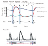
The diastolic phase is composed of four phases; isovolumic relaxation, early rapid ventricular filling, diastasis, and atrial contraction (Fig. 1). LV filling is not a passive process.10) During LV contraction, myocardium is compressed and twisted.11) Relaxation of the myocardial contraction and twist unloads this energy, and this process begins before the end of LV ejection. LV pressure falls rapidly during the isovolumic relaxation period and produces an early diastolic pressure gradient between the left atrial (LA) and LV that sucks blood out of the LA and fills the LV rapidly. Thus, in the normal heart, myocardial relaxation (e') and suction precede the onset of LV passive filling (E). In contrast, the failing ventricle shows reduction of passive ventricular filling and elevation of LA pressure,12)13) so blood is pushed rather than sucked into the LV. In this setting, myocardial diastolic motion (e') reflecting cardiac movement during diastole may be secondary to filling (E).14) The distinction in the mode of LV filling (and thereby e') explains the different behaviour of E/e' with impaired and preserved LV function.

Echocardiographic methods in the estimation of LV filling pressure

A number of echocardiographic techniques can be used to measure LV filling pressure. Increased LA size on 2-dimensional echocardiography is an indicator of increased LV filling pressure.15) The presence of enlarged LA, while non-specific, provides evidence of long-standing elevation LV filling pressure elevation, and LA volume is a more sensitive marker than LA diameter.16)17) However, the process of reverse remodelling of the LA may not be rapid, so that LA enlargement may be a legacy of previously increased LV filling pressure.
Mitral inflow velocity (E) correlates well with LV filling pressure in heart failure, but in the broader community, abnormalities are non-linear because the measurement is affected by both myocardial relaxation and filling pressure. Transmitral inflow is proportionate to the ratio between LA pressure and the relaxation time constant, tau, whereas e' is inversely proportionate to tau only, leading the ratio E/e' to be proportionate to LA pressure. The use of E/e' is generally the most a feasible as well as among the most reproducible method for estimation of filling pressure. Several prominent validation studies have confirmed the correlation of this ratio with filling pressure, and the prediction of normal and abnormal filling pressure is most reliable when the ratio is < 8 or > 15.18)19) However, the examiner should use these as a combination of other Doppler variables and should check for the presence of other clinical conditions that can influence these variables.19)20) Recently, E/e' has been correlated with ambulatory measurement of LA pressure in 60 simultaneous studies, with an area under the receiver operating curve > 0.9.21)
The correlation of E/e' with LA pressure compares favorably with the low correlation of LA pressure with type B natriuretic peptide. Moreover, while the correlation of E/e' with LA pressure is best in the setting of impaired LV systolic function, it holds true with preserved systolic function, and despite changes of loading, for example in aortic stenosis and exercise.22) Although high heart rates may present a challenge because of fusion of the E and A waves, the relationship appears to hold true in atrial fibrillation. Finally, the measurement of E/e' has been shown to correlate with outcome in patients following myocardial infarction, in aortic stenosis and post exercise.23)24)

Pitfalls of E/e'

Despite favorable correlations, some investigators have proposed that the method is an unreliable means of assessing filling pressure. In decompensated advanced systolic heart failure, E/e' showed a poor correlation with intracardiac filling pressures, especially in large LV volumes, worse cardiac indices, and in the presence of cardiac resynchronization therapy.25) These results are likely driven by significant mitral regurgitation (present in 22% of the patients) as well as half of the patients with a broad QRS and consequent abnormal septal motion.21) The study should remind us of the pitfalls of measuring E/e'.

Technical considerations

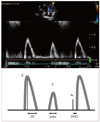
The measurement of E velocity is derived from pulsed-wave (PW) Doppler, usually in the apical 4-chamber view. Using color flow imaging is helpful for the optimal location of sample volume of the Doppler beam. A 1-3-mm sample volume is placed between the mitral leaflet tips during diastole. Mitral inflow waveforms can be recorded clearly after optimization of spectral gain and wall filter setting. Mitral inflow velocities should be measured at end-expiration with a higher sweep speed (100 mm/sec) (Fig. 2). Errors may arise from use of inappropriate location, sample volume or respiratory state. Measurements should be averaged over more than three consecutive beats.
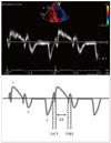
Annular pulsed wave Doppler tissue imaging is also obtained from the apical 4-chamber view, using a 1- to 2-mm size sample volume (Fig. 3). Angulation between the ultrasound beam and the plane of cardiac motion should be minimized (< 20°). Averaging of e' velocity from the septum and lateral side of the mitral annulus is desirable. Errors may arise from excessive Doppler gain, which may cause spectral broadening - if this occurs, a modal velocity should be measured.
Table 1 lists situations where the ratio of E/e' should be interpreted with caution. Tissue e' may be reduced in situations where the mitral annulus might be tethered by calcium or prosthetic rings.26) Caution should be used when using E/e' in LV disorders such as hypertrophic cardiomyopathy27) and myocardial infarction,28) as the downward movement during diastole can be influenced by upward movement during systole.29)30) Conversely, the transmitral E measurement may be the source of misleading information in the setting of moderate to severe mitral regurgitation (MR)31) and severe LV dysfunction.25) In patients with constrictive pericarditis, LV diastolic properties are usually well preserved despite increased LV filling pressure, 32) and the positive correlation of e' with LV filling pressure leads to an inverse relationship between E/e' and LV filling pressure.33)

Alternative approaches

The E/e' ratio is an imperfect marker that should be supplanted or supplemented by other echocardiographic and even invasive measurements under certain circumstances. Other echocardiographic methods include measurement of pulmonary venous waveforms, duration of Ar velocity and the time difference between Ar and A-wave duration (Ar-A; with increased LV filling pressure, Ar velocity and duration increase).20) Flow propagation velocity of mitral inflow (Vp) is evaluated as the slope of the first aliasing velocity during early ventricular filling. It is measured from the mitral valve plane to 4 cm distally into the LV cavity and > 50 cm/s is considered normal.34) Vp can be used to predict LV filling pressure by combination with E velocity (E/Vp)34) or IVRT {LV end-diastolic pressure = 4.5 × [103/(2 × IVRT + Vp) - 9]}.35) In patients with depressed LVEF, a E/Vp ratio > 2.5 predicts LV end-diastolic pressure >15 mm Hg,36) and an E/Vp ratio > 1.5 can be used as a prognostic marker in the prediction of in-hospital heart failure and survival after an acute myocardial infarction.37) However, this parameter can be influenced by many factors including elastic recoil of the LA and LV, the diastolic properties of the LV, and LA pressure, so patients with normal LV systolic function (normal LV volumes and EF) and elevated LV filling pressures can have normal Vp.
Recently, global and regional diastolic function has been analyzed using strain and strain rate derived from speckle tracking and velocity vector imaging. These techniques do not have the limitation of angle dependency and have been validated with sonomicrometry,38) and applied clinically.39) Global diastolic strain rate during IVR (SRIVR) by 2-dimensional speckle tracking imaging is a preload independent parameter. E/SRIVR can predict LV filling pressure in patients with normal LVEF and regional dysfunction.40) Because diastolic untwisting of the LV represents elastic recoil, a reduced rate of untwisting indicates the presence of diastolic dysfunction.41) Decreased left atrial longitudinal strain during systole (< 30%) is associated with increased LV filling pressure.42) However, the evidence base for using deformation imaging techniques in the evaluation of diastolic dysfunction requires further study.
In conclusion, the ratio between transmitral inflow and tissue velocity is a robust marker in the prediction of LV filling pressure. However, it is imperfect and should be interpreted with consideration of many situations that can affect this value. For this reason, this parameter should be supplemented by other echocardiographic and even invasive measurements under certain circumstances.

Figures and Tables

Fig. 1
Diagram of intracardiac pressures according to the cardiac cycle. During diastole, pressure gradient between left ventricle and left atrium derives blood flow.
jcu-19-169-g001
Fig. 2
Mitral inflow velocities obtained by pulsed wave Doppler technique and their schematic diagram. Peak mitral inflow velocity during early diastole (E wave), peak mitral inflow velocity at atrial contraction (A wave), mitral deceleration time (DT), duration of A wave (Adur), and interval between aortic valve closure (Ac) and start of mitral inflow (IVRT) are labelled. IVRT: isovolumic relaxation time.
jcu-19-169-g002
Fig. 3
Mitral annular velocities obtained by tissue Doppler echocardiography. s' velocity: systolic velocity, e' velocity: early diastolic velocity, a' velocity: late diastolic velocity, IVCT: isovolumic contraction time, ET: ejection time, IVRT: isovolumic relaxation time.
jcu-19-169-g003
Table 1
Situations where the use of E/e' may be unreliable
jcu-19-169-i001

References

1. McMurray JJ. Clinical practice. Systolic heart failure. N Engl J Med. 2010. 362:228–238.
2. Jessup M, Brozena S. Heart failure. N Engl J Med. 2003. 348:2007–2018.
crossref
3. Paulus WJ, Tschöpe C, Sanderson JE, Rusconi C, Flachskampf FA, Rademakers FE, Marino P, Smiseth OA, De Keulenaer G, Leite-Moreira AF, Borbély A, Edes I, Handoko ML, Heymans S, Pezzali N, Pieske B, Dickstein K, Fraser AG, Brutsaert DL. How to diagnose diastolic heart failure: a consensus statement on the diagnosis of heart failure with normal left ventricular ejection fraction by the Heart Failure and Echocardiography Associations of the European Society of Cardiology. Eur Heart J. 2007. 28:2539–2550.
crossref
4. Liang HY, Cauduro SA, Pellikka PA, Bailey KR, Grossardt BR, Yang EH, Rihal C, Seward JB, Miller FA, Abraham TP. Comparison of usefulness of echocardiographic Doppler variables to left ventricular enddiastolic pressure in predicting future heart failure events. Am J Cardiol. 2006. 97:866–871.
crossref
5. Kuroda T, Shiina A, Suzuki O, Fujita T, Noda T, Tsuchiya M, Yaginuma T, Hosoda S. Prediction of prognosis of patients with idiopathic dilated cardiomyopathy: a comparison of echocardiography with cardiac catheterization. Jpn J Med. 1989. 28:180–188.
crossref
6. Cheng CP, Noda T, Nozawa T, Little WC. Effect of heart failure on the mechanism of exercise-induced augmentation of mitral valve flow. Circ Res. 1993. 72:795–806.
crossref
7. European Study Group on Diastolic Heart Failure. How to diagnose diastolic heart failure. Eur Heart J. 1998. 19:990–1003.
8. Vasan RS, Levy D. Defining diastolic heart failure: a call for standardized diagnostic criteria. Circulation. 2000. 101:2118–2121.
9. Little WC, Oh JK. Echocardiographic evaluation of diastolic function can be used to guide clinical care. Circulation. 2009. 120:802–809.
crossref
10. Little WC. Diastolic dysfunction beyond distensibility: adverse effects of ventricular dilatation. Circulation. 2005. 112:2888–2890.
11. Bell SP, Nyland L, Tischler MD, McNabb M, Granzier H, LeWinter MM. Alterations in the determinants of diastolic suction during pacing tachycardia. Circ Res. 2000. 87:235–240.
crossref
12. Cheng CP, Freeman GL, Santamore WP, Constantinescu MS, Little WC. Effect of loading conditions, contractile state, and heart rate on early diastolic left ventricular filling in conscious dogs. Circ Res. 1990. 66:814–823.
crossref
13. Rovner A, Greenberg NL, Thomas JD, Garcia MJ. Relationship of diastolic intraventricular pressure gradients and aerobic capacity in patients with diastolic heart failure. Am J Physiol Heart Circ Physiol. 2005. 289:H2081–H2088.
crossref
14. Oki T, Tabata T, Yamada H, Wakatsuki T, Shinohara H, Nishikado A, Iuchi A, Fukuda N, Ito S. Clinical application of pulsed Doppler tissue imaging for assessing abnormal left ventricular relaxation. Am J Cardiol. 1997. 79:921–928.
crossref
15. Appleton CP, Galloway JM, Gonzalez MS, Gaballa M, Basnight MA. Estimation of left ventricular filling pressures using two-dimensional and Doppler echocardiography in adult patients with cardiac disease. Additional value of analyzing left atrial size, left atrial ejection fraction and the difference in duration of pulmonary venous and mitral flow velocity at atrial contraction. J Am Coll Cardiol. 1993. 22:1972–1982.
crossref
16. Tsang TS, Barnes ME, Gersh BJ, Bailey KR, Seward JB. Left atrial volume as a morphophysiologic expression of left ventricular diastolic dysfunction and relation to cardiovascular risk burden. Am J Cardiol. 2002. 90:1284–1289.
crossref
17. Moller JE, Hillis GS, Oh JK, Seward JB, Reeder GS, Wright RS, Park SW, Bailey KR, Pellikka PA. Left atrial volume: a powerful predictor of survival after acute myocardial infarction. Circulation. 2003. 107:2207–2212.
crossref
18. Ommen SR, Nishimura RA, Appleton CP, Miller FA, Oh JK, Redfield MM, Tajik AJ. Clinical utility of Doppler echocardiography and tissue Doppler imaging in the estimation of left ventricular filling pressures: a comparative simultaneous Doppler-catheterization study. Circulation. 2000. 102:1788–1794.
crossref
19. Nagueh SF, Mikati I, Kopelen HA, Middleton KJ, Quiñones MA, Zoghbi WA. Doppler estimation of left ventricular filling pressure in sinus tachycardia. A new application of tissue doppler imaging. Circulation. 1998. 98:1644–1650.
crossref
20. Nagueh SF, Appleton CP, Gillebert TC, Marino PN, Oh JK, Smiseth OA, Waggoner AD, Flachskampf FA, Pellikka PA, Evangelisa A. Recommendations for the evaluation of left ventricular diastolic function by echocardiography. Eur J Echocardiogr. 2009. 10:165–193.
crossref
21. Ritzema JL, Richards AM, Crozier IG, Frampton CF, Melton IC, Doughty RN, Stewart JT, Eigler N, Whiting J, Abraham WT, Troughton RW. Serial Doppler echocardiography and tissue Doppler imaging in the detection of elevated directly measured left atrial pressure in ambulant subjects with chronic heart failure. JACC Cardiovasc Imaging. 2011. 4:927–934.
crossref
22. Dalsgaard M, Kjaergaard J, Pecini R, Iversen KK, Køber L, Moller JE, Grande P, Clemmensen P, Hassager C. Left ventricular filling pressure estimation at rest and during exercise in patients with severe aortic valve stenosis: comparison of echocardiographic and invasive measurements. J Am Soc Echocardiogr. 2009. 22:343–349.
crossref
23. Hillis GS, Møller JE, Pellikka PA, Gersh BJ, Wright RS, Ommen SR, Reeder GS, Oh JK. Noninvasive estimation of left ventricular filling pressure by E/e' is a powerful predictor of survival after acute myocardial infarction. J Am Coll Cardiol. 2004. 43:360–367.
crossref
24. Chang SA, Park PW, Sung K, Lee SC, Park SW, Lee YT, Oh JK. Noninvasive estimate of left ventricular filling pressure correlated with early and midterm postoperative cardiovascular events after isolated aortic valve replacement in patients with severe aortic stenosis. J Thorac Cardiovasc Surg. 2010. 140:1361–1366.
crossref
25. Mullens W, Borowski AG, Curtin RJ, Thomas JD, Tang WH. Tissue Doppler imaging in the estimation of intracardiac filling pressure in decompensated patients with advanced systolic heart failure. Circulation. 2009. 119:62–70.
crossref
26. Sengupta PP, Mohan JC, Mehta V, Arora R, Pandian NG, Khandheria BK. Accuracy and pitfalls of early diastolic motion of the mitral annulus for diagnosing constrictive pericarditis by tissue Doppler imaging. Am J Cardiol. 2004. 93:886–890.
crossref
27. Nagueh SF, Bachinski LL, Meyer D, Hill R, Zoghbi WA, Tam JW, Quiñones MA, Roberts R, Marian AJ. Tissue Doppler imaging consistently detects myocardial abnormalities in patients with hypertrophic cardiomyopathy and provides a novel means for an early diagnosis before and independently of hypertrophy. Circulation. 2001. 104:128–130.
crossref
28. Alam M, Wardell J, Andersson E, Samad BA, Nordlander R. Effects of first myocardial infarction on left ventricular systolic and diastolic function with the use of mitral annular velocity determined by pulsed wave doppler tissue imaging. J Am Soc Echocardiogr. 2000. 13:343–352.
crossref
29. Yip G, Wang M, Zhang Y, Fung JW, Ho PY, Sanderson JE. Left ventricular long axis function in diastolic heart failure is reduced in both diastole and systole: time for a redefinition? Heart. 2002. 87:121–125.
crossref
30. Popovic ZB, Desai MY, Buakhamsri A, Puntawagkoon C, Borowski A, Levine BD, Tang WW, Thomas JD. Predictors of mitral annulus early diastolic velocity: impact of long-axis function, ventricular filling pattern, and relaxation. Eur J Echocardiogr. 2011. 12:818–825.
crossref
31. Yesildag O, Koprulu D, Yuksel S, Soylu K, Ozben B. Noninvasive assessment of left ventricular end-diastolic pressure with tissue Doppler imaging in patients with mitral regurgitation. Echocardiography. 2011. 28:633–640.
crossref
32. Garcia MJ, Rodriguez L, Ares M, Griffin BP, Thomas JD, Klein AL. Differentiation of constrictive pericarditis from restrictive cardiomyopathy: assessment of left ventricular diastolic velocities in longitudinal axis by Doppler tissue imaging. J Am Coll Cardiol. 1996. 27:108–114.
crossref
33. Ha JW, Oh JK, Ling LH, Nishimura RA, Seward JB, Tajik AJ. Annulus paradoxus: transmitral flow velocity to mitral annular velocity ratio is inversely proportional to pulmonary capillary wedge pressure in patients with constrictive pericarditis. Circulation. 2001. 104:976–978.
34. Garcia MJ, Ares MA, Asher C, Rodriguez L, Vandervoort P, Thomas JD. An index of early left ventricular filling that combined with pulsed Doppler peak E velocity may estimate capillary wedge pressure. J Am Coll Cardiol. 1997. 29:448–454.
crossref
35. Gonzalez-Vilchez F, Ares M, Ayuela J, Alonso L. Combined use of pulsed and color M-mode Doppler echocardiography for the estimation of pulmonary capillary wedge pressure: an empirical approach based on an analytical relation. J Am Coll Cardiol. 1999. 34:515–523.
crossref
36. Rivas-Gotz C, Manolios M, Thohan V, Nagueh SF. Impact of left ventricular ejection fraction on estimation of left ventricular filling pressures using tissue Doppler and flow propagation velocity. Am J Cardiol. 2003. 91:780–784.
crossref
37. Møller JE, Søndergaard E, Seward JB, Appleton CP, Egstrup K. Ratio of left ventricular peak E-wave velocity to flow propagation velocity assessed by color M-mode Doppler echocardiography in first myocardial infarction: prognostic and clinical implications. J Am Coll Cardiol. 2000. 35:363–370.
crossref
38. Pirat B, Khoury DS, Hartley CJ, Tiller L, Rao L, Schulz DG, Nagueh SF, Zoghbi WA. A novel feature-tracking echocardiographic method for the quantitation of regional myocardial function: validation in an animal model of ischemia-reperfusion. J Am Coll Cardiol. 2008. 51:651–659.
crossref
39. Pislaru C, Bruce CJ, Anagnostopoulos PC, Allen JL, Seward JB, Pellikka PA, Ritman EL, Greenleaf JF. Ultrasound strain imaging of altered myocardial stiffness: stunned versus infarcted reperfused myocardium. Circulation. 2004. 109:2905–2910.
crossref
40. Wang J, Khoury DS, Thohan V, Torre-Amione G, Nagueh SF. Global diastolic strain rate for the assessment of left ventricular relaxation and filling pressures. Circulation. 2007. 115:1376–1383.
crossref
41. Fuchs E, Müller MF, Oswald H, Thöny H, Mohacsi P, Hess OM. Cardiac rotation and relaxation in patients with chronic heart failure. Eur J Heart Fail. 2004. 6:715–722.
crossref
42. Wakami K, Ohte N, Asada K, Fukuta H, Goto T, Mukai S, Narita H, Kimura G. Correlation between left ventricular end-diastolic pressure and peak left atrial wall strain during left ventricular systole. J Am Soc Echocardiogr. 2009. 22:847–851.
crossref
TOOLS
Similar articles