

 PDF
PDF ePub
ePub Citation
 Print
Print


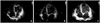
A 67-year-old man was admitted to the cardiology department because of exertional dyspnea and orthopnea aggravated for 10 days. Transthoracic echocardiography showed dilated left ventricle (LV) with global hypokinesia with severely decreased systolic function (ejection fraction was estimated to be about 15%). Two oscillating masses with smooth, spherical contour were found in the LV apex (Fig. 1A). The size of masses were estimated 22 by 29 mm and 14 by 18 mm respectively. The patient received unfractionated heparin intravenously for 5 days, followed by oral warfarin therapy. On the 8th hospital day, follow-up echocardiography revealed partial resolution of thrombi with resultant highly movable friable remnants (Fig. 1B). One the 18th hospital day, follow-up echocardiography revealed nearly complete resolution of thrombi (Fig. 1C). The patient was discharged home uneventfully.
Ball shaped-masses in LV may be thrombi, vegetations or tumors. Although echocardiography is the procedure of choice for the diagnosis of cardiac mass, differentiation between myxoma and thrombus may be very difficult if the mass size is small, contours are smooth, or attachment site is atypical or ill-defined.1) Short-term anticoagulation therapy, which can differentiate thrombi from tumors, makes unnecessary surgical procedure avoidable.
 XML Download