

 PDF
PDF ePub
ePub Citation
 Print
Print


Introduction
Carcinoid heart disease occurs in about one third of patients affected by carcinoid tumours (especially, ileal carcinoid) with hepatic metastases.1) It may be a part of carcinoid syndrome and is a cause of cardiac impairment characterized by plaque-like fibrous endocardial thickening and valve incompetence, usually concerning the tricuspid valve only and/or pulmonary valve. The left heart involvement does not occur in these patients, except for those with bronchial carcinoids or right-left shunts. The carcinoid tumors with hepatic metastases may exhibit a constellation of symptoms (called as carcinoid syndrome) due to the excessive serum release of serotonin (5-HT), and other some vasoactive substances (histamine, tachykinins, and prostaglandins also released by the metastatic hepatic cells).2)3) It includes: flushing and telangectasias, most commonly occurring in the face and caused by the release of tachykinin. Diarrhea, frequently accompained by abdominal cramps and pain and related to 5-HT secretion. Tachycardia and decreased blood pressure are also frequently found. In addition, bronchospasm (related to the secretion of bradykinin or 5-HT), and pellagra (caused by a deficiency of tryptophan) may be manifest too. Cardiac involvement (also named as carcinoid heart disease) is often present in patients with carcinoid syndrome. It includes tricuspid and/or pulmonary valves insufficiency, or right heart failure symptoms with swelling (oedema) in the extremities and enlargement of the heart. On the contrary, the left side of the heart is usually not affected in these patients because the lungs can break down 5-HT.
In the present report, we illustrated a case of carcinoid heart disease due to primitive ileal tumour with hepatic metastases.
Case
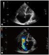
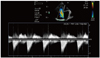
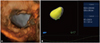
A 72-year-old man with a previous hystory of ileal carcinoid disease and hepatic metastases was admitted to our Department for severe dyspnoea, peripheral oedema at lower extremities, diarrhea, episodic flushing and bronchospasm. The urinary level of 5-Hydroxyindoleacetic acid (5-HIAA) (the main urinary metabolite of 5-HT), resulted elevated (368 µmol/L). A systolic murmur was auscultated on IV parasternal space. Interna jugular systolic pulsations were elevated. Atrial fibrillation with a mean frequency of 72 beats/min was recorded at E.C.G. Right axis deviation and low voltage in both peripheral and precordial derivations were also evidenced. A-V time-interval was normal (0,15"); QRS-width was 110 ms. without ischemic changes of S-T. Arterial blood pressure was 140/80 mmHg. 2-D transthoracic echocardiography performed in B-Mode from apical 4 chambers approach revealed dilated right atrium and right ventricle The tricuspid valve leaflets were thickened, retracted with incomplete coaptation. Endocardial plaques of the subvalvar apparatus are also notable (Fig. 1A). Colour flow Doppler imaging shows a severe tricuspid regurgitation through a wide regurgitant orifice (Fig. 1B). Continuous wave Doppler of tricuspid valve showed the "dagger shaped" spectrum (Fig. 2). In order to better define right RV function, three-dimensional echocardiography (3-DE) was also performed. Short axis 3-D transthoracic echocardiography evidenced the adhesion of tricuspid valve leaflets to the right ventricular walls (Fig. 3A). The shape of systo-diastolic RV and its main hemodynamic parameters are reported in Fig. 3B. End diastolic volume (EDV) resulted of 104.4 mL; end systolic volume (ESV) was 70.3 mL; stroke volume (SV) was 34.0 mL, and RVEF% resulted of 32.6%. Despite ileo-cecal resection, chemotherapy and treatment with somatostatin, the disease progressively advanced. Six months later, the patient underwent laparatomy for small bowel resection. Hepatic metastatic carcinoid disease was treated with arterial hepatic embolization. Administration of octreotide (that blocks hormone release) was also performed whereas, right-sided heart failure was treated with diuretics, ACE-inhibitors and long-acting nitrates. But, patient died three years later for hepatic failure and primitive ileal tumor growth.
Discussion
Carcinoid heart disease is a rare condition, that may be present in patients suffering from ileal tumors and hepatic metastases. Its pathogenesis remains incompletely understood, although a growing body of evidence points towards 5-HT playing a key role.4) The substance has been shown to increase synthesis and upregulate tissue growth factor-β, as well as stimulating collagen synthesis by heart wall interstitial cells, producing tricuspid and/or pulmonary valve insufficiency with plaques of fibrous tissue due to the deposition of fibrous tissue on the endocardial surfaces of the heart. The excessive amounts of the hormone 5-HT contribute to the pathophysiology, because 5-HT receptors are present in human heart valves. In fact, 5-HT has been shown to stimulate collagen synthesis by heart valve interstitial cells.5) Patients with carcinoid tumors may have relatively few signs or sympoms in the early stages with high urinary levels of 5-HIAA.6) Successively, the gradual decline in right ventricular function and increasing severity of tricuspid insufficiency lead to right heart failure and a poor outlook if treated medically. Echocardiography is the choice investigation. Classically, tricuspid valve leaflets and its subvalvular apparatus is thickened; excursion of the leaflets become retracted, fixed, and noncoapting, leading to the valve remaining in a semiopen position. A "dagger-shaped" continuous wave-doppler profile resulting from severe tricuspid regurgitation with elevated right atrial pressure (that causes early peak pressure and its rapid decline) is seen at continuous wave-doppler record.7) Because the blood cannot be adequately ejected through the pulmonary valve, the right ventricle work increases. The right atrium and ventricle are enlarged becomes volume overloaded. But, RV function seemingly remains intact until quite late in the disease course. The heart's lesions may cause right-sided heart failure. Three-DE provided more detailed anatomic informations about the tricuspid valve. In addition, it seems to be more useful for the assessment of RV size and function in comparison to two-dimensional echocardiography because it is not based on geometrical assumptions. The systo-diastolic RV shape highlights the unhomogeneous RV contractility related to the degree of its dysfunction. RV volumes and RVEF% evaluated with 3-DE were significantly increased (volumes) and decreased (ejection fraction) respectively, in comparison to the normals,8) and are well correlated with MRI estimated as reference method.9) 3DE slightly overestimated RV end diastolic and end systolic volumes, although the degree of overestimation was not significant. On the contrary, RVEF was underestimated in respect to MRI. Possibile reasons for these differences between 3-DE and MRI include difficulties in defining the endocardial borders, artifacts induced by the respiration movements and some uncertainties in to precisely identify valvular planes.
Conclusively, while 2-D echocardiography is the choice method for define the valvular involvements in carcinoid heart disease, the 3-D echocardiography seems able to provide more detailed and precise anatomic and hemodynamic informations about RV size and function and valvular anatomic and functional changes.10)11)
 XML Download