

 PDF
PDF ePub
ePub Citation
 Print
Print


Introduction
Stress induced cardiomyopathy (SCMP) is characterized by a transient left ventricular (LV) dysfunction with akinesis and systolic expansion of apical segments and hyperkinesis or normokinesis of the basal segments. Various emotional or physical stressors have been described as triggering factors of this syndrome. Although the prognosis of SCMP is generally known to be excellent, several fatal complications have been described. Here, we report a rare case of SCMP complicated by LV thrombosis and multiple cerebral infarctions in a patient with essential thrombocythemia (ET).
Case
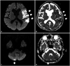
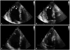
A 75-year-old female presented with sudden onset of mental deepening, hemiparesis, and dysarthria which developed 1 hour ago. She had a history of dural sinus thrombosis and deep vein thrombosis 2 years ago, and the cause of venous thrombosis was ET. She had been treated with 0.5 mg of anagrelide and 500 mg of hydroxyurea daily for ET at hematologic department for last 2 years. Before 1 day of admission, she had experienced severe emotional stress due to arguments with her husband for economic problems, and thereafter chest tightness developed and persisted. Brain magnetic resonance imaging revealed acute infarctions of left middle cerebral and right posterior inferior cerebellar arterial territories (Fig. 1). Electrocardiogram (ECG) at admission showed convex ST segment elevation and T-wave inversions in precordial leads with QT prolongation. Laboratory studies showed thrombocytosis with platelet count of 665 × 103/mm3 and mildly elevated cardiac troponin I with the level of 0.65 ng/mL. Other laboratory findings were unremarkable. Transthoracic echocardiography (TTE) was performed to evaluate cardiac abnormalities relevant to the abnormalities of ECG and cardiac enzymes, and cardiac source of cerebral embolism. TTE revealed akinesia and systolic bulging of the apical segments with hyperkinetic basal wall motions, and the estimated ejection fraction was 51.8%. About 3.3 × 2.4 cm sized mural thrombus was observed within the dyskinetic apical segments of the LV (Fig. 2A and B). Coronary angiography was performed to exclude acute myocardial infarction (AMI) and revealed no significant stenotic lesions in both coronary arteries. The most probable diagnosis in the present case was SCMP with apical mural thrombi complicated by multiple cerebral infarctions in the setting of ET. Anticoagulation with intravenous heparin and general managements for heart failure and cerebral infarctions were done without discontinuation of anagrelide and hydroxyurea. The symptoms and signs of cerebral infarctions including motor and speech disturbances were improved gradually. The wall motion abnormalities of the LV apex were improved, and the thrombus was disappeared on follow-up TTE (Fig. 2C and D). The patients was discharged with medications including anagrelide and hydroxyurea and managed at out-patient clinic without any clinical events.
Discussion
SCMP, also known as apical ballooning or Takotsubo cardiomyopathy, is a syndrome of transient cardiac dysfunction associated with acute emotional or physical stress, but the pathogenesis of SCMP is not well established. Catecholamine-mediated multivessel epicardial or microvascular coronary vasospasm or direct catecholamine-mediated myocyte injury has been proposed as possible pathophysiological mechanisms.1) Because clinical characteristics of SCMP are quite similar to those of AMI, the differential diagnosis between SCMP and AMI is very difficult and usually requires coronary angiography.2) Coronary angiography was also performed in the present case to rule out AMI as a cause of LV dysfunction and revealed normal coronary circulation. Provocation test for vasospasm was not performed, because vasoactive drugs including intravenous nitrates were prescribed already. Therefore, the possibility of vasospasm as a cause of wall motion abnormality could not completely rule out in the present case.
Emotional stress, as in most of the previously reported cases, was the most probable predisposing cause of SCMP in the present case, but the role of anagrelide should also be considered. Anagrelide inhibits platelet aggregation through the selective inhibition of type III cyclic adenosine monophosphate phosphodiesterase on the megakaryocyte cell lineage, and thus anagrelide used in the treatment of ET to prevent the incidence of thromboembolic events. Because it may have positive inotropic and chronotropic activity, and directly induce vasospasm of the coronary arteries, several serious cardiovascular side effects including congestive heart failure, arrhythmia, acute coronary syndrome, and SCMP has been described.3) These side effects of anagrelide developed usually within 1 month of medication and decreased with improved tolerance to treatment over time in the previous report.3)4) Because anagrelide had been prescribed for last two years without any side effects in the present case, it is assumed that the role of anagrelide in the development of SCMP might be negligible in the present case. Therefore, anagrelide and hydroxyurea were prescribed continuously in the present case during admission and after discharge. Because the platelet count of the patient was maintained in acceptable range with the combination therapy of relatively low dose of anagrelide (0.5 mg) and hydroxyurea, we did not change the dosage of anegrelide. If the platelet count is not controlled within acceptable range, it would be reasonable to adjust the dosage of anegrelide. In case of confirmed acute coronary syndrome, the discontinuation of anagrelide has been recommended.5)
Although the prognosis of SCMP is known to be excellent, various complications, including severe heart failure with pulmonary edema, shock and inhospital death have been reported. The present case presented with multiple cerebral infarction presumably caused by the embolization of the LV thrombi, and cerebral infarction associated with LV thrombosis is a rare complication of SCMP. In a recent systematic review of de Gregorio,6) LV thrombosis occurred approximately 5% of the patients with SCMP, and one third of them experienced thromboembolic events. The study of Haghi et al.7) and Mitsuma et al.8) also demonstrated that LV thrombosis and subsequent embolism was a possible complication of SCMP. Recent meta-analysis showed that LV thrombus is a significant complication and systemic embolism is 2nd common causes of mortality in SCMP.9) Therefore, it is suggested that the early initiation of anti-coagulation therapy to prevent LV thrombosis and subsequent embolic complications would be a reasonable approach in patients with SCMP.
The LV thrombosis in patients with SCMP is probably caused by low blood flow in the left ventricle as well as LV systolic dysfunction. ET is known to be associated with bleeding or thromboembolic complications. Arterial thromboses occurring in coronary, cerebral or peripheral arteries are regarded as more typical than venous thromboembolic events in patients with ET. The present case had a tendency of thrombosis evidenced by the previous history of dural sinus and deep vein thrombosis associated with ET, and thus ET might play an important additional role in the thrombus formation within the LV in the present case, besides SCMP itself. Baker et al.10) also reported a similar case of LV mural thrombus in a patient with thrombocytosis and agnogenic myeloid metaplasia.
In conclusion, we report a rare case of SCMP complicated by LV thrombosis and multiple cerebral infarctions in a patient with ET. Early initiation of anti-coagulation therapy should be considered in patients with SCMP, especially who had combined coagulation disorders such as ET.
 XML Download