

 PDF
PDF ePub
ePub Citation
 Print
Print


Introduction
Vasoactive substances from carcinoid tumors, which are uncommon malignancies arising from enterochromaffin cells, underlie the systemic manifestations of carcinoid syndrome. Primary ovarian carcinoid tumors are rare, accounting for 0.5% of all carcinoid tumors,1) and carcinoid syndrome and carcinoid heart disease are believed to complicate fewer than 10% of these rare ovarian neoplasms.2) Such tumors are usually suspected on detection of a pelvic mass and the presence of carcinoid syndrome, which is characterized by facial flushing and diarrhea, or typical carcinoid heart disease (right-sided valvular involvement).3) Most carcinoid tumors have to metastasize to the liver to result in symptoms of carcinoid syndrome; however, primary ovarian carcinoid tumors are unique in this regard because the venous drainage of the ovaries bypasses the portal venous system.4) We report the case of carcinoid heart disease associated with a surgical biopsy-confirmed primary ovarian carcinoid tumor.
Case
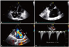
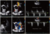
A 60-year-old woman was admitted to our hospital with a three-month history of pitting edema of both lower extremities, worsening dyspnea on exertion, and easy fatigability. She had no specific cardiovascular risk factors except for hypertension. On physical examination, heart sounds were regular, and a grade III-IV pansystolic murmur was heard along the left lower parasternal border. In addition to pitting edema of the lower legs, facial flushing with telangiectatic skin changes was apparent in her face and legs. Routine laboratory examination including complete blood cell counts and liver or renal function tests did not reveal any abnormalities. Cardiomegaly was apparent on chest X-ray, and transthoracic echocardiography showed a normal left ventricular (LV) ejection fraction (56%), normal LV systolic and diastolic dimensions, and trivial mitral regurgitation. However, on right-sided heart evaluation, the tricuspid valve (TV) leaflet was severely retracted and shortened, and its chordae were also small and deformed (Fig. 1A and B). The TV leaflets showed very restricted movement and poor coaptation during the systolic phase. Accordingly, severe tricuspid regurgitation (TR) was noted on color Doppler imaging, and the TR velocity was measured at 2.7 m/s (Fig. 1C and D). Deformation was also apparent in the pulmonary valve (PV); the PV leaflets shrunk in size and had a motion limitation (Fig. 2A). Thus, moderate regurgitation was noted on color Doppler images (peak velocity, 1.5 m/s) (Fig. 2B and C). Together with regurgitation, color flow was accelerated through mild stenosed PV (peak velocity, 1.8 m/s) because of retracted PV annulus and stiffened leaflets (Fig. 2D, E and F). In addition to this valvular dysfunction, the right ventricle was enlarged, and apical contraction was depressed on echocardiography.
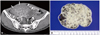
We sought systemic or hormonal etiologies for the exclusively right-sided valve involvement in this patient. The 24-hour urinary level of 5-hydroxyindoleacetic acid (5-HIAA) was measured, and abdominal computed tomography (CT) imaging was performed. On CT images, an intra-abdominal mass was detected on the right ovary (Fig. 3A), and we confirmed the increased level of 5-HIAA (68.2 mg/24 hr, normal range; 2.0-8.0 mg/24 hr) in 24-hour urine collection. The mass was surgically removed; it was 80 × 124 × 78 mm in size with an irregular multinodular shape and solid in nature (Fig. 3B). Histologically, the sections from the ovarian tumor displayed uniform, polygonal cells arranged in solid sheets with a trabecular pattern (Fig. 4A, 100×, hematoxylin-eosin staining). The trabeculae were lined with one to two layers of cells. The cells showed central, monotonous, and round nuclei with no mitoses or atypia (Fig. 4B, 400×). The amount of cytoplasm of the constituent cells was large, and it was strong acidophilic. Immunohistochemical analysis of the tumor cells showed strong granular cytoplasmic positivity for chromogranin A (Fig. 4C, 100×, chromogranin staining). Finally, the patient was diagnosed as having a primary ovarian carcinoid tumor. She was treated with adjuvant chemotherapy including paclitaxel and carboplatin and was subsequently discharged without any worsening complication. Treatment with diuretics and angiotensin converting enzyme inhibitors was continued to relieve right heart failure (HF) symptoms, with slight improvement of pitting edema and dyspnea. Follow-up echocardiography 3 months later showed no improvement of the valve deformity or TR severity; surgical correction, i.e., the replacement of the deformed valve using mechanical or bioprosthetics, was therefore considered to reduce right HF symptoms and to substantially delay the deterioration of right ventricular dysfunction. However, the patient wanted to defer surgical correction of the severe TR and to remain on conservative medical therapy because of the stated satisfaction with the clinical response and tolerability. Three years after diagnosis, she remains clinically stable; she has mild dyspnea on exertion in daily activities, and she is regularly followed-up at the outpatient clinic.
Discussion
Carcinoid heart disease can manifest in more than half of patients with carcinoid syndrome. However, primary ovarian carcinoid tumors, accounting for 0.5% of all carcinoid tumors, are very rare, and fewer than 10% of primary ovarian carcinoid tumors are associated with carcinoid syndrome.1)2)
Patients with carcinoid heart disease exhibit fibrotic valvular plaques and myofibroblast proliferation that may cause valvular thickening and retraction leading to dysfunction.5) Most patients present with right-sided heart valve dysfunction because of the venous drainage of a vasoactive substance that is metabolized and secreted by the liver; left-sided valve dysfunction, which is usually accompanied by coronary vasospasm, is present as the result of a patent foramen ovale in less than 10% of individuals with carcinoid heart disease.6-8)
Despite an unusual manifestation of myocardial infiltrative pattern,9) echocardiography of patients with carcinoid heart disease has characteristic findings of thickened, shortened, and retracted TV and PV leaflets, changes that lead to incomplete coaptation, resulting in moderate or severe TV/PV regurgitation. Moreover, the PV is thickened and retracted, which also causes pulmonary regurgitation. Most findings can be explained by the systemic effects of 5-HIAA, which is activated via hepatic metabolism and drains into the right-sided heart system.8)10)11) Thus, importantly, severe TR alone without mitral valve involvement should raise suspicion of carcinoid heart syndrome as the cause of right-sided valvular heart disease.
There is no consensus on the appropriate treatment of carcinoid heart syndrome to improve mortality. In general, early detection and surgical removal of primary carcinoid tumors seems to be the only effective treatment that results in favorable clinical outcomes; two-year survival with surgical removal has been reported to be 40%, whereas that of medical treatment without surgery is only 8%.12) Because of systemic or hormonal effects, surgical removal would not be sufficient to treat carcinoid heart disease or to promote the regression of valvular lesions. With the recent decrease in valvular perioperative mortality, surgical correction of TV dysfunction could be generally undertaken, even in mildly symptomatic patients, to prevent further HF progression, right HF symptoms or signs, such as right ventricular enlargement or decline in right ventricular systolic function, or in anticipation of hepatic surgery.13) In the case of carcinoid syndrome with echocardiographic evidence of cardiac involvement, the three-year survival is reported to be 31%.14) Thus, individualized surgical correction of TV and right HF treatments are also needed to achieve better outcomes.
In conclusion, this case suggests that, in the differential diagnosis, we should consider a rare condition such as carcinoid heart disease; this condition is characterized by isolated, severe TR without significant left-sided valvular dysfunction, which could be easily assessed by transthoracic echocardiography.

 XML Download