

 PDF
PDF ePub
ePub Citation
 Print
Print


Introduction
Right ventricular (RV) apical pacing allows for safe and stable long term pacing. However, RV apical pacing inevitably induces non-physiologic left ventricular (LV) activation with alteration of the intraventricular contraction sequence, which delays LV activation.1) This delay is accompanied with LV dyssynchrony, and the development of LV dyssynchrony was reported to be associated with deterioration of heart failure symptoms and the systolic LV dysfunction.2) To overcome this potential limitation, other pacing sites including the RV outflow tract, the interventricular septum, and the his bundle have been tried.3) However, contradictory results have been reported in the literature4)5) and the impact of different RV pacing sites on LV dyssynchrony has not been seriously investigated. In this study, we sought to evaluate the acute changes of LV dyssynchrony according to the RV pacing sites.
Methods
Study populations
This study is a multicenter, prospective observational study. The study population consisted of the patients who needed permanent pacemaker implantation for sick sinus syndrome (SSS) or high degree atrio-ventricular (AV) block. Of these, we excluded the patients who had a temporary or permanent pacemaker prior to the study, and those with poor image quality, significant mitral or aortic steno-insufficiency, underlying left bundle branch block, or advanced LV systolic dysfunction (ejection fraction < 40%). Seventy nine patients with pacemaker implantation and 15 age-matched healthy controls were included.
Pacemaker implantation
Pacemaker leads were inserted through the subclavian vein using standard implantation techniques. The RV leads were positioned in the RV apex (n = 45) or interventricular septum (n = 34) under fluoroscopic guidance. The decision for the lead positioning was determined at the electrophysiologists' discretion.
Echocardiography
All the patients underwent comprehensive echocardiographic evaluation before and after (< 7 days) pacemaker implantation. All the images were obtained with a standard ultrasound machine (Vivid 7, GE Vingmed, Horton, Norway) with a 2.5 MHz probe. Standard techniques were used to obtain M-mode, two-dimensional, and Doppler measurement in accordance with the American Society of Echocardiography guidelines. The tissue Doppler derived peak systole (S') were measured at the septal mitral annulus. The averaged peak systolic tissue velocity (Sm) was also calculated from 6 basal segments. S' and Sm were considered to represent the LV longitudinal function. The LV end-systolic, end-diastolic volume, and ejection fraction were calculated by the Simpson's methods from the apical four and two chamber views.
Assessment dyssynchrony
Digital loops with one cycle of fundamental 2D image and three cycles of the color coded tissue Doppler imaging (TDI) were acquired from a parasternal short axis view at the mid-papillary and three apical views for off-line analysis of LV dyssynchrony using Echopac (BT07, GE, Vingmed). All the images were transferred to one center and analyzed by one observer (GY Cho), who was blinded to the clinical data and the other echocardiographic information.
Atrio-ventricular dyssynchrony
A delay in the LV ejection can be reflected in the LV filling time, which is measured by the mitral inflow velocity. The atrio-ventricular dyssynchrony was measure as the LV filling time as the ratio of the RR interval.6)
Inter-ventricular dyssynchrony
Using pulsed-wave Doppler, we measured the difference between the pre-ejection intervals from the QRS onset to the beginning of ventricular ejection at the right and left ventricular outflow tract.7)
Intra-ventricular dyssynchrony
A) M-mode echocardiography: The septal to posterior wall motion delay (SPWMD) was assessed using M-mode echocardiography at the parasternal window.8) The interval between the maximal thickening of the septum and posterior wall was calculated.
B) Conventional Doppler imaging technique: We measured the pre-ejection interval from the QRS onset to the beginning of ventricular ejection at the LV outflow tract by using pulsed-wave Doppler for the assessment of global intra-ventricular dyssynchrony.
C) Tissue Doppler imaging technique: The peak myocardial velocity during the ejection phase and the time to the peak myocardial velocity (Ts) were measured with reference to the QRS complex. If the peak velocity could not be defined because of the noise signal or flat velocity contour, then the sample volume (12 × 6 mm) was gradually moved to the apex or base until clear signal intensity could be obtained. Intra-ventricular dyssynchrony was assessed by measuring the difference of Ts between the basal septum and basal lateral segment (Ts-SL)9) or by measuring the standard deviation of Ts of 12 basal and mid segments (Ts-SD).10) For strain analysis, the longitudinal strain curves were obtained from 12 basal and mid segments. The region of interest with a 12 × 6 mm oval shaped was placed in the middle of the respective segments from the three apical views and maintained same position during the cardiac cycle by manually tracking to avoid blood or pericardial contamination. The minimal frame rate was 130 frames per second. The time to peak strain (Tε) with reference to the QRS complex were measured. The time difference of Tε between basal septum and basal lateral segment (Tε-SL) or standard deviation in time to peak strain among the 12 segments (Tε-SD) was obtained for the strain derived dyssynchrony.11) The timing of events, such as aortic valve opening and closure, was obtained from color-coded M-mode of anterior mitral valve from the apical windows.12)
D) 2D speckle strain: Radial strain using speckle tracking was assessed on LV short axis at the mid-papillary muscle level (frame rate varied from 60 to 80 frames per second). Endocardium was traced manually at the end-systolic frame. The traced endocardium was automatically divided into 6 segments. The strain curves for each segment were constructed. We measured the time to peak radial strain of each segment. The absolute time interval of peak strain between anteroseptum and posterior segment was calculated.13) In addition, the time interval between the earliest and latest segment (maximal temporal difference) was also measured.
Statistical methods
Data are presented as the mean ± standard deviation for continuous variables and as proportion for the categorical variables. The mean values of continuous variable were compared by t-test or ANNOVA, and the differences in the prevalence between the groups were compared via χ2-test. All the analyses were performed with SPSS version 13.0 (SPSS Inc., Chicago, IL, USA) and p < 0.05 was considered to be statistically significant.
Results
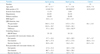
The baseline characteristics and echocardiographic measurements are summarized in Table 1. Age, pre-pacing QRS duration and LV ejection fraction were comparable between the two groups (Table 1). After pacemaker implantation, LV volume and ejection fraction did not significantly change. The QRS duration was significantly increased in both groups after pacing, but the difference between the pre- and post-pacing QRS duration was significantly higher in apical pacing group (57.1 ± 28.3 versus 32.8 ± 40.5 msec).
The echocardiographic variables immediately after pacemaker implantation are demonstrated in Table 2. The patients with RV apical pacing showed a lower S' (5.3 ± 1.3 versus 5.7 ± 1.5 cm/sec) and Sm (4.2 ± 1.0 versus 4.9 ± 1.3 cm/sec) than those with septal pacing. Aortic pre-ejection time and SPWMD in patients with a pacemaker were longer compared to those of normal controls, but there was no significant difference. Both TDI velocity and strain analysis showed no difference of dyssynchrony indices between the two groups, despite that there was a higher tendency of Doppler strain dyssynchrony indices in RV apical pacing group compared with those of the control (p = 0.063 for Tε-SL and p = 0.026 for Tε-SD).
Discussion
In this study, we demonstrated that both septal and apical pacing produce LV dyssynchrony, but septal pacing is superior to RV apical pacing in terms of LV longitudinal function. However, there was no significant difference in LV dyssynchrony between septal and apical pacing.
Pacing and LV dysfunction
In the past several years, there has been increasing recognition of the deleterious clinical effects of RV apical pacing, both in patients with pacemakers and in those with ICDs. In patients with a permanent pacemaker, every 1% incremental of RV pacing increases the risk of atrial fibrillation by 1% and the risk of heart failure hospitalization by 5.4%.14) Several studies have reported that RV apical pacing is associated with regional perfusion defects,15) adverse LV remodeling,16) a decrease in LV ejection fraction,17-19) and heart failure.2) More recently, several studies have reported that dyssynchronous LV contraction results from RV apical pacing.2)20)21)
The DAVID (Dual Chamber with VVI Implantable Defibrillator) trial suggested that RV apical pacing was associated with an increased risk of death and hospitalization for heart failure in patients with an implantable defibrillator.22) In that study, a higher cumulative percent of ventricular pacing was manifest in a significantly prolonged QRS duration at 6 months after pacemaker implantation and therefore, RV pacing might produce electrical dyssynchrony. Dual-chamber minimal ventricular pacing, as compared with conventional dual-chamber pacing, reduces ventricular desynchronization and moderately reduces the risk of persistent atrial fibrillation in patients with sinus node disease.23) The cumulative percent of ventricular pacing is associated with heart failure hospitalization and atrial fibrillation.14)22) Furthermore, in the patients with SSS, DDD pacing but not AAI pacing induces significant LV desynchronization and reduction of LV ejection fraction.24) Therefore, unnecessary RV pacing induces dyssynchronous LV contraction, which results in deterioration of LV systolic function and therefore, it can induce clinical heart failure. In our study, a dramatic increase of the QRS duration and SPWMD immediately after pacemaker implantation was demonstrated, and this suggests the potential detrimental long-term effect on the LV performance. Although the LV EF did not change immediately after implantation, the development of heart failure might depend on the pacing duration and so longer clinical observation is warranted.
By speckle tracking analysis, more than 50% of the patients showed dyssynchrony after RV pacing, which results in deteriorated LV systolic function and a worsened NYHA functional class.21) The development of LV dyssynchrony after permanent pacing is an important mechanism of LV function deterioration.25) Therefore, an alternative pacing mode or alternative pacing sites have been tested in order to prevent LV dyssynchrony and hemodynamic deterioration. Several studies have showed that either RV outflow tract (RVOT) pacing26-28) or RV septal pacing29)30) might have an advantage over classic RV apical pacing, but controversial results have also been reported.4) We could not demonstrate any significant difference of the LV dyssynchrony indices between the RV apical and septal pacing. According to the PROSPECT trial, no single echocardiographic measure of dyssynchrony may be recommended because of the poor reproducibility and moderate sensitivity of cardiac resynchronization therapy response.31) In this study, we used various kinds of mechanical dyssynchrony parameters. However, none of the echocardiographic measures of dyssynchrony showed a significant difference according to the pacing site.
One interesting finding of our study is that RV septal pacing showed better longitudinal systolic movement than did RV septal pacing. Although the resting LV EF was similar between the groups, this difference might affect the long-term LV performance, which should be tested by another study.
Study limitations
The LV mechanical function and dyssynchrony could be evaluated by a recently introduced speckle-tracking imaging technique, which might provide other indices including LV twist and 2-dimensional radial strain.32) Using this relatively new technique, it might be interesting to test whether LV mechanical function or dyssynchrony indices show significant difference according to the different pacing sites. Finally, although longitudinal function was better in the septal pacing group, we could not rule out the possibility that the difference of age and sex between two groups might affect our results.

 XML Download