

 PDF
PDF ePub
ePub Citation
 Print
Print


Introduction
Left ventricular (LV) pseudoaneurysm is defined as a partial cardiac rupture that is contained by the adherent pericardium or scar tissue, and this results in the formation of a false aneurysm that maintains communication with the LV cavity.1) If a LV pseudoaneurysm ruptures, which is a rare complication but one of the most severe complications following mitral valve replacement (MVR), then this may require emergency surgery that can lead to a fatal outcome.2) Left ventricle-coronary sinus fistula, which is another rare complication after MVR, should be differentiated from LV pseudoaneurysm. A variety of methods can be used to diagnose these complications. In most cases, transthoracic echocardiography (TTE), computed tomography (CT) and angiography are used to detect the structure. We describe here a case of an outpouching lesion of the atrioventricular (AV) groove that was difficult to differentiate between a LV pseudoaneurysm and a left ventricle-coronary sinus fistula by echocardiography. Performing cardiac CT (CCT) angiography enabled us to diagnose a LV pseudoaneurysm in an asymptomatic female patient who received MVR more than 5 years previously.
Case
A 27-year-old female visited our clinic for a regular checkup after receiving MVR in the other hospital 5 years previously. The patient had undergone MVR due to infective endocarditis. Since then, the patient had continued taking daily medication of warfarin 6 mg. Physical examination revealed a blood pressure of 110/70 mmHg, a pulse rate of 74 beats per minute, a respiratory rate of 16 breaths per minute and a body temperature of 36.4℃. No murmur was heard on auscultation. On the blood analysis, the prothrombin time international normalized ratio (PT INR) was prolonged to 2.19. On chest radiography, a prosthetic mitral valve with a normal size heart and three sternal wire sutures were shown. Normal sinus rhythm was observed on the electrocardiogram.
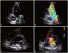
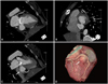
The TTE showed good LV systolic function (ejection fraction: 65%) without regional wall motion abnormality. The prosthetic mitral valve function was good without any paravalvular leakage. Mild resting pulmonary hypertension (43 mmHg) was observed, but the right ventricle was not dilated. There was a 25×23 mm sized outpouching lesion with a neck distance of 6 mm on the postero-interior wall of the LV below the mitral annulus. TTE also revealed draining flow and redundant motion (Fig. 1). However, it is difficult to distinguish between a left ventricle-coronary sinus fistula and a LV pseudoaneurysm as the opening site of the lesion was the postero-inferior wall where the coronary sinus drains. But the echocardiographic findings suggested that it was more likely a LV pseudoaneurysm because if it was a left ventricle-coronary sinus fistula, then volume overload to the right side of the heart may result in dilation of the right atrium and right ventricle. CCT was then done. It showed about a 30×25 mm sized outpouching lesion arising from a defect of the posteroinferior wall of the LV, just beneath the replaced mitral valve (Fig. 2A and B). The aneurysmal sac was observed at the AV-groove without a discernible wall, and it was considered to be a pseudoaneurysm (Fig. 2C). The pseudoaneurysm was compressing the opening of the coronary sinus, resulting in dilatation of the coronary sinus and the adjacent cardiac veins. The pseudoaneurysm and dilated coronary sinus were clearly demonstrated on the three-dimensional reconstruction image (Fig. 2D). According to the CCT findings, we were able to confirm a LV pseudoaneurysm. We recommended surgical intervention, but the patient refused to undergo any further evaluation and treatment. The patient is regularly visiting the outpatient department with no definite symptoms.
Discussion
LV pseudoaneurysms are known to rarely occur as a serious complication after MVR. Their incidence is very low and they are reported to occur in 0.02% to 2% of all MVRs.3) The cause of this complication is not clear, but its potential is present whenever there is early separation of the mitral annulus from the fibrous skeleton of the heart. Reoperation, endocarditis and an oversized prosthesis have been reported to predispose a patient to its formation. Minimal damage of the annulus or partial thickness endomyocardial disruptions of the LV may eventually cause a full-thickness defect late after MVR and this can develop into a LV pseudoaneurysm.4) It differs from a true aneurysm because the walls of the pseudoaneurysm consist of fibrous tissue and pericardium, and not myocardium. So it has a high risk for rupture. The clinical course of asymptomatic LV pseudoaneurysm has not yet been clearly defined.
Proper anatomic delineation is important to plan appropriate therapy. Unlike in the postmyocardial infarction cases, these pseudoaneurysms following MVR tend to be subannular in location.5) The close proximity of pseudoaneurysms to vascular structures and the artifacts associated with the prosthetic valve make imaging them more difficult. Cardiac catheterization and echocardiography (transthoracic and transesophageal) have been successfully used for diagnosing this complication.6) However, CCT resulted in excellent delineation of the anatomic details in this patient.
TTE is generally the initially used technique for making the diagnosis of this serious complication. When there is a clinical suspicion of a pseudoaneurysm, the physician should carefully examine the AV junction with taking care to identify a paravalvular leak or an area of discontinuity of the posterior ventricular muscle adjacent to the prosthetic valve. The demonstration of an echo-free space posterolaterally is not specific and it might be the result of a localized pleural effusion, hematoma or cyst.7) Although the echocardiographic features in this current case suggested it was pseudoaneurysm, the lesion's relation to the adjacent vessels and bronchus could not be clearly identified. CCT well delineated the lesion's close proximity to the left lower pulmonary artery and left lower bronchus. Thus, CCT provided detailed information about the extension of the pseudoaneurysm. Anatomical information about the coronary sinus and AV groove were obtained to differentiate the pseudoaneurysm from other pathologies such as left ventricle-coronary sinus fistula. Cardiac catheterization and coronary angiography showed the compression on the left circumflex artery, which was not clear either on echocardiography or CT angiography. However, no additional knowledge about the other anatomic relations was obtained with these modalities.
When comparing all the pre-operative image modalities, CCT has a definite complementary role in delineating the anatomical relations of LV pseudoaneurysm following MVR.8) To emphasize, complications such as LV pseudoaneurysm and left ventricle-coronary sinus fistula are unusual and they may be lead to a poor outcome. In our case, the female patient stayed in asymptomatic state for 5 years after MVR and an outpouching lesion was incidentally found on TTE. CCT was performed to properly diagnose and treat the outpouching lesion, which initially thought to be a postoperative complication. We were able to confirm the LV pseudoaneurysm by performing CT angiography.
 XML Download