

 PDF
PDF ePub
ePub Citation
 Print
Print


Introduction
Though epicardial fat is avisceral thoracic fat accumulated along the heart, it has not been studied thoroughly and relatively neglected component of the heart.1) Like other visceral adipose tissue, it might function as an endocrine organ secreting hormones and as an inflammatory tissue secreting cytokines and chemokines. Moreover, it is located proximally to the adventitia of the coronary arteries, it could play significant roles in the pathogenesis of coronary artery disease (CAD).1-4) With technical improvement, epicardial fat can be measured with the transthoracic echocardiography (TTE) and it is correlated well with abdominal adipose tissue by the magnetic resonance imaging.5) Because TTE can be used repeatedly without giving additional harm to the patients, it may be the best method to compare the effects of treatment.
Recently, the use of statins has been associated with decreased adipokine release from visceral adipose tissue.6) We hypothesized that the use of statins can be associated with a decrease in epicardial fat thickness (EFT). By using TTE, we evaluated the effects of statins on the epicardial fat tissue in patients with coronary artery disease underwent percutaneous coronary intervention (PCI) and compared the effects of statins.
Methods
This study is a retrospective cohort study. We included patients with chronic stable angina who were scheduled to undergo PCI. All patients were included in the prospective study in our institution. EFT was measured consecutively in the patients underwent successful PCI and scheduled to be taken the follow up coronary angiography after six to eight months from March 2007 to June 2009. The EFT was calculated twice on the time of PCI and the follow up angiography. All patients were medicated with any type of statins without any limitations and targeted to lower the low density lipoprotein (LDL)-cholesterol less than 70 mg/dL. Patients who were treated with one of two statins [atorvastain (20 mg) or simvas-tatin/ezetimibe (10/10 mg)] were included in this study.
The exclusion criteria in this study were the same as the exclusion criteria of the prospective studies. Also, patients without follow-up echocardiographic examinations and patients with inadequate echocardiographic images in which EFT could not be measured were also excluded.
Plasma lipid profiles, high sensitive C-reactive protein (hs-CRP), blood urea nitrogen level and serum creatinine were measured. Clinical profiles were acquired from the medical records. This study protocol was approved by our institutional review board, and all patients gave their written informed consent for use of their medical records.
Measurement of echocardiographic epicardial fat thickness
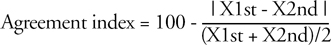
Two-dimensional transthoracic echocardiography (GE Vivid 7 with 4.0 MHz transducer; GE medical systems, Andover, MA, USA) was used to calculate EFT. We recorded three cycles of the two-dimensional parasternal long-axis view and the parasternal short-axis view at the basal left ventricular level. After increasing the depth of each view until the aortic and mitral valves were positioned lowest on the screen to allow for better visualization and accurate estimation of EFT. EFT was calculated on the free wall of the right ventricle (RV) in the still images obtained at end diastole on both parasternal long-axis and short-axis views. The anterior echolucent space between the RV outer wall and the linear echodense parietal pericardium was considered to be epicardial fat (Fig. 1). We excluded mediastinal fat, presenting as an echolucent area above the parietal pericardium, in the measurement of EFT. The mean value of two images obtained in the parasternal long-axis and short-axis views was calculated. To compare its change more objectively, we used standard images of the heart and tried to find as similar images of the previous study.
Statistical analysis
We used a commercial program, SPSS version 12.0 (SPSS Inc., Chicago, Illinois, USA), for statistical analysis. All continuous variables are expressed as mean ± SD and categorical variables are expressed as number and percentage. Intra- and inter-observer agreements in the EFT were tested using baseline echocardiography of 12 patients according to the statistical methods proposed by Bland and Altman.7)
Inter-and intra-observer agreements-All measurements were transformed to an equivalent percentage scale of agreement, according to the following formula;
in which X1st and X2nd are measures obtained in twice-repeated evaluation using same technique in the same patient. The measure of reproducibility was 2 SD of the intraobserver and interobserver agreement indexes. Therefore, these coefficient of variations (COV) were equal to 2 SD of  . The intraobserver agreement was 93.8% and the COV was 3.2% (p < 0.001). The interobserver agreement was 93.2% and the COV was 4.6% (p < 0.001).
. The intraobserver agreement was 93.8% and the COV was 3.2% (p < 0.001). The interobserver agreement was 93.2% and the COV was 4.6% (p < 0.001).We compared the clinical characteristics of patients who were medicated with atorvastatin and simvastatin with ezetimibe. Between two groups, comparisons of continuous variables were performed using the independent sample t-test and categorical variables by using χ2 test. The effects of statins were analyzed by using paired sample t-test. A p-value less than 0.05 considered statistically significant.
Results
During the study period, 181 patients were scheduled to have the follow up coronary angiography as participants of clinical trial after six to eight months later. They were medicated with any type of cholesterol lowering agents. Eighty-eight, 65, and 23 patients were medicated with 20 mg of atorvastatin, 10 mg of simvastatin plus 10 mg of ezetimibe, and other statins, respectively. The remaining 5 patients were not treated with statins due to complications associated with statin use.
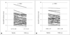
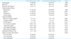
After exclusion of 36 patients (23 patients were treated with other statins, 8 patients without follow-up echocardiographies with optimal images, and 5 patients not treated with statins for any reason), we included 145 patients (58 females; mean, 63.5 ± 9.5 years). Of the 145 patients, 82 received 20 mg of atorvastatin (atorvastatin group) and 63 medicated with 10 mg of simvastatin with 10 mg of ezetimibe (simvastatin/ezetimibe group). Their baseline clinical characteristics were listed on the Table 1. There were tendencies of higher concentration of total cholesterol, triglycerides and hs-CRP in the simvastatin/ezetimibe group. However, there was no significant statistical difference. The pattern of coronary artery disease was similar in the two groups.
With statin treatments, total cholesterol concentration (189.1 ± 36.1 to 143.3 ± 36.5 mg/dL, p < 0.001), triglycerides (143.5 ± 65.5 to 124.9 ± 63.1 mg/dL, p = 0.005), LDL-cholesterol (117.4 ± 33.0 to 74.7 ± 30.6 mg/dL, p < 0.001), hs-CRP (3.8 ± 5.3 to 2.5 ± 2.9 mg/L, p = 0.034) and EFT (4.08 ± 1.37 to 3.76 ± 1.29 mm, p < 0.001) were significantly decreased. However, high density lipoprotein (HDL)-cholesterol (42.0 ± 9.5 to 42.3 ± 9.5 mm, p = 0.723) and body mass index (BMI)(25.1 ± 3.1 to 24.9 ± 3.3 kg/m2, p = 0.272) were not changed.
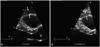
Atorvastatin and simvastatin/ezetimibe showed similar improvements in the cholesterol profiles. Their effects of statins were listed on Table 2. In the atorvastatin group, there was significant reduction of triglyceride concentration (136.4 ± 63.4 to 116.4 ± 58.4 mg/dL, p = 0.002). However, there was insignificant reduction of triglyceride concentration in the simvastatin/ezetimibe group (152.1 ± 69.6 to 135.6 ± 67.6 mg/dL, p = 0.387). Moreover, atorvastatin was associated with decreased EFT more significantly than simvastatin/ezetimibe (EFT change; 0.47 ± 0.65 in the atorvastatin vs. 0.12 ± 0.52 mm in the simvastatin/ezetimibe group; p = 0.001)(Fig. 2). The type of statin was the only independent factor associated with the EFT change as a result of multivariate analysis (Table 3).
Discussion
In this study, we demonstrated the effects of statins on epicardial fat in patients with CAD underwent PCI. Despite atorvastatin and simvastatin/ezetimibe equally lowered the cholesterol profiles in them, atorvastatin significantly decreased the EFT more than that of simvastatin/ezetimibe.
The epicardium, a visceral layer of the pericardium, is composed with mesothelial cells.8) Epicardial fat, accumulated adipose tissue covers 80% of the heart's surface, is present along the course of the coronary arteries, over the RV, anterior surface and at the apex. It is closely contacted to the myocardium and supplied by branches of the coronary arteries.9) Mature adipocytes are located in the epicardial fat especially anterior to the right ventricle (RV) and these may act as more readily available, direct sources of free fatty acid for cardiomyocytes.10)11) The reported mean value of the EFT in patients with CAD is about 4.0 mm,4) which is similar to our result.
Recently, the scientific and clinical interest in epicardial fat is rapidly growing.12-14) Iacobellis and Sharma14) showed that echocardiographic assessment of epicardial fat can serve as a new index of cardiac and visceral adiposity. Moreover, the epicardial fat has known to be clinically correlated with abdominal visceral adiposity measured by magnetic resonance imaging,15) presence of atherosclerosis,16)17) and coronary artery disease.4)18)
Though it is unknown how much visceral fat loss is needed to reduce cardiometabolic risk presently, the reduction of visceral adipose tissue is known to be associated with a significant improvement of the metabolic profile.19) Busetto et al.20) reported that visceral adipose tissue loss in the phase of rapid weight loss after laparoscopic adjustable silicone gastric banding.
Recently, the epicardial adipose tissue can be regarded as a therapeutic target during clinical interventions modulating the adipose tissue.21) Iacobellis et al.22) reported that significant reduction of epicardial fat after weight loss during sixmonth duration with very low calorie diet program in severely obese patients. Moreover, the reduction of epicardial adipose tissue was higher than changes of waist circumference and BMI. The reduction of visceral adiposity was related with the improvement of cardiac diastolic function.22) We demonstrated similar result of epicardial fat reduction by using statins within relatively short duration in patients with significant CAD underwent PCI. Though mean EFT was decreased 7.0% during the study period, the EFT was significantly decreased in the atorvastatin group than in the simvastatin/ezetimibe group (10.0 ± 15.0% in the atorvastatin group vs. 3.1 ± 13.3% in the simvastatin/ezetimibe group, p = 0.001). Statins inhibits HMG CoA reductase (rate-limiting step in cholesterol biosynthesis) in the liver, increases LDL-receptor expression and activity, and enhances removal of lipoprotein by LDL-receptor. It also reduces hepatic release of lipoprotein into the blood circulation.23) Despite of lipid lowering effects, statins have other pleiotrophic effects including inhibition of migration and proliferation of arterial myocytes, inhibition of macrophage growth, inhibition of metalloproteinase secretion, and Inhibition of cell adhesion.24) Currently, there are no studies which have demonstrated the statin effect (decrease) on the thickness of adipose tissues. However, metabolic syndrome is associated with increased visceral adiposity and increased production of pathologic adiopokines,25) and the use of atorvastatin and fenofibric acids is associated with decreased adipokine release from visceral adipose tissue.6) The use of statin might decrease the production of pathologic adipokines in the epicardial adipose tissue, and the reduced adiopokine production might subsequently be associated with a decrease in EFT.
There are many kinds of commercially available statins. Though statins share similar properties, there are several differences. Atorvastatin decreased the progression of carotid intima-media thickness in the familial hypercholesterolemia patients.26) However, simvastatin/ezetimibe combination therapy did not result in a significant difference in changes of carotid intima-media thickness.27) This different effects of statins on the EFT might be due to statin difference. In this study, the degree of cholesterol reduction and hs-CRP change were similar in the two groups. However, the exact mechanism of the EFT lowering was unknown. This might be originated from cholesterol independent mechanisms of statins.
Albeit this study provides original and intriguing findings, it may have also some limitations. First, this is a retrospective cohort study. The use of statin was not assigned randomly. Moreover, the patients without follow up echocardiography and inadequate echocardiographic image quality were excluded in this study. To evaluate this difference of the statins on the EFT, more large randomized trial will be needed. Second, the sample size in this study was relatively small. Though there is statistically significant difference in the lowering EFT, we believe further studies in a larger population are necessary to confirm these data. Third, the echocardiography might not be the optimal technique for quantification of epicardial fat. Because echocardiography measures EFT linearly, echocardiographic EFT may not reflect the total epicardial fat volume exactly. However, echocardiographic examination is relatively accurate, easier and reliable method. Moreover, it is more accessible than the magnetic resonance imaging or computerized tomographic scanning, and can be performed without giving any hazards including radiation exposure or toxicity of radiocontrast agents.
In conclusion, we demonstrated that statin, especially atorvastatin, use can be associated with significant reduction in the EFT, a marker of visceral adiposity, in patients with significant coronary artery disease.


 XML Download