

 PDF
PDF ePub
ePub Citation
 Print
Print


Introduction
Although a right atrial and inferior vena caval thrombus is an extremely rare condition, it can result in highly dangerous complications. The condition manifests mainly through its symptoms, which result from the occlusion of the tricuspid valve or from a pulmonary embolism. We encountered a rare thrombus case in which the patient came to the hospital showing dyspnea, was suspected, based on echocardiography, of having a myxoma in the right atrium and inferior vena cava, and was treated through surgical excision. The following is a detailed report of the case accompanied by a review of previous studies.
Case
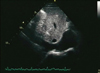
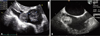
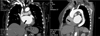
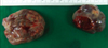
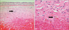
A 66-year-old female patient came to the hospital complaining of dyspnea that began recently and was becoming increasingly worse. She had previously been diagnosed with atrial fibrillation at the same hospital and was under follow-up observation while being treated with aspirin. When she came in for an appointment two months prior, she did not complain any particular symptoms besides mild dyspnea. The echocardiography conducted four years earlier had not shown a right atrial mass, although it did show mild aortic stenosis, mild mitral stenosis, and moderate mitral regurgitation (Fig. 1). The chest X-ray taken during the current admission showed mild cardiomegaly. The electrocardiography showed atrial fibrillation, as was found previously. Laboratory tests were all normal. Autoimmune tests and hypercoagulability tests were all normal. During the echocardiography, two large, well-defined lobulated masses with echo-free space were discovered in the right atrial septal wall and the inferior vena cava. The masses measured 3.2×2.9 cm (right atrium) and 3.1×3.0 cm (inferior vena cava) (Fig. 2). Chest computed tomography (CT) showed two well-defined masses in the right atrium and inferior vena cava (Fig. 3). The findings from echocardiography led to a diagnosis of myxoma, and the patient was transferred to the department of cardiothoracic surgery to undergo a procedure to excise the masses. During surgery, we can observe smooth-surfaced round masses (Fig. 4). Histologic examination revealed the tumor to be an organized thrombus without neoplastic tissue (Fig. 5). The co-excised atrial tissue did not have an any inflammatory change in the endocardium and myocardium. She had an uneventful postoperative course and was discharged with warfarin in good general condition. At present, warfarin therapy has been continued after discharge and she has remained asymptomatic.
Discussion
Echocardiography is routinely utilized to assess cardiac function and chamber size. It has been become a valuable tool with which to diagnose intra-cardiac masses in patients with atrial fibrillation. The differential diagnoses of intraatrial masses include atrial myxoma, thrombus related to atrial fibrillation, vegetation, metastatic tumors, and primary benign or malignant tumors.1) Atrial myxomas are the most common cardiac tumors. Although they are typically found in the left atrium, they may arise in the right atrium in a substantial proportion of patients.1) They are usually large mobile ovoid masses, and occur in the setting of a normal sized right atrium. In addition, they often arise from the interatrial septum. Although a thrombus usually shows up differently in an echocardiographic image than does a myxoma,2) on echocardiography the thrombus in our case had features similar to those of a myxoma. Therefore, it was not until the pathological diagnosis was established that we could differentiate a thrombus from a myxoma. The recent study indicates that myxomas and thrombi can be accurately differentiated using CT by assessing the distinguishing features of size, origin, shape, mobility and prolapse.3)
Thrombi are rare in the structurally normal heart, except for catheter related thrombi, and can be found in patients with hypercoagulability, malignant tumors,4) ulcerative colitis,5) and Behcet's disease.6) Right atrial thrombi are also seen in low-output states, cardiomyopathy, and cardiac arrhythmias.7) Thrombi that originate within the heart generally occur in the left heart and seldom in the right heart. Wartman and Hellerstein reported that of 2,000 autopsies, only 14 cases were found to right side thrombi.8)
When a right sided intracardiac mass is found, it is important to differentiate the type and the shape of the mass by transesophageal echocardiography. Transesophageal echocardiography allows accurate observation of the atrial septum and is useful in understanding the shape of masses in the right atrium.9)
However, in many cases, it is difficult to make an accurate preoperative diagnosis. Right sided thromboses are associated with an increased risk of pulmonary embolism and must be surgically removed to be accurately diagnosed. If the thrombus is not securely attached to the atrial septum, it may produce a pulmonary embolism.
Patients with a right atrial thrombus have several therapeutic options. For the patient at high risk for surgery, anticoagulation with heparin followed by an ongoing regimen of warfarin may be the treatment of choice.10) Thrombolytic therapy is also reported to be successful in some patients,11) although the possibility of a pulmonary embolism caused by a fragmented thrombus due to thrombolytic therapy is a serious concern. Therefore, whenever the patients are able to sustain cardiopulmonary bypass they should undergo surgical intervention and thereby avoid a poor outcome.
 XML Download