

 PDF
PDF ePub
ePub Citation
 Print
Print


Introduction
Papillary fibroelastomas have been reported to arising from almost any cardiac structure including papillary muscle, chordae tendineae, and endocardium, however the majority, approximately 80%, occur on the cardiac valves.1) The majority of fibroelastomas have been described on the left side of the heart involving the aortic and mitral valves. We present a rare case of a papillary fibroelastoma occurring on the pulmonic valve.
Case
Recently, we experienced a 62 year-old female patient who had a pulmonary stenosis with mobile mass on pulmonary valve. She visited the emergency room for abdominal discomfort and dyspepsia for 5 days. Although she was already diagnosed to have pulmonary stenosis several years ago, she was not treated.
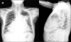
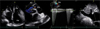
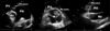
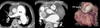
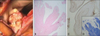
On physical examinations, we could hear grade V systolic ejection murmur at pulmonic area. Her liver was palpated about three finger. Her chest films revealed marked cardiomegaly with prominent pulmonary artery and subsegmental atelectasis on left upper lung field (Fig. 1). On echocardiography, right atrium, right ventricle, pulmonary artery and inferior vena cava were marked dilated. Mild to moderate pulmonary valve stenosis and regurgitation were revealed (Fig. 2). There was hyper-mobile frond-like structure on pulmonary valve leaflet in parasternal view unusually. The 1.2×0.9 cm sized echogenic mass arose from the right cusp of the pulmonary valve and protruded into the pulmonary artery during systole (Fig. 3). In computer tomography scan, there was markedly dilated right side heart chamber and nodular structure on pulmonary valve and no evidence of pulmonary thromboembolism (Fig. 4). Because of the fronds of the lesion and combined stenotic valve, vegetations could not be excluded. The presumptive diagnosis was infective endocarditis despite of negative result of blood culture. But, she had no history of fever and no abnormal laboratory findings such as leucocytosis or elevated acute phase reactants. Although the mass was located on right side valve, we decided surgical removal to prevent its potential thromboembolic risks and to relieve stenotic symptoms. She was replaced the pulmonary valve and reconstructed the right ventriclular outflow tract. Surgical pathology revealed papillary fibroelastoma of the pulmonary valve (Fig. 5). She recovered from abdominal discomfort and dyspepsia after surgery. We concluded she had presented right heart failure symptom due to papillary fibroelastoma on pulmonic valve.
Discussion
A mass attached to a cardiac valve can be a tumor, or thrombus, or vegetation. Tumors on the cardiac valves are mostly benign and include fibroelastoma, myxoma, lipoma, and Lambl's excrescences. Fibroelastomas are therefore exceedingly rare primary cardiac tumors and affect the aortic, mitral, tricuspid, and pulmonary valve in this order.2) Papillary fibroelastomas are usually single, but multiple tumors have been reported. The pathogenesis of papillary fibroelastoma is unknown. Papillary fibroelastomas sometimes exhibit a surface lamination, a finding consistent with the concept of growth by successive organization of fibrin deposits. The papillary fibrous surface is considered to result from exposure to the hemodynamic stress of flowing blood.3)4) Lambl excrescences origin was suggested, but most authors believe that papillary fibroelastoma is a separate entity.5) Other theories suggest that papillary fibroelastoma represent neoplasms, hamartomas, and inflammatory nodules.6)7)
Histologically fibroelastomas are encased by endothelium which envelopes a core of connective tissue. This loose connective tissue contains a mucopolysaccharide acid matrix, smooth muscle cells, collagen, elastin fibers, and occasional cysts and areas of hemorrhage.8)9) Despite their benign histology, it should be excised because of their embolic complications. Fibroelastomas have been reported as a cause of distal embolic disease and stroke.10)11) Surface thrombus is common with these tumors, but warfarin administration is not a protective measure, since patients still present with transient ischemic attacks despite warfarin therapy. Emboli may originate either from fragments of the tumor or from a thrombus formed around the tumor. Papillary fibroelastoma is a friable tumor, and aggressive manipulation may result in fragmentation and further embolism.12) Although these tumors are benign and rarely cause valvular dysfunction, the most appropriate therapy for papillary fibroelastomas is surgical resection due to this potential for embolic complications. Surgery should be performed with minimal manipulation of the tumor and inspection of all four cardiac chambers to check if the tumor is multifocal and to provide adequate exposure for complete resection.13)14) Anticoagulation was still controversy.15)
Papillary fibroelastomas of the pulmonic valve have been infrequently reported. This is a relatively rare case of pulmonary valve fibroelastoma mimicking vegetation treated with surgical resection.
 XML Download