

 PDF
PDF ePub
ePub Citation
 Print
Print


Introduction
Cardiac myxoma is the most frequent benign cardiac tumor.1) Its various clinical presentations are mainly related to the location of the mass. Classic presentations include obstructive cardiac signs, embolic events and constitutional manifestations. ECG abnormalities are not uncommon and most frequent findings are left atrial hypertrophy. But atrial flutter is known to be very rare.2)
Cases
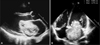
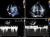
A 41-year-old man presented with palpitation and dyspnea on exertion for 3 days. He had no history of diabetes, hypertension and smoking. He felt mild dyspnea 3 years ago, but ignored it. Initial vital signs were: blood pressure of 110/70 mmHg, heart rate of 162 beats/min, and respiratory rate of 20 breaths/min. On physical examination, heart murmurs were rarely audible due to rapid heart rate and other findings were unremarkable. On laboratory findings, BNP level was 445 pq/ml, and CRP level was 3.4 mg/dl (reference range 0-1 mg/dl). The initial electrocardiogram (ECG) revealed atrial flutter with 4:1 AV block and 75 beats/min of ventricular rate (Fig. 1). After 30 minutes, the patient's blood pressure dropped to 90/60 mmHg and the heart rate rose to 150-160 beats/min on ECG monitoring. We performed trans-thoracic echocardiography (TTE) for evaluation of dyspnea, which revealed a large round mobile mass in the left atrium (Fig. 2). The mass was so large that it nearly obliterated the left atrial cavity and it had central necrosis. And grade IV/IV mitral regurgitation and grade II/IV tricuspid regurgitation were showed. Peak velocity of tricuspid regurgitation was 4.2 m/s, estimated right ventricular systolic pressure was 80 mmHg (Fig. 3). We thought that the mass was likely to be a myxoma and performed transesophageal echocardiography (TEE) and a chest computerized tomography (CT) for further evaluation. On TEE, the mass was attached to inter-atrial septum without stalk, and no other mass was founded. Chest CT scan also showed similar results. Then the patient was sent to the operating room. The mass, which was attached to the fossa ovalis and 7×4×3 cm in size, was removed and the septal defect was repaired. Histological findings were compatible with myxoma (Fig. 4). After surgery, the patien's rhythm on ECG converted to normal sinus rhythm (Fig. 1). Follow-up echocardiography showed neither remnant mass nor shunts. Mitral regurgitation decreased to mild and peak velocity of tricuspid regurgitation also decreased to 1.67 m/s (Fig. 3). The patient was discharged in stable condition.
Discussion
Clinical features of myxoma vary in each patient. Electrocardiographic abnormalities associated with myxomas are also variable. Left atrial hypertrophy is the most frequent finding; ST-segment abnormalities or nonspecific ECG abnormalities, and ventricular hypertrophy can occur. But atrial flutter and conduction disturbances are rare. Keeling et al. reported that myxomas in the context of rhythms other than sinus and arising from the left atrium were associated with a significant risk of embolism.3) Pinede et al. reported that ECG abnormalities are associated with systemic manifestations and cardiac signs, but not with embolic or neurological symptoms.2) However, Nadeem et al. reported a left atrial myxoma case presenting with ventricular fibrillation, which had significant association with embolic symptoms;4) histologic finding showed surface thrombus and extensive myxoid background.
In this case, the initial presentation was atrial flutter, a very unusual finding, which was converted to normal sinus rhythm successfully after surgical resection; the atrial flutter may be due to overloading of the left atrium by both the large tumor mass itself and secondary mitral regurgitation. Atrial flutter is known as a right atrial rhythm and a feature that renders this case of left atrial myxoma presenting with atrial flutter very unusual. The functional mitral stenosis resulting from the large myxoma probably underlies atrial flutter. On occasion, atrial flutter develops in the setting of mitral stenosis due to severe pulmonary hypertension induced right atrial dilatation. In mitral stenosis with atrial flutter, PMV (Percutaneous Mitral Valvuloplasty) can eliminate atrial flutter. In this case, atrial flutter converted to sinus rhythm after removal of myxoma.
Another possible etiology for atrial flutter may be related to inflammatory processes. There are several reports on increased interleukin-6 (IL-6) levels in atrial flutter patients and decreased IL-6 levels after curative atrial flutter ablation.5) Myxoma is well known for its paracrine properties including increased secretion of inflammatory cytokines such as IL-6.6) Surgical removal of the myxoma decreased IL-6 levels, which in turn was associated with elimination of the atrial arrhythmia. Recent research documented that the inflammatory process is also important in atrial fibrillation.7) Unfortunately however, we could not measure IL-6 levels before surgery.
The precise mechanism of atrial flutter in this case could not be evaluated because of very large left atrial mass and the fact that atrial arrhythmia could increase thrombogenicity and the likelihood of an embolic episode during an electrophysiology study.


 XML Download