

 PDF
PDF ePub
ePub Citation
 Print
Print


Jung-Won Hwang, MD1, Un-Jung Choi, MD1, Sung-Gyun Ahn, MD1 , Hong-Seok Lim, MD1, Soo-Jin Kang, MD1, Byoung-Joo Choi, MD1, So-Yeon Choi, MD1, Myeong-Ho Yoon, MD1, Gyo-Seung Hwang, MD1, Seung-Jea Tahk, MD1, Joon-Han Shin, MD1, Doo-Kyung Kang, MD2
, Hong-Seok Lim, MD1, Soo-Jin Kang, MD1, Byoung-Joo Choi, MD1, So-Yeon Choi, MD1, Myeong-Ho Yoon, MD1, Gyo-Seung Hwang, MD1, Seung-Jea Tahk, MD1, Joon-Han Shin, MD1, Doo-Kyung Kang, MD2
Abstract
Background
Several studies suggested that epicardial adipose tissue (EAT) might be associated with metabolic syndrome and coronary atherosclerosis. But, little had been studied whether the thickness of EAT on echocardiography could represent the whole amount of EAT. The purpose of this study was to identify the best echocardiographic methods reflecting total amount of EAT.
Methods
Sixty subjects (32 women, mean: 58±12 years-old) who underwent 64-slice multidetector computed tomography (MDCT) were consecutively enrolled. All CT scanning was performed one Brilliance CT-64-channel configuration scanner (Philips, Cleveland, USA) and axially contiguous 10-mm-thickeness sections were obtained from aortic valve to diaphragm level. EAT area was manually traced in each slice and summed up. The EAT thickness was measured as the echo-lucent or echo-dense space between epicardium and pericardium at parasternal long-axis, modified 4-chamber, and apical 4-chamber view.
Results
The EAT thickness at parasternal long-axis and modified 4-chamber view and the sum of EAT thickness from each views (median thickness: 1.0, 2.8, 1.1 and 5.0 mm, respectively) were all correlated with total EAT area on MDCT. Among echo parameters, the EAT thickness measured on parasternal long-axis view during diastole correlated best with total EAT area on MDCT (r=0.572, p<0.001).
Conclusion
The echocardiographic EAT measurement might be easily accessible and less harmful method representing whole amount of EAT. The measurement of the thickness of EAT on parasternal long-axis view during diastole by echocardiography might be feasible and reliable in the studying field of EAT.
Figures and Tables
Fig. 1
Measurement of total epicardial fat by computed tomography. EAT was manually traced on each MDCT slice from aortic valve to diaphragm level and then EAT area (area which was colored) was calculated automatically through PACS system (PI view STAR, version 5025 Infinitt, Seoul, Korea). The total EAT area was defined as the sum of each EAT area. EAT: epicardial adipose tissue, MDCT: multidetector computed tomography, PACS: picture archiving and communication system.

Fig. 2
Measurement of epicardial fat thickness by Echocardiogram. The thickness of EAT, MAT, and PAT was measured at parasternal long axis, modified 4-chamber, and apical 4-chamber view. EAT thickness was defined as distance from outer side of myocardium to pericardium, MAT thickness from pericardium to inferior side of sternum, and PAT thickness from outer side of myocardium to inferior side of sternum. EAT: epicardial adipose tissue, MAT: mediastinal adipose tissue, PAT: pericardial adipose tissue, PLX: parasternal long axis view, M4C: modified 4-chamber view, A4C: apical 4-chamber view, LV: left ventricle.
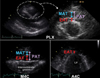
Fig. 3
Correlation of the thickness of epicardial adipose tissue by transthoracic echocardiography at parasternal long axis view and the area of epicardial adipose tissue by multidetector computed tomography. EAT: epicardial adipose tissue, MDCT: multidetector computed tomography, PLX: parasternal long axis view.
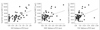
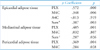
Table 2
Measurement of parameters using transthoracic echocardiography & multidetector computed tomography
References
1. Hyun DW, Kim KS. Change in coronary blood blow and coronary blood flow reserve after a single high-fat and low-fat meal in patients with ischemic heart disease assessed by transthoracic echocardiography. J Kor Soc Echo. 2003. 24–30.

2. Iacobellis G, Sharma AM. Obesity and the heart: redefinition of the relationship. Obes Res. 2007. 8:35–40.
3. Dagenais GR, Yi Q, Mann JF, Bosch J, Pogue J, Yusuf S. Prognostic impact of body weight and abdominal obesity in women and men with cardiovascular disease. Am Heart J. 2005. 149:54–60.
4. Kwon KY, Han JH. Clinical significance of visceral adipose tissue. J Korean Acad Fam Med. 2007. 28:729–747.
5. Iacobellis G, Assael F, Ribaudo MC, Zappaterreno A, Alessi G, Mario UD, Leonetti F. Epicardial fat from echocardiography: a new method for visceral adipose tissue prediction. Obese Res. 2003. 11:304–310.
6. Jeong GH, Kim SK, Chung JO, Cho DH, Chung DJ, Chung MY. Association between ultrasonographic visceral fat indices and cardiovascular risk factors in type 2 diabetes patients. Korean J Intern Med. 2007. 73(6):618–630.
7. Sacks HS, Fain JN. Human epicardial adipose tissue: a review. Am Heart J. 2007. 153:907–917.
8. Ahn SG, Lim HS, Joe DY, Kang SJ, Choi BJ, Choi SY, Yoon MH, Hwang GS, Tahk SJ, Shin JH. Relationship of Epicardial Adipose Tissue by Echocardiography to Coronary Artery Disease. Heart Heart. 2008. 94:e7.
9. Baik SH, Ahn SG, Choi JH, Koh BR, Yoo JH, Kang SJ, Choi BJ, Choi SY, Yoon MH, Tahk SJ, Shin JH. The relationship of epicardial adipose tissue to metabolic syndrome and cardiovascular risk factors. Korean J Med. 2007. 72:245–252.
10. Jeoung JW, Jeoung MH, Yun KH, Oh SK, Park EM, Kim YK, Rhee SJ, Lee EM, Lee J, Yoo NJ, Kim NH, Park JC. Echocardiographic epicardial fat thickness and coronary artery disease. Circ J. 2007. 71:536–539.
11. Flüchter S, Haghi D, Dinter D, Heberlein W, Kühl HP, Neff W, Sueselbeck T, Borggrefe M, Papabassiliu T. Volumetric assessment of epicardial adipose tissue with cardiovascular magnetic resonanace imaging. Obesity. 2007. 15:870–878.
12. Iacobellis G, Ribaudo MC, Assael F, Vecci E, Tiberti C, Zappaterreno A, Di Mario U, Leonetti F. Echocardiographic epicardial adipose tissue is related to anthropometric and clinical parameters of metabolic syndrome: a new indicator of cardiovascular risk. J Clin Endocrinol Metab. 2003. 88:5163–5168.
13. Mazurek T, Zhang L, Zalewski A, Mannion JD, Diehl JT, Arafat H, Sarov-Blat L, O'Brien S, Keiper EA, Johson AG, Murtin J, Goldstein BJ, Shi Y. Human epicardial adipose tissue is a source of inflammatory mediators. Circulation. 2003. 108:2460–2466.
14. Baker AR, Silva NF, Quinn DW, Harte AL, Pagano D, Bonser RS, Kumar S, McTernan PG. Human epicardial aidpose tissue expresses a pathogenic profile of adipocytokines in patients with cardiovascular disease. Cardiovasular Diabetology. 2006. 5:1–8.
 XML Download