

 PDF
PDF ePub
ePub Citation
 Print
Print


Abstract
In this study, we examined whether melatonin promotes apoptotic cell death via p53 in prostate LNCaP cells. Melatonin treatment significantly curtailed the growth of LNCaP cells in a dose- and time-dependent manner. Melatonin treatment (0 to 3 mM) induced the fragmentation of poly(ADP-ribose) polymerase (PARP) and activation of caspase-3, caspase-8, and caspase-9. Moreover, melatonin markedly activated Bax expression and decreased Bcl-2 expression in dose increments. To investigate p53 and p21 expression, LNCaP cells were treated with 0 to 3 mM melatonin. Melatonin increased the expressions of p53, p21, and p27. Treatment with mitogen-activated protein kinase (MAPK) inhibitors, PD98059 (ERK inhibitor), SP600125 (JNK inhibitor) and SB202190 (p38 inhibitor), confirmed that the melatonin-induced apoptosis was p21-dependent, but ERK-independent. With the co-treatment of PD98059 and melatonin, the expression of p-p53, p21, and MDM2 did not decrease. These effects were opposite to the expression of p-p53, p21, and MDM2 observed with SP600125 and SB202190 treatments. Together, these results suggest that p53-dependent induction of JNK/p38 MAPK directly participates in apoptosis induced by melatonin.
References
1. Ben Sahra I, Laurent K, Giuliano S, Larbret F, Ponzio G, Gounon P, Le Marchand-Brustel Y, Giorgetti-Peraldi S, Cormont M, Bertolotto C, Deckert M, Auberger P, Tanti JF, Bost F. Targeting cancer cell metabolism: the combination of metformin and 2-deoxyglucose induces p53-dependent apoptosis in prostate cancer cells. Cancer Res. 2010; 70:2465–2475.

2. Yu CX, Zhang XQ, Kang LD, Zhang PJ, Chen WW, Liu WW, Zhang JY. Emodin induces apoptosis in human prostate cancer cell LNCaP. Asian J Androl. 2008; 10:625–634.
3. Lee DH, Kim C, Zhang L, Lee YJ. Role of p53, PUMA, and Bax in wogonin-induced apoptosis in human cancer cells. Biochem Pharmacol. 2008; 75:2020–2033.
4. Logan IR, McNeill HV, Cook S, Lu X, Lunec J, Robson CN. Analysis of the MDM2 antagonist nutlin-3 in human prostate cancer cells. Prostate. 2007; 67:900–906.
5. Sainz RM, Mayo JC, Tan DX, León J, Manchester L, Reiter RJ. Melatonin reduces prostate cancer cell growth leading to neuroendocrine differentiation via a receptor and PKA independent mechanism. Prostate. 2005; 63:29–43.
6. Tam CW, Mo CW, Yao KM, Shiu SY. Signaling mechanisms of melatonin in antiproliferation of hormone-refractory 22Rv1 human prostate cancer cells: implications for prostate cancer chemoprevention. J Pineal Res. 2007; 42:191–202.
7. Cucina A, Proietti S, D'Anselmi F, Coluccia P, Dinicola S, Frati L, Bizzarri M. Evidence for a biphasic apoptotic pathway induced by melatonin in MCF-7 breast cancer cells. J Pineal Res. 2009; 46:172–180.
8. Martín-Renedo J, Mauriz JL, Jorquera F, Ruiz-Andrés O, González P, González-Gallego J. Melatonin induces cell cycle arrest and apoptosis in hepatocarcinoma HepG2 cell line. J Pineal Res. 2008; 45:532–540.
9. Xi SC, Tam PC, Brown GM, Pang SF, Shiu SY. Potential involvement of MT1 receptor and attenuated sex steroid induced calcium influx in the direct anti-proliferative action of melatonin on androgen-responsive LNCaP human prostate cancer cells. J Pineal Res. 2000; 29:172–183.
10. Lupowitz Z, Zisapel N. Hormonal interactions in human prostate tumor LNCaP cells. J Steroid Biochem Mol Biol. 1999; 68:83–88.
11. Moretti RM, Marelli MM, Maggi R, Dondi D, Motta M, Limonta P. Antiproliferative action of melatonin on human prostate cancer LNCaP cells. Oncol Rep. 2000; 7:347–351.
12. Xi SC, Siu SWF, Fong SW, Shiu SYW. Inhibition of androgen sensitive LNCaP prostate cancer growth in vivo by melatonin: association of antiproliferative action of the pineal hormone with MT1 receptor protein expression. Prostate. 2001; 46:52–61.
13. Joo SS, Yoo YM. Melatonin induces apoptotic death in LNCaP cells via p38 and JNK pathways: therapeutic implications for prostate cancer. J Pineal Res. 2009; 47:8–14.
14. Galluzzi L, Morselli E, Kepp O, Tajeddine N, Kroemer G. Targeting p53 to mitochondria for cancer therapy. Cell Cycle. 2008; 7:1949–1955.
15. Califice S, Waltregny D, Castronovo V, van den Brûle F. Prostate carcinoma cell lines and apoptosis: a review. Rev Med Liege. 2004; 59:704–710.
16. Shieh SY, Ikeda M, Taya Y, Prives C. DNA damage-induced phosphorylation of p53 alleviates inhibition by MDM2. Cell. 1997; 91:325–334.
17. Kim YS, Lee HJ, Jang C, Kim HS, Cho YJ. Knockdown of RCAN1.4 Increases Susceptibility to FAS-mediated and DNA-damage-induced Apoptosis by Upregulation of p53 Expression. Korean J Physiol Pharmacol. 2009; 13:483–489.
18. Bulavin DV, Saito S, Hollander MC, Sakaguchi K, Anderson CW, Appella E, Fornace AJ Jr. Phosphorylation of human p53 by p38 kinase coordinates N-terminal phosphorylation and apoptosis in response to UV radiation. EMBO J. 1999; 18:6845–6854.
19. Huang C, Ma WY, Maxiner A, Sun Y, Dong Z. p38 kinase mediates UV-induced phosphorylation of p53 protein at serine 389. J Biol Chem. 1999; 274:12229–12235.
20. She QB, Chen N, Dong Z. ERKs and p38 kinase phosphorylate p53 protein at serine 15 in response to UV radiation. J Biol Chem. 2000; 275:20444–20449.
21. Sayed M, Kim SO, Salh BS, Issinger OG, Pelech SL. Stress-induced activation of protein kinase CK2 by direct interaction with p38 mitogen-activated protein kinase. J Biol Chem. 2000; 275:16569–16573.
22. Hu MC, Qiu WR, Wang YP. JNK1, JNK2 and JNK3 are p53 N-terminal serine 34 kinases. Oncogene. 1997; 15:2277–2287.
23. Kwon YW, Ueda S, Ueno M, Yodoi J, Masutani H. Mechanism of p53-dependent apoptosis induced by 3-methylcholanthrene. J Biol Chem. 2002; 277:1837–1844.
Fig. 1.
Viability of melatonin-treated LNCaP cells. LNCaP cell viability was determined using the Cell Counting Kit-8 assay (A) 48 hr after exposure to melatonin at varying doses and (B) at varying times after exposure to 3 mM melatonin. In (A) and (B), results for cells not treated with melatonin are shown for comparison. Results are the means of 3 independent experiments (bars represent SD). ∗∗p<0.01, ∗∗∗p<0.001 vs. control.
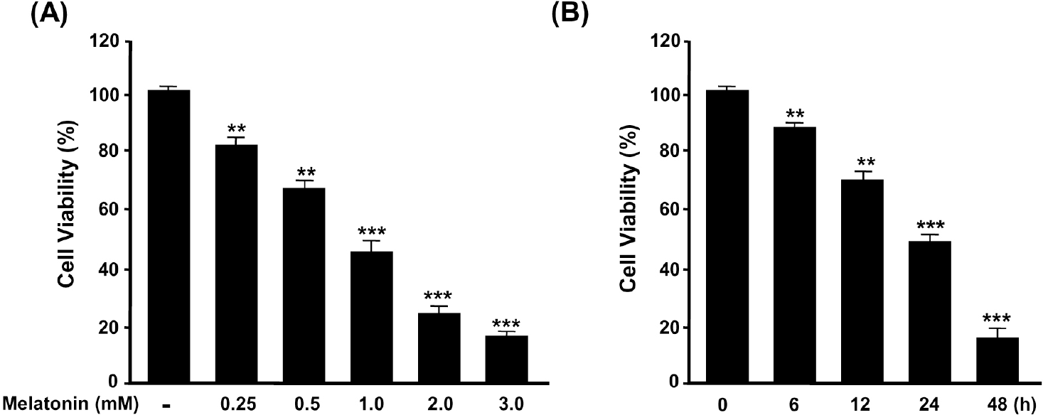
Fig. 2.
Induction of LNCaP cell apoptotic cell death by melatonin. LNCaP prostate cancer cells were cultured in DMEM containing 10% FBS and then treated with melatonin at varying doses for 48 h. (A) Cell lysates prepared at the indicated culture times were separated by 10% SDS-PAGE and immunoblotted with antibodies to PARP, caspase-3, –8, –9, and GAPDH. (B) Cell lysates prepared at the indicated culture times were separated by 12% SDS-PAGE and immunoblotted with antibodies to Bax, Bcl-2, and GAPDH. (C) The relative amounts of Bax and Bcl-2 were quantified as described in Materials and Methods. ∗∗∗p<0.001 vs. control. ††p<0.01, †††p<0.001 vs. control.
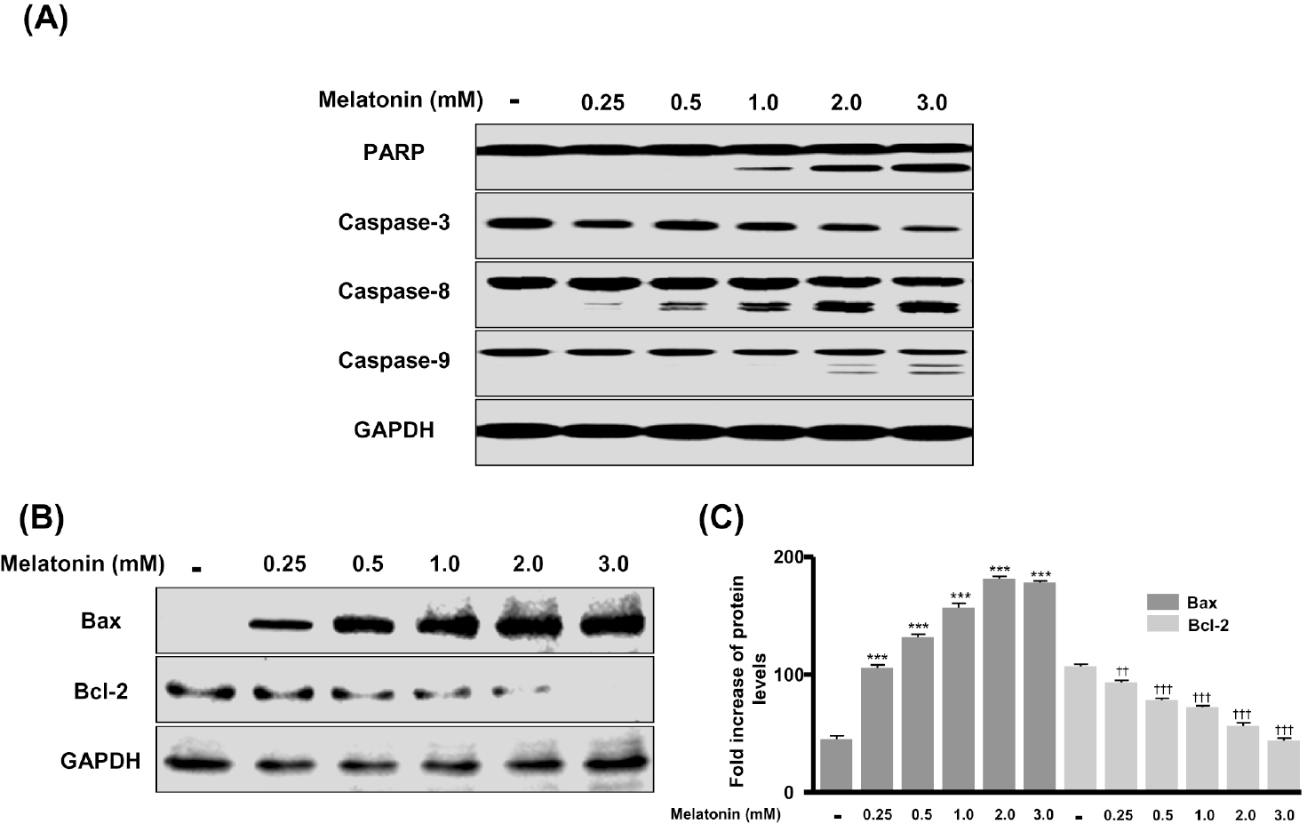
Fig. 3.
Activation of p53, p21, and p27 in melatonin-treated LNCaP cells. Cells were cultured in DMEM medium containing 10% FBS and then treated with melatonin at varying doses for 48 h. (A) Cell lysates prepared at the indicated culture times were separated by 12% SDS-PAGE and immunoblotted with antibodies to p53, p21, p27, and GAPDH. The relative amounts of p53 (B), p21 (C), and p27 (D) were quantified as described in Materials and Methods. ∗∗p<0.01, ∗∗∗p<0.001 vs. control.
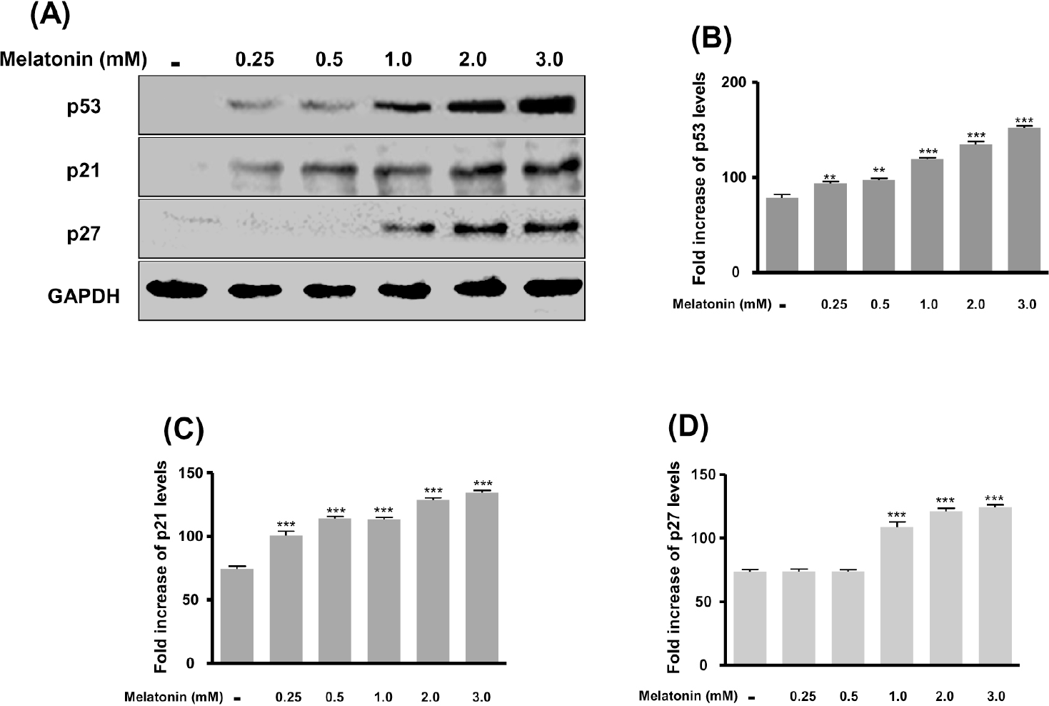
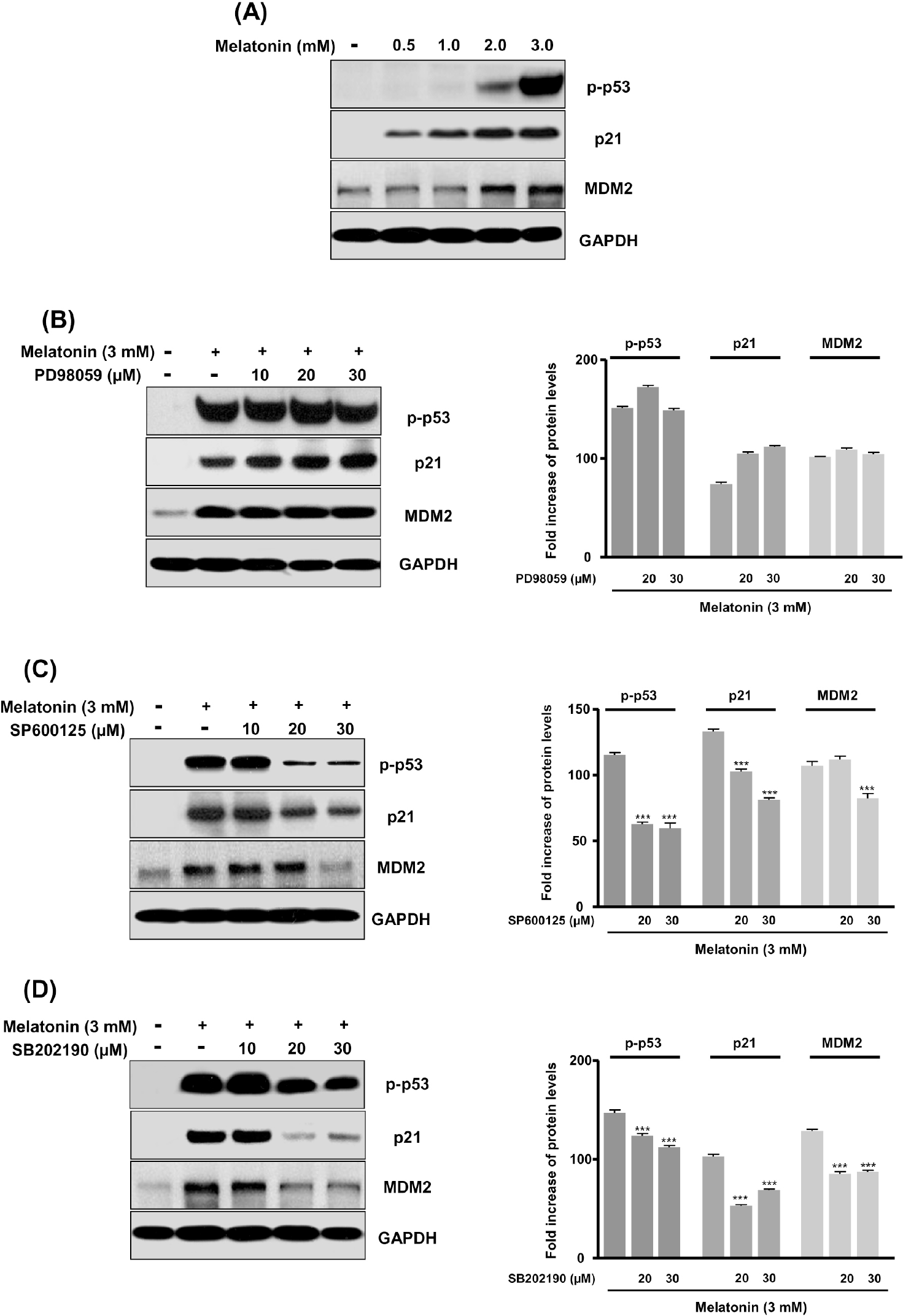
Fig. 4.
Activation of p-p53, p21, and MDM2 by blockade with PD98059, SP600125, and SB202190 inhibitors on melatonin-induced apoptotic cell death. Inhibitor concentrations of 10 to 30 μM for PD98059, SP600125 and SB202190 were added to cells 1 hr before melatonin (3 mM) treatment for 48 hr. (A) LNCaP cells were cultured with melatonin at varying doses. (B), (C), and (D) Cells were treated with 3 mM melatonin for 48 hr in the presence of various concentrations of PD98059 (B), SP600125 (C), or SB202190 (D). Cell lysates prepared at the indicated culture times were separated by 12% SDS-PAGE and immunoblotted with antibodies to p-p53, p21, MDM2, and GAPDH. ∗∗∗p<0.001 vs. control.
 XML Download