

 PDF
PDF ePub
ePub Citation
 Print
Print


Introduction
The occurrence of diabetes mellitus (DM), especially type 2 DM, is increasing at an epidemic rate worldwide [1]. Controlling blood glucose is one of the major goals for DM treatment [2]. Although enormous advances have been made in the development and clinical application of oral hypoglycemic agents, most current hypoglycemic agents have undesirable side effects and reduced efficacy over time [3]. Therefore, there have been extensive searches for naturally-derived antidiabetic agents with fewer side effects. Quercetin (QE), a flavonoid antioxidant, is a leading potential candidate for treating DM [4-8]. The long-term consumption of QE appears to control blood glucose levels in streptozotocin (STZ)-induced diabetic animals [4-8]. It has been suggested that QE protects the pancreas against oxidative stress in STZ-treated animals, improving hyperglycemia [4,5].
It has also been reported that QE inhibits α-glucosidase activity in vitro [9,10]. α-Glucosidase inhibitors are oral hypoglycemic agents that inhibit the digestion of carbohydrates in the small intestine, delaying increases in blood glucose after a meal. They are used to control both fasting and postprandial hyperglycemia in patients [11]. Since both fasting and postprandial glucose are important in determining overall glycemic control [12], α-glucosidase inhibitors are useful in achieving optimal blood glucose control in patients with type 2 DM. Plant extracts with α-glucosidase inhibitory activity in vitro, such as pine [13] and chestnut skin [14], have a flattening effect on postprandial glucose levels in STZ-treated rats, an animal model of type 1 DM, and their chronic administration leads to overall control of fasting glucose levels in animal models of type 2 DM.
Therefore, QE is expected to be effective in reducing postprandial glucose response. However, its inhibitory activity against α-glucosidase has not been fully investigated in vivo. We examined the acute effects of QE on postprandial hyperglycemia in STZ-induced diabetic rats using a carbohydrate-loading test and compared its effect with that of acarbose, a competitive α-glucosidase inhibitor. We also examined the chronic effect of QE on fasting hyperglycemia and intestinal maltase activity in db/db mice, an animal model of type 2 DM, to evaluate its potential as a hypoglycemic agent.
Materials and Methods
Reagents
We used a glucose assay kit from Yeongdong Co. (Seoul, Korea), an insulin assay kit from Linco Co. (St. Charles, MO, USA), and a glycated hemoglobin (HbA1C) assay kit from BioSystems (Barcelona, Spain). Casein, L-cysteine, mineral mixture, and vitamin mixture were purchased from ICN Pharmaceuticals Inc. (Costa Mesa, CA, USA). Tert-butyl hydroquinone was purchased from Fluka Co. (Milwaukee, WI, USA). Cornstarch was acquired from Daesang Co. (Seoul, Korea), and sucrose and soybean oil from Cheiljedang Co. (Seoul, Korea). Acarbose was obtained from Bayer Korea Ltd. (Seoul, Korea). QE, STZ, Alphacel, choline bitartrate, and all other chemical reagents used in this study were purchased from Sigma Chemical Co. (St. Louis, MO, USA).
Measurement of control of postprandial hyperglycemia in STZ-induced diabetic rats
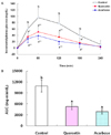
Male Sprague-Dawley rats weighing 240-260 g (Bio Genomics, Inc., Seoul, Korea) were housed under standard laboratory conditions (24 ± 5℃ and 55 ± 5% relative humidity, with a 12-hr light:12-hr dark cycle). The rats were fed a commercial chow (Samyang Co., Seoul, Korea) ad libitum for 2 weeks after arrival. The animals were injected intraperitoneally with STZ (65 mg/kg) in citrate buffer at pH 4.5 [15]. After one week, fasting glucose was measured by tail-vein sampling using a glucometer (Glucotrend, Roche Diagnostics, UK). Animals with fasting blood glucose levels between 200 and 400 mg/dL were selected and randomly assigned to one of 3 groups (n = 18). After an overnight fast, rats were administered a soluble starch (1 g/kg, control group), starch with QE (100 mg/kg), or acarbose (40 mg/kg) via gastric intubation [16]. Tail tip blood samples were obtained 0, 30, 60, 120, 180, and 240 min afterward and were centrifuged at 1,500 g for 15 min. Plasma glucose was measured enzymatically [17] using a commercial kit; levels were expressed relative to the baseline, and areas under the response curves (AUCs) were calculated using the trapezoidal rule.
Measurement of control of fasting hyperglycemia in db/db mice
To study the chronic effect of QE on fasting hyperglycemia, five-week-old male C57BL/KsJ-db/db mice (n = 18) were obtained from Japan SLC, Inc. (Hamamatsu, Japan). After 1 week of adaptation, during which time the animals had free access to commercial chow, they were randomly divided into three groups. The animals were fed ad libitum for 7 weeks with an AIN-93G diet, a diet containing QE at 0.08%, or a diet containing acarbose at 0.03%. The AIN-93G diet was composed of 39.8% cornstarch, 20% casein, 13.2% dextrinized cornstarch, 10% sucrose, 7% soybean oil, 5% Alphacel, 3.5% mineral mixture, 1% vitamin mixture, 0.3% l-cystine, 0.25% choline bitartrate, and 0.0014% tert-butyl hydroquinone [18]. At the end of the experimental period, the mice were sacrificed by heart puncture after an overnight fast. Samples of the small intestine were collected for further assay. The duodenum was removed and discarded. The proximal one-third of the remaining jejunoileum was excised and used for maltase assays. The segment was cut longitudinally, washed in saline on ice, and then blotted on tissue paper. The mucosa was scraped off with a glass slide and homogenized in four volumes of cold saline with a teflon homogenizer. The homogenates were centrifuged at 12,000 g for 30 min, and the supernatants were stored at 70℃ until further analysis.
Blood HbA1C was measured with a chromatographic assay kit [19]. Plasma glucose was measured as described above, and insulin levels were determined using a radioimmunoassay kit [20]. Maltase activity of the intestinal mucosa homogenate was determined according to the method described by Dahlqvist [21], using 0.056 M maltose in 0.1 M maleate buffer (pH 6.0) as a substrate. Maltase activity was determined by measuring the amount of glucose released from maltose. Protein concentration was determined using the method described by Lowry et al. [22], using bovine serum albumin as a standard. Enzyme activity was expressed as specific activity (U/g protein), which was defined as µmoles of maltose hydrolyzed per minute per gram of protein. All animal experiments were done according to the guidelines of the Animal Resource Center at Inje University, Korea.
Results
Effect of QE on postprandial glucose in STZ-induced diabetic rats
Consumption of QE (100 mg/kg) by STZ-treated rats significantly suppressed the elevation of plasma glucose at 30 (P < 0.05), 60, 120 (P < 0.01), and 180 min (P < 0.05) after a single oral dose of starch and reduced the AUC of the glucose response curve (5,261 ± 796 mg·min/dL) compared to that of the control group (10,875 ± 1,625 mg·min/dL, P < 0.01, Fig. 1). Incremental plasma glucose levels at 30, 60, 120 (P < 0.01), and 180 min (P < 0.05) and the AUC of the acarbose (40 mg/kg) group (3,863 ± 629 mg·min/dL, P < 0.01) were also significantly decreased compared to those of the control group. There were no significant differences between the incremental glucose levels at any time point or between the AUCs of the QE- and acarbose-treated groups.
Effect of chronic feeding of QE on fasting hyperglycemia in db/db mice
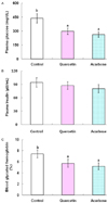
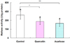
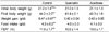
Body weight, food intake, and feed efficiency ratio (FER) of the control, QE, and acarbose groups were not significantly different (Table 1). QE consumed at 0.08% of diet or acarbose consumed at 0.03% of diet significantly decreased fasting plasma glucose levels (298 ± 34 mg/dL and 262 ± 29 mg/dL, respectively) compared with the control group (437 ± 51 mg/dL, P < 0.01, Fig. 2). There was no significant difference between the plasma glucose levels of the QE and acarbose groups. Plasma insulin levels of the QE group (87.1 ± 8.4 µU/mL) were not significantly different from those of the control (94.2 ± 10.6 µU/mL) or acarbose group (80.3 ± 9.1 µU/mL). Blood HbA1C levels of the QE and acarbose groups (5.7 ± 0.7% and 5.2 ± 0.6%, respectively) were significantly lowered compared with the control group (7.4 ± 0.7%, P < 0.01). Blood HbA1C levels in the QE group were not significantly different from those of acarbose group. Consumption of QE (280.1 ± 51.0 U/g protein) or acarbose (249.6 ± 56.0 U/g protein) significantly suppressed the maltase activities of the proximal part of the jejunoileum compared with the control group (388.3 ± 68.6 U/g protein, Fig. 3).
Discussion
QE has been shown to inhibit rat intestinal α-glucosidase in vitro with an IC50 of 0.48-0.71 mM [9,10]. In this study, QE administered at a dose of 100 mg/kg reduced the AUC of the postprandial glucose response by 51.6% compared to the control group, an effect comparable to that of acarbose at a dose of 40 mg/kg (64.5% reduction). Previous studies have shown that acarbose administered at a dose of 40 mg/kg effectively suppresses the blood glucose response after carbohydrate loading in STZ-treated rats [23,24]. Postprandial glucose is known to be an independent risk factor for cardiovascular complications associated with DM [25]. The effective control of blood glucose elevation by QE after starch loading suggests that it is a candidate agent for alleviating postprandial hyperglycemia.
QE consumed at 0.08% of a total diet decreased fasting plasma glucose and HbA1c without influencing insulin levels in db/db mice. These results were not significantly different from those of animals that consumed acarbose at 0.03% of a total diet. The average intake of QE and acarbose was calculated to be 101.5 and 39.0 mg/kg/d respectively, based on food intake and average body weight.
Chronic consumption of QE (0.1% of diet) decreased blood glucose in STZ-treated rats [6,7]. QE protected pancreatic β cells from oxidative stress and damage, resulting in increased insulin secretion in STZ-treated rats [5]. Ishida et al. [26] suggested that a reduction in oxidative stress could preserve the pancreatic β cell mass in db/db mice, thereby decreasing hyperglycemia. From our data, it is not clear whether QE could exert a protective effect on the pancreas of db/db mice, as these mice have hyperinsulinemia and hyperglycemia in early life [27]. The insulin levels of these animals begin to decrease due to degenerating pancreatic islet cells after 5-6 months of age [28]. It might be useful to elucidate the effect of QE on insulin secretion in the db/db mice after that age.
It has been demonstrated that acarbose can decrease the requirement for insulin by controlling postprandial hyperglycemia [29], and that it can chronically reduce glucose and insulin levels after meals, improving insulin sensitivity [30]. In addition, it has been suggested that QE could improve insulin signaling and therefore insulin sensitivity in rats with insulin resistance [31]. QE could improve fasting hyperglycemia by enhancing insulin sensitivity via α-glucosidase inhibition and enhanced insulin signaling in db/db mice.
DM has been reported to induce intestinal disaccharidase activities, which could increase the digestion and absorption of carbohydrates [32-34]. Lee et al. [33] reported that the administration of α-glucosidase inhibitor (Bay-o-1248) for 7 days decreased maltase activities in the jejunoileum of db/db mice. Acarbose given to STZ-induced diabetic rats for 5 weeks lowered small intestine maltase activities [34]. Ramachandra et al. [7] demonstrated that long-term administration of QE (0.1% of diet) decreased the activities of small intestinal maltase and sucrase in STZ-treated rats. They suggested that the decreased activities of disaccharidases by QE could play a major role in the amelioration of diabetes. A reduction of maltase activities by QE could reduce digestion of dietary carbohydrates and contribute to the control of hyperglycemia in db/db mice.
Clinically, acarbose is used as an antidiabetic agent, alone or in combination with other oral hypoglycemic agents. However the chronic use of acarbose could be limited by unfavorable gastrointestinal side effects, such as flatulence, abdominal cramping, and diarrhea [35]. QE is a well-documented bioflavonoid that is abundant in fruits and vegetables and has been marketed as a nutraceutical in several countries, including the United States [36]. QE could potentially be alternative hypoglycemic agent without side effects.
In conclusion, dietary QE alleviated fasting and postprandial hyperglycemia in an animal model of DM, at least in part by inhibiting α-glucosidase activity. Thus, QE may be useful for improving overall glycemic control in the management of DM.
 XML Download