

 PDF
PDF ePub
ePub Citation
 Print
Print


Introduction
Rabies continues to be a major fatal zoonotic disease in various countries. In Indonesia, the disease is endemic to the major islands and the number of infected areas is increasing at an unpredictable rate. Its transmission to formerly free areas, including the islands of Bali and Nias, was reported from 2008 to 2010, with rabies being confirmed in Bali for the first time in November 2008. The disease is distributed in all districts of Bali and was responsible for 135 human deaths from 2008 to 2011. Accordingly, rabies is now a major public health problem in Bali [34]. The origin of the rabies virus (RABV) that has spread to Bali is currently unclear, although its hypothetical geographic origin is considered to be Sulawesi or Kalimantan [2324].
Rabies is a fatal neurotropic disease of humans and other mammals caused by RABV, which belongs to the genus Lyssavirus of the family Rhabdoviridae [1122]. As a member of the genus Lyssavirus, RABV has a non-segmented single-stranded RNA genome of approximately 12 kb, which encodes five proteins: nucleoprotein (N), phosphoprotein (P), matrix protein (M), glycoprotein (G), and RNA polymerase or large protein (L) [1927]. The nucleotide sequence of the N gene fragment has been studied extensively and used as a tool to clarify the geographic distribution patterns of RABV [9293233]. All dog-related and vampire-bat-related rabies viruses in Brazil were successfully distinguished using reverse transcription (RT)-PCR with specific primers developed to hybridize with the gene sequence [1718].
Being an archipelagic nation with highly varied inhabitant density, the spread of RABV in Indonesia can be discordant to the expected distribution. As the most densely populated island and the location where rabies was first reported [16], it is plausible that Java Island is the only origin of RABV found in other islands. However, the close distance between islands might also complicate the understanding of disease epidemiology. Moreover, domestic dogs are believed to be the main carriers of rabies transmission to other animals and humans. However, no quantitative analyses have been conducted to support this notion. Clarifying the phylogenetic relationships among RABVs based on geography and species is important for tracing the dynamics of rabies infections. This approach allows the historical reconstruction of events leading to the introduction of rabies into a specific area [29], enabling a better understanding of how to control the disease, especially in an archipelagic setting in which canine rabies is the only circulating virus. Here, we present the phylogenetic relationships of the most recent RABVs strains in Indonesia based on the nucleotide sequences of the N fragment to understand the chain of their transmission throughout Indonesia.
Materials and Methods
Sample collection
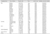
Brain samples from different animal species were collected on the islands of Bali, Sumatra, Java, Kalimantan, Sulawesi, and Flores. The specimens were obtained from the collection of The Animal Disease Investigation Centers in Medan (North Sumatra), Bukittinggi (West Sumatra), Yogyakarta, Banjarbaru (South Kalimantan), Maros (South Sulawesi), and Denpasar (Bali). The specimens were stored at -80℃ without preservatives. A total of 27 brain samples from 20 dogs, three cows, two goats, a cat, and a pig were used for the phylogenetic analysis in this study (Table 1).
Case confirmation and virus inactivation
The samples were diagnosed as RABV positive with a direct fluorescent antibody test using a standard protocol [12] conducted at the aforementioned Animal Disease Investigation Centres. The samples were suspended in phosphate-buffered saline (PBS) with 1% sodium dodecyl sulfate to inactivate the virus and transported on dry-ice in a closed container.
Nucleotide sequencing
The RT-PCR products were purified with a QIAquick PCR Purification Kit (Qiagen, Germany). The purified products were sequenced directly using the same primer applied in RT-PCR. A cycle sequencing reaction was conducted with the BigDye Terminator ver. 3.1 Cycle Sequencing Kit (Applied Biosystems, USA). The sequencing products were obtained with the ABI PRISM 3100 Genetic Analyzer (Applied Biosystems) at the Eijkman Institute, Jakarta. The sequencing results were aligned using ClustalW in the Mega4 software [35] and RABV was identified by BLAST analysis [1]. The N gene target sequence was 609 bp, corresponding to the nucleotides at positions -22 to 587. All nucleotide sequences have been submitted to GenBank and assigned accession numbers (Table 1).
Phylogenetic analysis and geographic projection
The sequence data of this study were aligned with the database of Indonesian RABVs available in GenBank (National Center for Biotechnology Information, USA). A RABV sequence from China, Guizhou_Qx5 (accession no. DQ666296), was also included as a reference. The phylogenetic reconstruction was inferred using the BEAST software package ver. 1.7 [13] with the BEAGLE library [3]. A location-annotated XML file was generated for discrete analysis using the Bayesian Evolutionary Analysis Utility (BEAUti) software [13]. The best tree model was selected by applying various clock and substitution models, such as strict, log normal uncorrelated relaxed, and exponential uncorrelated relaxed clocks, and Hasegawa-Kishino-Yano, general time reversible (GTR), and Tamura-Nei (TN93) substitutions. The other parameters used were three codon partitions, a symmetric substitution model with Bayesian stochastic search variable selection social network, and a continuous time Markov chain rate reference [20] for prior distribution. The length of the chain was 60,000,000 and the log parameter was programmed every 10,000 chains. The best tree model was selected by calculating the Bayes factor (BF) between models using Tracer ver. 1.5 [25]. The maximum clade credibility (MCC) phylogeny was inferred with Bayesian Markov chain Monte Carlo (MCMC) using TreeAnnotator and visualized with the FigTree ver. 1.4.0 software. To trace the geographic dispersal of the virus, a Keyhole Markup Language file of the resulting tree was created using the SPREAD software [4] and visualized in Cartographica (GBP Software, USA).
Results
The RABV N genes were amplified successfully from all 27 rabid animal brain samples from endemic areas in Indonesia by RT-PCR. The length of the readable cDNA sequences for the 17 RABVs was 534 bp. The cDNA sequences obtained in this study have been submitted to GenBank under the accession numbers listed in Table 1.
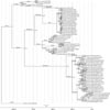
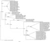
The location-annotated MCC phylogeny for the N gene fragments of recent RABVs, analyzed together with the sequence data for some viruses in the Indonesian RABV database, is shown in Fig. 1. The species annotated MCMC tree is shown in Fig. 2. The trees were generated using the best model of TN93+G+I with a log normal uncorrelated relaxed clock. The model generated the posterior and prior effective sampling sizes of 313.752 and 272.22, respectively. These results show that all RABVs currently circulating in Indonesia are grouped into two major clusters, group 1, which consists of viruses from Java and Sumatra, and group 2, which contains the majority of RABVs from Kalimantan and all the RABVs from Sulawesi, Flores, and Bali. The viruses recently introduced into Bali evolved from the Kalimantan KL00-18, while viruses from Nias originated in Sumatra. We identified three lineages of RABV in Kalimantan. Lineage 1 (KL97-03) is related to the Sumatran viruses, Lineage 2 (KL177-09 and KL268-09) is related to RABV from Flores (FL007) and Lineage 3 (KL00-18) is related to all RABVs from Bali. The RABVs from Flores form a cluster with the viruses from Sulawesi (Makasar, Pangkep, and Menado). Fig. 2 demonstrates that the RABVs from other animal species originated from dog.
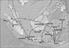
The geographic dispersal of the Indonesian RABVs is shown in Fig. 3. The origin of RABVs in Indonesia was the ancestor of the Javanese RABVs. This ancestor was transmitted to Kalimantan, then to Sumatra, Flores, and Bali. The Flores descendant was subsequently transmitted to Sulawesi (Makasar, Pangkep, and Menado) and back to Kalimantan.
Discussion
Phylogenetic reconstruction can be used to support incidence data from the field to determine the epidemiology of RABV. Information regarding the origin of RABV entering an area could be important for successful rabies control and should be invaluable for preventing such transmission in the future.
To obtain a complete picture of the inter-island and interspecies transmission of RABV across the Indonesian Archipelago, we used the most current RABVs from various islands and different host species in Indonesia, together with previously published data. We identified the ancestor of the Indonesian RABVs as a strain circulating in Java. Its descendant was transmitted to Kalimantan, then spread to Sumatra, Flores, and Bali. All available Sulawesi RABVs are derived from the Flores RABV. Some strains were still circulating in the same islands, including Sumatra and Java, approximately 10 years after their first identification [33]. In addition to descendants of the formerly circulating virus, RABV seems to have been reintroduced into Kalimantan from Flores.
We previously reported that the ancestor of the Bali RABVs descended from the Kalimantan 00-18 strain, but did not differ significantly from the RABVs of Sulawesi and Flores [23]. Using a different dataset and a discrete analysis, we showed that the Bali strain originated from a relative of a Kalimantan RABV. However, there is still some phylogenetic uncertainty in this study. The long branch from KL00-18 to the Bali strains shows that the exact origin of the Bali outbreak was not via a direct descendent of strain KL00-18, which was identified in Samarinda [33]. The direct parent of the outbreak must have evolved somewhere in Kalimantan or beyond, before it was transported to Bali in early 2008 [23].
This study brings new insight to the dispersal of RABV in Indonesia. In this archipelago, rabies has been reported since the 19th century in West Java and Sulawesi, while it was identified in Sumatra and Kalimantan in 1959 and 1974, respectively [16]. Flores has been infected since the 1990s, following the transportation of dogs from Sulawesi. The virus circulating in Flores was predicted to have originated from Sulawesi [38], which was confirmed by genetic analysis [33]. We found that the oldest virus in Indonesia was from Java. The location with the highest probability, based on all Indonesian clades, is Cirebon. Contrary to earlier findings, the origin of Flores RABV was not Sulawesi [3338], but Kalimantan, while the Sulawesi (Makasar, Pangkep, and Menado) viruses were introduced from Flores.
Lack of surveillance and collection of specimens are the most likely reasons for this discrepancy. Because Kalimantan is the largest but most sparsely inhabited island in Indonesia, rabies might have been present there for centuries. There was a population influx into the island after the 1960s, when the timber and mining industries flourished. A proportion of the incoming people were also residents of Java and Bali that were participating in the so-called "transmigration", a government program to move people from densely populated areas (Java and Bali) to sparsely inhabited islands, such as Sumatra, Kalimantan, and Sulawesi.
This is not only true for Indonesia. RABV surveillance in endemic and developing countries has very low detection probabilities of less than 10% [36]. However, there have been few studies conducted to collect isolates and develop reliable databases. As a result, the origins of epidemics cannot be traced, which has prevented the efficient targeting of future outbreaks.
We believe that the method used here to infer the historical background and geographic diffusion of RABV in Indonesia is highly accurate. The MCMC algorithm has provided evidence of spatial and temporal patterns in the evolution of several viruses, including foot and mouth disease virus [10], RABV [737], human immunodeficiency virus [2], and other agents. This approach allows the incorporation of environmental and epidemiological data [10], and offers a unique opportunity to explore viral evolution in greater detail [14] at the local, regional, and international scales. In this study, we selected the best tree model after extensive runs and BF analyses applying various clock and substitution models. We found that the best model was TN93+G+I with a log normal uncorrelated relaxed clock. The GTR+G+I log normal analysis resulted in higher BF than the selected model, although it generated a log file with invalid estimates of the posterior and prior effective sampling sizes (< 200), even when the length of the chain was increased. The BF calculation is a standard approach to model selection on a Bayesian phylogenetic platform [283031], and a BF value of > 20 should mean strong support for the favored model [30]. The Tracer software provides a tool with which to perform the calculation [25].
The most obvious finding of this study is that the inter-island transport of infected animals, predominantly dogs, has been responsible for the introduction and reintroduction of rabies into many islands in Indonesia. There are many examples of the introduction and spread of rabies owing to human-mediated movement of reservoir animals [561538]. Dogs transported on fishing boats have been implicated as carriers in the inter-island transmission of RABV to Terengganu in Malaysia [21]. This study presents clear evidence that the Nias outbreak was introduced from Sumatra, while the Bali outbreak can be attributed to an introduction from Kalimantan. The divergent viruses circulating in Kalimantan include both the descendants of the traditional strain and newly reintroduced strains from Flores. The variety of Flores viruses, which are related to both Kalimantan and Sulawesi viruses, must be attributable to their reintroduction from these areas. Alternatively, the Flores strains might have been reintroduced back into the corresponding areas. The rabies outbreak in Flores Island was claimed to have occurred after introduction of an infected dog from Sulawesi [38]. Although it contradicts the major findings of this study, it is also possible that dogs were transported from Sulawesi to Flores, rather than from Kalimantan to Flores. Voyages between islands of Indonesia can be direct or interrupted en route by a short anchorage at any island for fuel, logistical reasons, or to avoid extreme weather.
The paradigm of rabies epidemiology is compartmentalization of the circulating virus by the host species and its geographic area. The phylogenetic analysis performed in this study showed that all RABVs identified within the Indonesian cluster are distributed according to their geographic origins. However, there was no pattern of genetic relationship among the Indonesian RABVs collected in 2008-2010 from domestic animals. This finding is consistent with the phylogenetic analyses of previous studies, in which the RABV variants were generally grouped according to their geographic origins [6833]. Therefore, isolates tend to cluster in the same geographic area [18]. Romijn et al. [26] also reported that RABVs clustered more closely together when they were isolated from the same host species.
We included samples from various animals other than dogs in this study. There are RABV sequences from various animals in GenBank, including cattle, deer, cats, tigers, and monkeys. The history of these sequences is unknown; however, they were probably collected from wild animals in captivity or zoos. We analyzed a species-annotated MCMC tree and found that the RABVs from other animal species originated from dog. These findings indicate that rabies predominantly affects dogs, with spill-over infections in livestock and wild animals. We also present direct evidence that the strains present in other animals (cats, goats, and pig) are similar to those found in dogs.
These results emphasize the risk of RABV introduction and reintroduction entailed by the inter-island transportation of infected dogs and the lack of rigor in our surveillance system. Government agencies must increase their preparedness because a RABV epidemic will likely occur in the future. Public awareness must be continuously maintained on neighboring islands that have not yet experienced the disease. The results presented herein will help prevent the spread of rabies in the future.
The results of this study indicate that circulation of RABVs in Indonesia is very dynamic. Some strains have remained in specific islands, including Sumatra and Java, whereas others have been continuously introduced and reintroduced into other islands, including Kalimantan, Sulawesi, and Flores. Recent outbreaks in formerly virus-free areas were caused by the introduction of infected dogs from neighboring islands, for example from Sumatra to Nias, and from Kalimantan to Bali. The risk of the introduction and reintroduction of RABV strains by the inter-island transportation of infected dogs remains high for newly free areas and intensified control areas such as Bali. The rigor of the surveillance system must be increased substantially to detect any possible epidemic. Finally, the control of rabies and all preparations for an epidemic must be enforced throughout Indonesia.
 XML Download