

 PDF
PDF ePub
ePub Citation
 Print
Print


Freiberg's infraction is an osteochondrosis of the heads of the metatarsal bones, mostly of the second or third metatarsal. Since it was initially described by Freiberg1) in 1914, various classifications and methods of management of Freiberg's disease have been developed.
Classifications are usually based on vascular influence and radiographs. Smillie2) classified the natural history of the metatarsal head in Freiberg's disease into five stages according to macroscopic appearance. Freiberg's disease can be managed by nonoperative3,4) or operative treatment. Surgical treatments for the disease include core decompression,5) debridement,6) perichondral grafting,2,7) metatarsal osteotomies,8-14) and arthroplasty.15,16) Among these surgical techniques, dorsiflexion osteotomy is advocated for early stages of the disease.17)
Weil osteotomy is a surgical treatment for metatarsalgia, a reliable procedure with good results.18) The aim of the osteotomy is to relieve excessive pressure under the metatarsal head which can be achieved by shortening or elevating the metatarsal head. For treatment of Freiberg's disease, we performed a modification of the Weil osteotomy, an intra-articular dorsal closing wedge osteotomy. The modified Weil osteotomy is composed of two components: shortening metatarsal osteotomy to offload the metatarsal head and dorsal closing wedge osteotomy of metatarsal bone to restore metatarsophalangeal (MTP) joint congruency.
The osteotomy was secured with a single screw to achieve reduced soft tissue irritation and relatively rigid fixation. These characteristics of the screw yield results similar to the study above in regards to range of motion (ROM) of the MTP joint.
The purpose of this study was to evaluate the clinical outcomes of modified Weil osteotomy fixed with a single screw in treatment of Freiberg's disease.
METHODS
Patients
Between November 2001 and July 2008, nineteen patients (twenty feet) underwent modified Weil osteotomy for Freiberg's disease. We included patients who complained of persistent pain in spite of more than 6 months of conservative treatment at any stage according to Smillie's classification system. Diagnosis was based upon clinical history, physical examination, and plain radiographs for all patients. The main complaints were pain upon walking or sports activities. This study was approved by the Institutional Review Board at our institute. Informed consent was waived because of the retrospective nature of the study.
Operative Technique
Under spinal anesthesia, patients were placed in the supine position. A pneumatic tourniquet was applied to the ipsilateral thigh and inflated to a pressure of 320 mmHg.
Via the dorsal longitudinal approach, the extensor digitorum longus tendon of the affected toe was retracted laterally, exposing the MTP joint. In the beginning, the joint was debrided and a synovectomy was carried out before osteotomy. If a loose body, peripheral osteophyte, periarticular spur or synovitis were observed, they were removed or a synovectomy was performed.
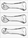
Afterwards, modified Weil osteotomy was done via the distal metaphysis. In the dorsal aspect, most of the damaged area of the metatarsal head and neck area was removed in the bone wedge. Healthy plantar cartilage was rotated to the center of the joint for forming a new articular surface. Finally, the osteotomy was stabilized using a single screw (Spin Screw; Integra LifeScience Co., Plainsboro, NJ, USA) with a low profile head and non threaded lag to the dorsal aspect of metatarsal neck (Fig. 1).
Postoperatively, patients were allowed to bear weight as tolerated on their heel in an open, hard-soled surgical shoe. Upon radiographic evidence of healing at the osteotomy site, transfer of weight to the forefoot in a regular shoe was permitted, usually 4 weeks later. The patients underwent periodic clinical and radiographic follow-up at 4 weeks, 3, 6, 12 months, and then annually.
Clinical and Radiologic Assessment
All clinical records and radiographs were reviewed retrospectively. Relief of pain was evaluated using visual analogue scale (VAS)19) rating from 0 to 10. The patients were examined using a standardized questionnaire based on the American Orthopaedic Foot and Ankle Society (AOFAS) lesser metatarsophalangeal-interphalangeal scale.20) This score includes clinical variables such as pain, restriction of footwear, painful callus, functional restriction of the MTP joint, ROM of MTP joint and alignment of the toes. In all cases, passive mobility of MTP joint was measured by an independent examiner. With the patient seated, the examiner stabilized the metatarsal with one hand while grasping proximal phalanx with the other hand. The examiner moved the toe cephalad and caudally in range of maximal area to assess dorsiflexion and plantarflexion. The patients' subjective satisfaction was also evaluated after surgery at last follow-up. Patients were asked to rate their result as excellent, good, fair or dissatisfied.21)
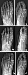
Although the Smillie's classification system is based upon inspection of the metatarsal head, several features may be interpreted from plain radiographs.2,17) We reviewed preoperative weight-bearing radiographs and intraoperative finding to classify the stage of Freiberg's disease according to the Smillie's classification system (Fig. 2).
We divided the patients again into early stage and late stage based on Smillie's classification system.11,12,17,22) The early stage included Smillie stage I to III and the late stage included the rest, stage IV and V. In this study, dorsal closing wedge osteotomy was performed in both early and late stage of Freiberg's disease.
In preoperative radiograph, the initial metatarsal length was measured. In postoperative radiograph, metatarsal shortening due to surgery was analyzed by the modification of Jones et al.23) In periodic radiographic follow-up, if bridging trabeculae across the osteotomy site emerged, it was considered as radiographic union.
Other complications were also reviewed preoperatively and postoperatively such as callus, floating toe, stiff toe, transfer metatarsalgia, fixation failure, fracture of metatarsal, subluxation of the MTP joint, soft tissue irritation, nerve injury and nonunion or delayed union.
All continuous data were expressed in terms of the mean and standard deviation. Wilcoxon signed-rank test and Mann-Whitney U-test were performed to test the hypotheses regarding effectiveness of the procedure. Null hypotheses of no difference were rejected if p-values were less than 0.05. Statistical analysis was performed using SPSS ver. 18.0.0 (SPSS Inc., Chicago, IL, USA).
RESULTS
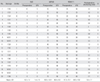
All of the lesions involved were in the second metatarsal bone. The average age of the patients was 33.6 years (range, 17 to 62 years), and the mean follow-up period was 72.6 months (range, 41 to 121 months) (Table 1).
The AOFAS score increased significantly after surgery from 63.3 ± 14.9 to 80.4 ± 5.6 (p < 0.0001). VAS and ROM of the MTP joint improved significantly after surgery. VAS improved from 6.2 ± 1.4 to 1.4 ± 1.5 at last follow-up (p < 0.0001), and ROM of the MTP joint increased from 31.3 ± 10.1 to 48.3 ± 13.0 degrees at last follow-up (p < 0.0001). All patients were satisfied, reporting excellent or good results except one patient who had joint stiffness. The patient had ROM limitation of 2nd MTP joint preoperatively and complained about no improvement of ROM after surgery, not being satisfied with the surgery.
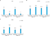
On plain radiographs, joint space widening and degeneration of the MTP joint were found. Four cases were classified as Smillie stage I, eleven cases as stage II, one case as stage III, two cases as stage IV, and two cases as stage V. There was no significant difference in variables such as improvement of VAS, AOFAS score and ROM of MTP joint after modified Weil osteotomy comparing early stages with late stages (Fig. 3).
Initially, 14 cases had a shorter metatarsal compared to opposite side and the average of initial metatarsal shortening was 1.0 ± 1.0 mm (range, 0 to 3.3 mm). Postoperatively, the mean metatarsal shortening due to operation was 3.4 ± 1.7 mm (range, 0.9 to 5.8 mm) without including initial shortening. Radiographic union was achieved at 8.2 ± 2.5 weeks (range, 4 to 12 weeks) after the osteotomy.
Postoperative complications included callus on the plantar area of the third metatarsal head in three cases (15%), floating toe in one case (5%) and stiff toe in one case (5%). One patient complained about transfer metatarsalgia with callus on the plantar area of the third metatarsal head (5%). There was no evidence of fixation failure, fracture of metatarsal, subluxation of the MTP joint, soft tissue irritation, nerve injury and nonunion or delayed union at final follow-up.
DISCUSSION
Freiberg's disease is not a common disease. Although various reports have described Freiberg's disease since 1914, classification and treatment methods thereof are not completely established.17) Smillie2) classified the appearance of the metatarsal head in Freiberg's disease into five stages in 1957.
In the early stages of the disease (Smillie stage I to III), fair evidence supports the use of the closing wedge osteotomy of the metatarsal head and neck.14,17) On the other hand, Kinnard and Lirette11,12) reported even with advanced cases there was sufficient plantar cartilage to perform the procedure. We divided our patients into early and late stages to compare the effectiveness of modified Weil osteotomy among the stages.11,12,17,22) Upon investigation, improvement of VAS, AOFAS score and ROM of the MTP joint did not significantly differ between early stages and late stages. Modified Weil osteotomy can be a good treatment option in both early stages and late stages of Freiberg's disease.
Modified Weil osteotomy involves open joint debridement and intra-articular dorsal closing wedge osteotomy. Open joint debridement allows removal of intraarticular pathology such as thickened synovium, loose bodies, delaminated articular cartilage and peripheral osteophytes and spurs.17)
Closing wedge osteotomy is a realignment osteotomy of metatarsal head and neck.18) The aim of closing wedge osteotomy is to redirect the articular surface and theoretically, dorsal closing wedge osteotomy can restore the blood supply to the metatarsal head, preventing further deformity and collaps.8,17,21)
Intra-articular dorsal closing wedge osteotomy enables less metatarsalgia than that of extra-articular osteotomy which often leads to excessive elevation of the metatarsal head.13) Dorsal closing wedge osteotomy redirects articular surface allowing the intact plantar cartilage to articulate with the proximal phalanx. Also, dorsal closing wedge osteotomy offers similar results to extra-articular osteotomy in ROM of the MTP joint. Kinnard and Lirette11) reported that dorsiflexion osteotomy gave excellent results with minimal loss of MTP joint motion and with an average of 2 to 5 mm metatarsal shortening.
Various fixation methods can be applied after completion of dorsiflexion osteotomy of the metatarsal, including cerclage wire,10) temporary pins,10-12) transosseous sutures,12) metal screw,18) dorsal T plate,9) polyglycolide pins.14) Gauthier and Elbaz10) suggested intra-articular dorsal wedge osteotomy fixed with cerclage wire in 1979. The cerclage wire may cause tendinitis and temporary pins have to be removed before motion exercise. Because the remaining intact portion of the metatarsal head was too small for wire fixation, Kinnard and Lirette11) modified the intra-articular dorsal closing wedge osteotomy fixated with absorbable suture. Transosseous sutures can bring about loss of reduction, whereas metal screw or plate fixation after extra-articular osteotomy is more rigid. However, the metal screw or plate may be bulky and can reduce the ROM of the MTP joint. Recently, absorbable polyglycolide pins have been used that do not decrease the ROM of the MTP joint. After performing dorsal closing wedge osteotomy with absorbable pin fixation, Lee et al.14) reported no loss and some gain in motion. Unfortunately, they are relatively weak and require multiple pinning.
In this study, we fixed metatarsal head with a single screw after dorsal closing wedge osteotomy. That being so, the ROM of MTP joint was significantly increased after surgery. Compared to other fixation devices such as temporary crossed Kirschner wires, patients were allowed to start motion exercise and bear weight relatively early. Also, the Spin Screw (Integra LifeScience Co.) with its low profile head, may reduce soft tissue irritation and has non-threaded lag for more compression. By placing the apex of the wedge as proximal as possible, a less shortened metatarsal and an enlarged distal fragment can be achieved.14)
Kinnard and Lirette11,12) underwent an intra-articular dorsal closing wedge osteotomy in fifteen patients with predominantly late stage Freiberg's disease. They reported a minimal loss of joint motion and an average metatarsal shortening of 2.5 mm. In this study, we experienced favorable improvement of ROM after operation and even a floating toe occurred in 1 case. We achieved moderate shortening of metatarsal by dorsal closing wedge osteotomy.
After screw fixation, foreign body reaction, fixation failure, perioperative fracture and displacement of osteotomy may occur. In our study, we could not find any complication of the sort; however, confirmation of such would require a more cases and longer follow-up. Limitations of this study include being a retrospective study, as well as no control group for being compared with dorsal closing wedge osteotomy. The number of participants was too small to draw any solid conclusions. Some prospective and long term follow-up study with more patients and control group can prove the efficacy of dorsal closing wedge osteotomy in Frieberg's disease.
Through the modified Weil osteotomy, we can expect offloading of the metatarsal head and restoration of MTP joint congruency. This study provides great improvement in pain and function after the procedure with a screw fixation for Freiberg's disease. Modified Weil osteotomy is believed to be a useful method for the treatment of Freiberg's disease in both early stages and late stages.
 XML Download