

 PDF
PDF ePub
ePub Citation
 Print
Print


INTRODUCTION
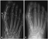
The hallux valgus angle (HVA) and first intermetatarsal angle (IMA) have been considered to be reliable indicators for the severity of a hallux valgus deformity.123456789 However, identification of the longitudinal axes of the proximal phalange, first metatarsal, and second metatarsal (which is essential for effective determination of the HVA and IMA) is subject to error. Accordingly, several studies have focused on decreasing the measuring error and increasing the reproducibility and validity of measurements for HVA and IMA.59 Despite these efforts, in the subluxated metatarsophalangeal joint (MTPJ), HVA measured by the conventional method do not reflect the true severity. Even though the distal metatarsal articular angle (DMAA) could adequately report the severity of deviation/subluxation of the first MTPJ, there is debate with regards to the accuracy, reproducibility, and validity of measurement of the DMAA.10 In the symptomatic hallux valgus patients with subluxated joint, bigger HVA would be related with clinical symptoms (Fig. 1).
Hence, the aims of this study are to 1) investigate new point-connecting measurements for HVA and IMA that can reflect clinical severity (such as degrees of subluxation of the first MTPJ) and 2) compare the validities of the conventional midline method and new point-connecting method for measuring HVA and IMA.
MATERIALS AND METHODS
Of the 60 feet from 57 patients with hallux valgus who underwent hallux valgus surgery (mean age, 54.4 years; range, 21 to 74 years) between June 2007 and June 2011 at our hospital, there were 20 cases classified as 'mild' (HVA<20; range, 17 to 19), 20 cases classified as 'moderate' (20<HVA<40), and 20 cases classified as 'severe' (HVA>40; range, 41 to 59). Among these 60 feet also, there were 26 feet with deviation of the first MTPJ and 34 feet with subluxated joints of the first MTPJ, as described by Piggott.11 These 60 feet were classified into two groups according to the type of metatarsal osteotomy. The two groups comprised patients that underwent either distal chevron metatarsal osteotomy (DCMO) (30 feet) or proximal chevron metatarsal osteotomy (PCMO) (30 feet). Radiographic assessments involved the collection of weight-bearing dorsal-plantar (DP) radiographs. Both HVA and IMA were measured preoperatively and postoperatively using both the conventional midline method and the new point-connecting method. The results of measurements for HVA and IMA between the conventional midline method and new point-connecting method were compared according to the severity of the cases, congruency of the first MTPJ, and the type of metatarsal osteotomy. The preoperative quality of life (QOL) was evaluated by the Short Form-36 (SF-36) questionnaire. The radiographic measurements of HVA/IMA between the new point-connecting method and conventional method were correlated with the SF-36. In addition, two authors evaluated the time required to measure the HVA and IMA using the new point-connecting method and the conventional method in 60 feet. The study protocols were approved by our ethics committee.
Radiographic analysis
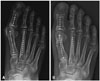
Digital versions of all radiographic images were obtained using the Picture Archiving Communication System (Petavision, Seoul, Korea). Weight-bearing foot DP radiographs were taken at a tube-film distance of 100 cm with the X-ray beam projecting vertically and centered to the middle of the third metatarsus of the patient with the knee in full extension. For the preoperative measurement of the HVA and IMA using the conventional midline method, the longitudinal axis of the first and second metatarsal was determined by connecting the centers of the metatarsal head and base (Fig. 2A), as previously described by Miller.12 Postoperatively, a connecting line between the center of the first metatarsal head and the proximal articular surface was used as the longitudinal axis of the first metatarsal to measure the HVA and IMA (Fig. 2B), as described by Shima, et al.9 The center of the proximal articular surface was defined as the midpoint between the medial and lateral edges.
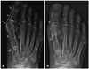
The new point-connecting method to measure the HVA and IMA involved the use of three connecting lines for the longitudinal axes of the proximal phalanges, first metatarsal, and second metatarsal. First, the longitudinal axis of the proximal phalanges was defined as the connecting line between the most medial prominent point of the proximal phalanges in the first interphalangeal joint and most medial prominent point of first metatarsal head. Second, the longitudinal axis of the first metatarsal was defined as the most medial prominent point of the first metatarsal head and most medial prominent and sclerotic point of first metatarsal base in the tarsometatarsal (TMT) joint, both preoperatively and postoperatively. Third, the longitudinal axis of the second metatarsal was defined as most medial prominent point of the second metatarsal head and the most medial prominent and sclerotic point of second metatarsal base in the TMT joint, both preoperatively and postoperatively (Fig. 3).
Inter- and intra-observer reliabilities were obtained for HVA and IMA between the conventional midline method and new point-connecting method by three foot and ankle surgeons (JY Ahn, Dimas RB, and JH Seo). To evaluate inter-observer reliability, each surgeon measured sixty DP images with no questions or discussions allowed during radiographic measurements. Before the start of the analysis, five samples of the weight-bearing foot DP images were evaluated by these three observers to ensure that they drew angles in the same manner. For intra-observer reliability, radiographic measurements were made by each observer during the two weeks after the initial measurements in a blind manner relative to previous measurement results. These two series of sequential measurements were then compared among the three observers.
Statistical analysis
The distribution of variables in each group was tested for normality using the Shapiro-Wilk test. A p value of <0.05 was considered significant. Statistical comparisons of the mean HVA and IMA values obtained using the conventional midline method and the new point-connecting method were assessed with an independent t-test among patient groups divided by severity, congruity and metatarsal osteotomies, both preoperatively and postoperatively. Inter- and intra-observer reliability were assessed on the basis of the intraclass correlation coefficient (ICC). Statistical comparisons of the outcomes between 2 measurements were assessed with independent t test. The correlations between the radiographic angles and SF-36 were assessed with a Spearman correlation analysis. Statistical analyses were performed using SAS statistical software (SAS Institute, Cary, NC, USA).
RESULTS
The mean preoperative and postoperative values for the HVA and IMA measurements between the conventional midline method and new point-connecting method are indicated in Table 1. For both the mean HVA and IMA, there were significant differences between the conventional midline method and new point-connecting methods when used either preoperatively or postoperatively (p=0.001). The mean differences between the conventional midline method and the new point-connecting method was 9.31 degrees (95% confidence interval, 8.99 to 9.63) for the preoperative HVA measurements, 2.56 degrees (95% confidence interval, 2.35 to 2.77) for the preoperative IMA, 4.04 degrees (95% confidence interval, 3.65 to 4.43) for the postoperative HVA, and 1.32 degrees (95% confidence interval, 1.08 to 1.55) for the postoperative IMA (Table 2). The new point-connecting method showed significantly higher mean values for the pre- and postoperative HVA and IMA (p=0.001) than for the corresponding preoperative values of conventional midline method.
Reliability statistics for inter- and intra-observer comparisons between the conventional midline method and new point-connecting method are listed in Table 3. Compared with the conventional midline method, the new point-connecting method showed a higher ICC for the inter- and intra-observer reliability of preoperative HVA/IMA and a similar/or higher ICC for the inter- and intra-observer reliability of postoperative HVA/IMA. For the DCMO group, the ICC for the inter- and intra-observer reliability of pre- and postoperative HVA/IMA determined using the new point-connecting method was higher than that determined using the conventional midline method. The PCMO group showed similar ICC levels for the inter- and intra-observer reliability of the pre- and postoperative HVA/IMA to those determined using the conventional midline method (Table 4 and 5).
The deviated group had a preoperative mean and HVA of 29.72±5.8 degrees (range, 17 to 45) and IMA of 12.57±3.1 degrees (range, 9 to 20) with the conventional midline method, and a mean preoperative HVA of 37.27±6.2 degrees (range, 26 to 54) and IMA of 15.20±3.2 degrees (range, 8 to 24) with new point-connecting method. The subluxated group had a preoperative mean HVA of 37.59±8.2 degrees (range, 21 to 59) and IMA of 15.69±3.0 degrees (range, 9 to 23) with the conventional midline method, and a mean preoperative HVA of 47.23±8.6 degrees (range, 30 to 72) and IMA of 17.90±3.3 degrees (range, 10 to 26) with the new point-connecting method (Table 6). Significant differences were found for the preoperative HVA and IMA between the conventional midline method and new point-connecting method in two groups (p=0.001). The significant mean difference between the conventional midline method and the new point-connecting method was 7.55 degrees (95% confidence interval, 6.98 to 8.11) in the deviated group and 9.64 degrees (95% confidence interval, 9.26 to 10.2) in the subluxated group for the preoperative HVA measurements (p=0.001). There were no significant differences for either the postoperative HVA or IMA between the conventional midline method and new point-connecting method in the two groups.
In the deviated group, mean physical component summary SF-36 was 43.2±11.2 and mental component summary SF-36 was 45.4±11.1. In the subluxated group, mean physical component summary SF-36 was 39.2±10.7 and mental component summary SF-36 was 34.3±12.5. There were significant differences in SF-36 between the deviated group and the subluxated group. While our results showed no significant correlation in the 60 feet as a whole, there was a significant negative correlation (p=0.001) in the 34 feet with subluxated joints of the first MTPJ (r=-0.67, p=0.001). In 26 feet with deviation of the first MTPJ, there were no significant correlation between radiographic angles and SF-36.
We checked the time required to measure HVA and IMA using the new point-connecting method and the conventional method. Mean necessary time to measure HVA with new point-connecting method was 5.9±0.2 seconds. Mean necessary time to measure HVA with conventional method was 12.3±0.6 seconds. Mean necessary time to measure IMA with new point-connecting method was 6.9±0.4 seconds. Mean necessary time to measure IMA with conventional method was 12.4±0.7 seconds. There was a significant difference in the time to measure the HVA/IMA between the new point-connecting method and the conventional method (p=0.001).
DISCUSSION
Preoperative radiographic measurements of the HVA and IMA have been essential for evaluating the severity of hallux valgus deformities and for the selection of an appropriate metatarsal osteotomy procedure to correct these deformities.3491314151617181920 There have been many efforts to increase the reliability and reproducibility of the measurement for HVA and IMA using many longitudinal axes of the first metatarsal from various start points to end points.1579212223 Nevertheless, physicians face difficulties in selecting one long axis of the metatarsus, because the long axes of the metatarsus could be made between the center of the proximal and distal metaphyseal bone. Especially, it is difficult to determine the center point of a displaced distal fragment after DCMO with bunionectomy (Fig. 2).2722 Although the conventional midline method can be used to select the long axis of the metatarsus with excellent reliability, preoperative measurements of the HVA and IMA cannot show the extent of protrusion of bunions in subluxated first MTPJs (Fig. 3). Using our new point-connecting method to measure the HVA and IMA will yield higher values of HVA in a subluxated first MTPJ, because it uses the medial margin as the axis of the metatarsal.
Along with these advantages, our new point-connecting method showed higher inter- and intra-observer reliability in a preoperative context and similar inter- and intra-observer reliability in a postoperative context, when compared with conventional midline measurements.
Our new point-connecting method also showed a higher ICC for the inter- and intra-observer reliability of pre- and postoperative HVA/IMA than the conventional midline method for the DCMO group than for the PCMO group. There was difficulty determining the center of the first metatarsal head because a DCMO displaced the metatarsal head with a bunionectomy in hallux valgus deformity, even though a conventional midline method, such as the Shima method, showed excellent measurement reliability. However, the Shima method was evaluated after proximal crescentic osteotomy, not after DCMO. Hence, our new point-connecting method could be more helpful in evaluating the severity of hallux valgus deformity after DCMO than after proximal metatarsal osteotomy or proximal crescentic osteotomy, given that using the visible most medial prominent point in the displaced metatarsal head could be less ambiguous than determining the longitudinal axis of proximal phalanges and metatarsal.
Subluxation of the first MTPJ can cause progression of hallux valgus deformity in spite of a similar HVA and IMA measurement using the conventional midline method.1124 Also, there is a high probability of cartilage degeneration with deterioration of symptoms caused by the larger sizes of bunions and higher pressure into the cartilage contact surface area in the subluxated first MTPJ.23242526 Similarly, osteoarthritis of the first MTPJ can occur because the pressure of the medial edge of the articular surface of the proximal phalanx may increase.2526 Hence, an evaluation of subluxation in the first MTPJ is important with regard to clinical prognosis in hallux valgus deformity cases. In our present study, there was, in general, more of a tendency to overestimate the HVA and IMA when using the new point-connecting method, compared with the conventional midline method, because the more medial axis was used to connect the medial margins along the longitudinal axis of the metatarsal. However, the new point-connecting method showed significantly greater mean differences in preoperative HVA and IMA depending on whether the first MTPJ was deviated or subluxated. This means that the point-connecting method generated higher HVA and IMA values preoperatively in the subluxated first MTPJ than the deviated first MTPJ. Similarly, the conventional midline method may underestimate the subluxated first MTPJ of a hallux valgus deformity with the HVA and IMA, even though the same HVA and IMA values are obtained for the deviated MTPJ and subluxated MTPJ. We thus believe that our new point-connecting method could better reflect the clinical severity of a hallux valgus deformity via parameters, such as the degrees of subluxation of the first MTPJ, with excellent reliability, especially preoperatively.
Some papers reported that the severity of HV (HVA/IMA) does not influence QOL.2728 In contrast, Menz, et al.29 and Lazarides, et al.30 demonstrated a progressive reduction in QOL with increasing severity of HV deformity. As the deformity progressed, the lateral displacement of the hallux interfered with the normal alignment and function of the lesser toes, resulting in hammer toe or claw toe deformities, altered weight-bearing patterns, and the development of plantar keratotic lesions.31 The impact of hallux valgus was not limited to the pain and physical function subscales of the SF-36, indicating that there was a significant downward trend in general health, vitality, social function, and mental health.29 Our results have shown that there was significant correlation between radiographic angles and SF-36 using new point-connecting method in the subluxated group of the first MTPJ, compared to the conventional method. Our new method might be better in the aspect of QOL in the subluxated group, compared to the conventional method, although further study is needed.
This study has four key limitations. First, we did not determine the normal range of the HVA and IMA values using our new point-connecting method; more cases must therefore be recruited to determine this. Second, we did not analyze correlations between radiographic parameters, including the HVA and IMA measured by the new point-connecting method, and clinical parameters such as bunion pain and the possibility of osteoarthritis in the first MTPJ. Third, the size of the bone may be a confounding factor for this new method. Last, the 60 cases in our study cohort might be an insufficient sample size to validate the clinical usability of the new point-connecting method for hallux valgus deformities or to compare the clinical effectiveness of the conventional and new methods. Hence, further analyses with additional cases will be useful for investigating the applicability of radiographic measurements of the HVA and IMA.
In conclusion, our new point-connecting method in measuring HVA and IMA in a subluxated first MTPJ may better reflect the severity of a hallux valgus deformity with a higher reliability than the conventional midline method. This method may also be especially useful for patients who have undergone DCMO, although it can also be used for patients who have undergone PCMO.


 XML Download