

 PDF
PDF ePub
ePub Citation
 Print
Print


Diabetes insipidus (DI) is a clinical syndrome characterized by the excretion of copious volumes of dilute urine combined with the persistent intake of abnormally large quantities of fluid. There are two general forms of DI: central (vasopressin-deficient) and nephrogenic (vasopressin-resistant). DI of central origin most often results from lesions in the hypothalamic-neurohypophyseal axis. The differential diagnosis of central DI should include the pathologic processes (e.g. traumatic, infectious or inflammatory, and neoplastic) that involve the structures normally found in the pituitary gland or contiguous structures. However, idiopathic central DI is diagnosed when central DI occurs in the absence of any alteration that is known to be responsible for DI (1).
MR imaging provides high-resolution images of the pituitary gland, pituitary stalk, and adjacent structures. The modality's multiplanar capability allows the diagnosis of various diseases that cause central DI, and thus helps guide its treatment.
In this article, we review the pathophysiology of central DI and the MR features of neurohypophysis, and demonstrate the MR imaging findings of various pathologic conditions that cause central DI.
The Pathophysiology of Diabetes Inspidus and the MR Features of Neurohypophysis
The neurohypophysis is divided into three parts: the posterior lobe, the pituitary stalk (infundibulum), and the median eminence. These structures derive from the downward outgrowth of diencephalic neuroectoderm and come into contact with the anterior lobe, which develops upward from stomodeal ectoderm. Antidiuretic hormone (ADH) and oxytocin are synthesized in the supraoptic and periventricular nucleiof the hypothalamus, are transported down along the pituitary stalk, and released by exocytosis into the posterior lobe. The functional state of the neural lobe is thus highly dependent upon the integrity of the pituitary stalk and the hypothalamus. Probably the most common causative defect in central DI is failure of ADH release in response to normal physiologic stimuli, caused by lesions involving the hypothalamic-neurohypophyseal axis.
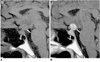
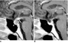
In most healthy individuals, a normal posterior lobe appears hyperintense on T1-weighted MR images, due to phospholipid or secretory granules contained in pituicytes (Fig. 1) (2). In longstanding DI, this high signal intensity of the posterior lobe is absent as a result of failure to synthesize, transport or store neurosecretory granules. After the administration of contrast material, the pituitary gland and pituitary stalk enhance homogeneously.
Traumatic Lesions
Transection of the Infundibular Stalk
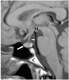
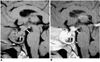
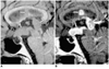
Post-traumatic central DI can be caused by any type of head trauma, in particular by motor vehicle accidents. Clinically, severe head trauma may cause anterior pituitary insufficiency and/or DI, and when this occurs, it is frequently associated with skull fractures and other neurological abnormalities. While the immediate onset of DI is believed to result from direct injury to the posterior lobe, its delayed onset is caused by transection of the stalk, since ADH stored in the posterior lobe is able to maintain its function over a period of time (3). In most cases, DI develops a few days after stalk transection and is commonly transient (Fig. 2). In addition to the absence of the high signal intensity normally seen in the posterior lobe, an ectopic bright spot in the proximal stump of the transected, retracted proximal stalk or hypothalamus can occasionally be identified.
Postoperative Sella
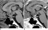
An association between transient DI and postoperative sella is not uncommon. The reported incidence of this is up to 60%, and it appears to represent selective damage to the posterior lobe without concurrent damage to the hypothalamic neurosecretory nuclei (4). Permanent DI after transsphenoidal hypophysectomy has been reported to occur in 0.4-3% of cases (5), and is usually caused by pituitary stalk injury. Fortunately, permanent DI is not a particularly disabling deficit because hormone replacement is a satisfactory treatment. MR imaging shows marked structural distortion and an area of high signal intensity in the pituitary fossa, with an associated mass effect on T1-weighted images within two weeks of surgery. This most likely represents methemoglobin accumulation due to hemorrhage in the surgical bed, a fairly typical feature of early postoperative sella (4).
Infectious or Inflammatory Lesions
Tuberculous Meningitis
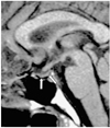
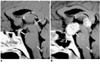
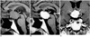
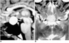
Tuberculosis is one of the common infective causes of central DI. The infiltrative nature of tuberculous meningitis can result in damage to the hypothalamic-neurohypophyseal axis leading to central DI (4). MR imaging reveals a uniformly thickened pituitary stalk, as seen in other granulomatous diseases, and diffuse meningeal enhancement, especially in the basal cistern (Fig. 3).
Epidemic Hemorrhagic Fever
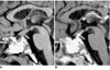
Korean hemorrhagic fever, more commonly known in the Western world as epidemic hemorrhagic fever, is an acute disease caused by viruses belonging to the genus Hantavirus (6). It is characterized clinically by fever, circulatory collapse, hemorrhage, and renal failure. In fatal cases, necrosis of the anterior lobe often develops, and there may be occasional hemorrhage at this location. The eventual result is atrophic change in the pituitary gland. Depending on the location and stage of the hemorrhage and the severity of the disease, the signal intensity of the hemorrhagic area varies (6). DI is a very uncommon symptom in patients with epidemic hemorrhagic fever, though it can result when atrophic change extends into the pituitary stalk (Fig. 4).
Lymphocytic Hypophysitis
In the last few years, a broad spectrum of presentation of the condition known as lymphocytic hypophysitis has been established. The entity is not confined to the anterior lobe but can involve the posterior lobe and pituitary stalk (7). Furthermore, both men and women may be affected, and the condition is thus not necessarily related to pregnancy. According to the anatomical site and severity of the inflammatory process, lymphocytic hypophysitis may be subclassified as lymphocytic adenohypophysitis, lymphocytic infundibuloneurohypophysitis, or necrotizing infundibulohypophysitis (8). The first and second of these are distinctly different entities, and are probably caused by different autoimmune processes (9). It has been reported that in lymphocytic infundibuloneurohypophysitis, inflammation is localized in the nerurohypophyseal system and forms a mass lesion in the posterior lobe and/or pituitary stalk, whereas MR imaging and histologic studies have shown that the anterior lobe is spared (9). However, simultaneous involvement of the anterior adenophysis has been reported (10), and in this situation, the term 'lymphocytic infundibulohypophysitis' is more appropriate than 'lymphocytic infundibuloneurohypophysitis' (Fig. 5) (10).
Granulomatous Inflammation
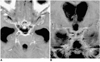
Granulomatous diseases such as sarcoidosis, Wegener's granulomatosis and Churg-Strauss syndrome can involve the hypothalamic-neurohypophyseal axis, and result in central DI. MR imaging reveals a uniformly thickened pituitary stalk, with occasional involvement of the adjacent hypothalamus or pituitary gland. The development of neurosarcoidosis is primarily leptomeningeal and vascular in nature and most commonly involves the meninges, cranial nerves, hypothalamus, infundibular stalk and pituitary gland. Wegener's granulomatosis is a disease characterized by necrotizing vasculitis and granulomatous inflammation of the upper and lower respiratory tracts, together with glomerulonephritis. DI is a very rare complication, the presumed mechanism of which is thought to be either hypothalamic vasculitis or direct granulomatous involvement, or both. Churg-Strauss syndrome, known as allergic granulomatosis and angiitis, is characterized by systemic vasculitis, extravascular granulomas, and eosinophilia, which occur in patients with bronchial asthma and allergy (11). MR imaging reveals diffuse swelling of the pituitary stalk, adjacent hypothalamus, and pituitary gland, which enhances strongly after the injection of contrast material (Fig. 6), a finding similar to that of other granulomatous diseases.
Neoplastic Lesions
Germinoma
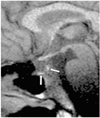
In Asian countries, germinoma is the most common intracranial tumor accompanied by central DI, especially in children. Since the hypothalamic-neurohypophyseal axis is one of the most common sites from which a germinoma originates, DI occurs frequently. Because a hypothalamic-pituitary area lesion is too subtle to be detected during the early stages of suprasellar germinoma, idiopathic central DI is sometimes diagnosed. In such cases, but where a germinoma is still suspected, follow-up MR imaging is therefore necessary, especially in teenage boys. In typical cases, MR imaging clearly demonstrates a thickened pituitary stalk with or without a lobulated pineal mass (Fig. 7). In patients with germinoma, the response to chemotherapy and radiotherapy is usually dramatic.
Langerhans Cell Histiocytosis
Although isolated Langerhans cell histiocytosis of the central nervous system is rare, central nervous system involvement as part of the systemic process is common. This usually takes the form of neuroendocrine disturbances involving the hypothalamic-neurohypophyseal axis. The absence of normal high signal intensity of the posterior lobe, associated with a thickened pituitary stalk, although non-specific for Langerhans cell histiocytosis, should, because of frequent systemic involvement, prompt further studies such as chest radiography, bone scanning, and temporal bone CT (Fig. 8) (12).
Metastasis
Metastases to the pituitary-hypothalamic axis account for 1% of sellar masses. Breast cancer metastases are most common, followed by those from gastrointestinal carcinoma (4). About 20% of these metastases to the pituitary-hypothalamic axis are diagnosed clinically, with DI as the main presenting symptom (13). DI of a transient nature may represent metastases to the posterior lobe of the pituitary, without significant encroachment on the supraoptic and periventricular nuclei of the hypothalamus. At MR, a destructive and inhomogeneously enhancing intrasellar and suprasellar lesion, with involvement of the adjacent structure, can be observed (Fig. 9).
Leukemia
DI is a rare complication of acute leukemia, despite the fact that perihypophyseal leukemic infiltrates are found at autopsy in 46% of such patients. In describing the pathogenesis of DI in patients with leukemia, two main histologic findings have been mentioned: diffuse leukemic infiltrates of the neurohypophysis, and thrombosis of the small vessels in the hypothalamic nuclei and neurohypophysis (14). MR may reveal thickening of the pituitary stalk, with or without adjacent hypothalamic enlargement (14).
Lymphoma
Primary lymphoma of the central nervous system is seen either in immunocompetent patients, or in those with human immunodeficiency virus (HIV) infection or in other immunocompromised states. It can cause DI by directly destroying hypothalamic ADH-producing neurons or seeding the third ventricle with lymphomatous cells (15). At MR, an enhancing mass can be seen in the pituitary stalk, hypothalamus, thalamus, basal ganglia, or periventricular white matter (Fig. 10). Lymphomatous spread to the cerebrospinal fluid is, however, rare.
Teratoma
Intracranial teratomas are defined as benign tumors which contain well-differentiated elements derived from the three germinal layers and have a predilection for the midline, that is, the pineal region, the third ventricle, and the infundibulochiasmal region. On rare occasions, intracranial suprasellar teratomas extend into the pituitary fossa, with enlargement of the sella turcica, thereby resulting in visual disturbance, DI, and hypopituitarism. At T1- and T2-weighted MR imaging, high signal intensities representing fat components can be seen in sellar and suprasellar areas.
Other Tumors
Pituitary adenoma and craniopharyngioma are less common causes of central DI. The former usually shows homogeneous signal intensity, with occasional hemorrhage. Rarely, pituitary adenoma can surround and compress the pituitary stalk, resulting in DI (Fig. 11). Several explanations have been proposed as to how ectopic pituitary tissue arises. Some have suggested that because the anterior lobe originates embryonically from Rathke's pouch, pituitary cells may be deposited along the route of embryologic development of the pituitary gland. Such remnants may become foci of a pituitary adenoma. A craniopharyngioma appears as a rather inhomogeneous mass commonly associated with cyst formation (Fig. 12). A Rathke cleft cyst has non-enhancing, well-defined walls, and the observed MR signal intensity, which depends on the intracystic protein concentration, varies. Most commonly, a Rathke cleft cyst shows a high signal intensity on T1-weighted images and iso- to low signal intensity on T2-weighted images, relative to gray matter, reflecting a high protein concentration in the cystic fluid (Fig. 13). Large hypothalamic/chiasmatic gliomas and meningiomas also can cause DI.
SUMMARY
The multiplanar capability of MR imaging plays an important role in the assessment of the hypothalamic-pituitary area and in determining the underlying cause of central DI. Although a wide spectrum of disease processes may cause central DI, MR imaging, particularly when coupled with clinical information, can help provide a more specific diagnosis.
 XML Download