

 PDF
PDF ePub
ePub Citation
 Print
Print


INTRODUCTION
Tinnitus is a perception of sound in the ear without corresponding external sound. Tinnitus may be pulsatile and synchronous with the patient's heartbeat or non-pulsatile and continuous. Pulsatile tinnitus is generally subjective but sometimes audible (objective) to the examiner by ear or by a stethoscope. Several vascular conditions can lead to pulsatile tinnitus, such as arterial bruit, venous hum, arteriovenous malformations, and vascular tumors. These sometimes result in objective tinnitus (1-3).
We report here an extremely rare case of a patient with unilateral objective tinnitus induced by an abnormally large, prominent mastoid emissary vein (MEV). To demonstrate and elucidate the mechanism of the pulsatile tinnitus, we performed Doppler ultrasound and imaging of the temporal bone and the posterior fossa.
CASE DESCRIPTION
A 44-yr-old female with tinnitus in the form of a high-pitched, 'whooshing' sound in the left ear visited our tinnitus clinic on April 11, 2012. The patient had a 6-month history of ear-ringing synchronous with the heart beat during physically stressful conditions such as fatigue, exhaustion, and emotional upset. It was heard about 50% of the time during the day and became louder when she experienced fatigue at her job in accounting. The patient described attenuation of tinnitus by manual compression of the left posterior auricular area. The patient did not complain of hearing impairment, fullness in the ear, hyperacusis, dizziness, or headache. She had no past head or neck trauma, no exposure to ototoxic drugs or extremely loud noise, chronic medical illness, or neuropsychiatric condition. The tinnitus was not audible even with a stethoscope around the left ear and mastoid area. The patient described that the Valsalva maneuver, or manual compression of the left internal jugular vein, reduced the loudness of the tinnitus, but it was unaffected by head position or compression of the right neck. The otoscopic examination and pure-tone audiogram were normal.
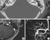
An axial temporal bone computed tomography with contrast enhancement revealed that the left MEV had a prominent opening to the sigmoid sinus and the large canal of the vein crossing the mastoid bone to the soft tissue, without other accessory MEVs (Fig. 1A). The outer foramen of the MEV was located on the posterior-superior mastoid region. The diameter of the inner foramen (at the sinus side) of the left MEV was approximately 4.5 mm whereas that of the right MEV was 1.3 mm. The magnetic resonance image (MRI, Gyroscan, Philips, the Netherlands) of the posterior fossa with contrast enhancement showed an outstanding figure of the MEV enhanced together with the sigmoid sinus, also demonstrated on an MR-venogram where no other significant abnormality was found in the venous structures except a size discrepancy between the left internal jugular vein and the right, with the left being slightly larger (Fig. 1B, C).
Doppler of the left mastoid region detected the origin of the pulsatile tinnitus and displayed pulsating outward venous flow (from the sigmoid sinus to the scalp) with approximately 0.5 m/s of peak velocity at the orifice of the MEV (Fig. 2A). However, no turbulence was detected by the Doppler waveform (confirmed by two professional sonographers) and the flow inside the mastoid bone was also not visible due to bony opacity. However, the Doppler recording seemed to be identical to the patient's tinnitus, which showed synchrony with her heartbeat and attenuation of the flow-signal by the Valsalva maneuver, which matched the timing of tinnitus-reduction described by the patient (Fig. 2B, C).
Although transfemoral venous embolization was recommended to obliterate the MEV, the patient preferred to adapt to the tinnitus and successfully modified her life style by avoiding stressful conditions and distracting attention from the tinnitus.
DISCUSSION
The current case report includes imaging studies revealing a large MEV and a synchronous Doppler sonogram with the pulsatile tinnitus. The original and outstanding point of this study is that objective pulsatile tinnitus can originate from a single large MEV. Based on the otoscopic examination and imaging studies, other possible vascular etiologies of pulsatile tinnitus were excluded, such as vascular tumors, arteriovenous malformation or fistula, aberrant carotid artery, and jugular vein variants.
Regarding the size of the MEV, the internal foramina of the sigmoid sinuses were measured in 50 normal cadaver skulls and their diameters were reported to be smaller than 2 mm in 60% and 3.5 mm in 85%, and the mean diameter of the MEV at the mastoid emissary foramen was reported by others as 3.5 (1.1 to 5.6) mm (4, 5). The diameter of the MEV at the inner foramen in our case was 4.5 mm, which is extraordinarily large, and the flow velocity in the left MEV was relatively high at 0.5 m/s, whereas flow in the right MEV (around the location of the presumed opening) was not detected at all.
An MEV is an embryonic residual venous tract that connects the sigmoid sinus and extracranial venous system, and may serve as an exit route for diploic venous blood (6). An MEV can become dilated by high-flow vascular malformations or when associated with severely hypoplastic jugular veins (7). In our case, however, we observed only a dilated MEV, with no other vascular abnormality seen in the posterior fossa or jugular venous system.
An MEV is a valveless vein that may allow bidirectional flow, suggesting the possibility of turbulence in the MEV, particularly when the dominant direction of blood flow is reversed. However, we observed no turbulent flow in the Doppler recording. Although the mechanism of tinnitus due to blood flow through the MEV was unclear in the present case, we presume that the tinnitus may be due to increased flow velocity or intracranial pressure (induced by exercise or stress) without developing turbulence. Enhanced flow or pressure could be transmitted into the large MEV and could present as audible tinnitus. Several factors may influence flow velocity, such as central or intracranial venous pressure, soft tissue conditions, and vessel wall tension. The Valsalva maneuver or compression of the jugular vein decreased the audibility of the tinnitus. Using MR venography, it has been found that breath holding (the Valsalva maneuver) decreased the flow-velocity in 65.4%-84.0% of normal sigmoid sinuses (8).
Forte et al. (4) reported a similar case of objective pulsatile tinnitus associated with an abnormal MEV. More recently, Chauhan et al. (9) presented multiple emissary veins of the posterior fossa and unusual origin of the left petrosquamosal sinus from a dilated mastoid emissary vein. Thus, our case report is probably the third account of MEV-related pulsatile tinnitus. Forte et al. (4) also objectively demonstrated pulsating tinnitus in a 13-year old girl whose MEV was surgically ligated. However, there was a high jugular bulb on the same side and the patient reported persistence of the tinnitus even after ligation of the MEV, suggesting partial contribution of the high jugular bulb in the tympanic cavity to the tinnitus (4). It was recently reported that a 30-year old woman's tinnitus was associated with a bony defect on the left mastoid area with a tortuous intramastoid bony canal forming a loop within the mastoid, which had two openings in the posterior fossa. Bilateral osseous canals in the lateral petrous temporal bones had originated from the enlarged mastoid emissary canal and from the transverse sinus, respectively. In addition, HRCT suggested an enlarged MEV orifice and the presence of condyloid and occipital emissary foramina (9).
The common feature of these MEV-associated tinnitus reports (including ours) is the left side tinnitus related to the abnormal MEV in female patients. The conspicuously different point of our case from the previous reports is that the pulsatile tinnitus was associated with a single large MEV, with no other vascular abnormalities present. We therefore conclude that a single large MEV can be the sole source of objective pulsatile tinnitus, and that Doppler imaging is a safe and essential diagnostic tool for identifying MEV-related tinnitus.

 XML Download