

 PDF
PDF ePub
ePub Citation
 Print
Print


Abstract
Purpose
To investigate the perception of 3-dimensional (3D) image after successful exotropia surgery and compare with Titmus stereo test.
Methods
A total of 23 children who underwent surgery for intermittent exotropia and 28 normal children were evaluated with a 3D laptop computer and Titmus stereo test.
Results
The mean minimal 3D scene depth level was 38.26 ± 19.69 in the exotropia group and 33.21 ± 16.11 in the normal group with no significant difference (p = 0.319). The mean stereoacuity was 60.97 ± 34.23 arc sec in the exotropia group and 76.69 ± 93.81 arc sec in the normal group with no significant difference (p = 0.444). The mean minimal 3D scene depth and mean stereoacuity showed statistically significant positive correlation in both the exotropia group (p = 0.024) and nor-mal group (p = 0.026).
References
1. Morrison D, McSwain W, Donahue S. Comparison of sensory outcomes in patients with monofixation versus bifoveal fusion after surgery for intermittent exotropia. J AAPOS. 2010; 14:47–51.

2. Stathacopoulos RA, Rosenbaum AL, Zanoni D. . Distance stereoacuity. Assessing control in intermittent exotropia. Ophthalmology. 1993; 100:495–500.
3. Adams WE, Leske DA, Hatt SR. . Improvement in distance stereoacuity following surgery for intermittent exotropia. J AAPOS. 2008; 12:141–4.
4. O'Connor AR, Birch EE, Anderson S, Draper H. FSOS Research Group. The functional significance of stereopsis. Invest Ophthalmol Vis Sci. 2010; 51:2019–23.
5. Park DC. Quality assessment and analysis of stereoscopic 3D tele-vision pictures. J Institute of Signal Processing and Systems. 2010; 11:278–88.
6. Fawcett SL. An evaluation of the agreement between con-tour-based circles and random dot-based near stereoacuity tests. J AAPOS. 2005; 9:572–8.
7. Romano PE, Romano JA, Puklin JE. Stereoacuity development in children with normal binocular single vision. Am J Ophthalmol. 1975; 79:966–71.
8. Takai Y, Sato M, Tan R, Hirai T. Development of stereoscopic acui-ty: longitudinal study using a computer-based random-dot stereo test. Jpn J Ophthalmol. 2005; 49:1–5.
9. Fox R, Patterson R, Francis EL. Stereoacuity in young children. Invest Ophthalmol Vis Sci. 1986; 27:598–600.
10. Hong SW, Park SC. Development of distant stereoacuity in visu-ally normal children as measured by the Frisby-Davis distance stereotest. Br J Ophthalmol. 2008; 92:1186–9.
11. Chung YR, Yang H, Lew HM. . The assessment of stereoacuity in patients with strabismus. J Korean Ophthalmol Soc. 2008; 49:1309–16.
12. Cho YA, Cho SW, Roh GH. Evaluation of criteria of stereoacuity for Titmus, Randot & TNO stereotests. J Korean Ophthalmol Soc. 1999; 40:532–7.
13. Park DJ, Kwak SH, Kim CM, Kim TG. Low-power discrete-event SoC for 3DTV active shutter glasses. J Institute of Electronics Engineers of Korea. 2011; 48:18–26.
14. Yu CB, Fan DS, Wong VW. . Changing patterns of strabismus: a decade of experience in Hong Kong. Br J Ophthalmol. 2002; 86:854–6.
15. Rah SH, Jun HS, Kim SH. An epidemiologic survey of strabismus among school-children in Korea. J Korean Ophthalmol Soc. 1997; 38:2195–9.
16. Yildirim C, Mutlu FM, Chen Y, Altinsoy HI. Assessment of central and peripheral fusion and near and distance stereoacuity in inter-mittent exotropic patients before and after strabismus surgery. Am J Ophthalmol. 1999; 128:222–30.
Figure 1.
(A) The patient watches the 3D image displayed by 3D laptop computer. (B) Animation is converted into 3D image by TriDef 3D media player.

Figure 2.
Comparison between exotropia group and normal group. (A) The mean minimal 3D scene depth level didn’t show sig-nificant difference (p = 0.319, independent samples t-test). (B) The mean stereoacuity by Titmus stereo test didn’t show significant difference (p = 0.444, independent samples t-test).
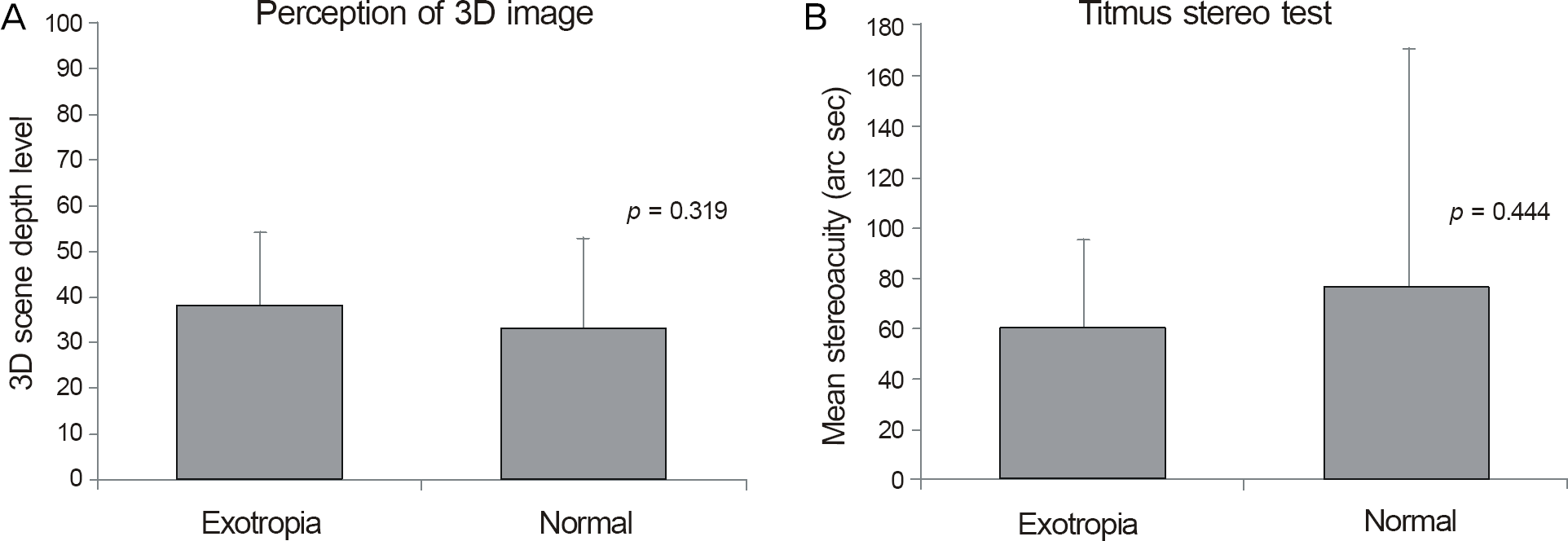
Figure 3.
Correlation between mean minimal 3D scene depth level and mean stereoacuity by Titmus test (A) in exotropia group (Pearson correlation coefficient 0.468, p = 0.024), (B) in normal group (Pearson correlation coefficient 0.421, p = 0.026).
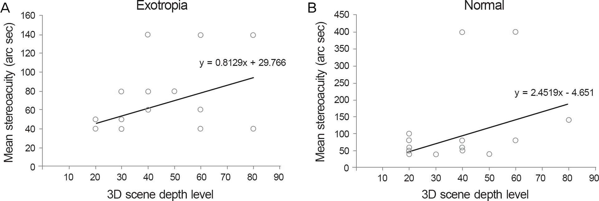
Table 1.
Characteristics of subjects
 XML Download