

 PDF
PDF ePub
ePub Citation
 Print
Print


INTRODUCTION
A giant aneurysm is one with the maximal diameter of 25 mm or more in the aneurysmal vessel. This entity represents approximately 5% of all intracranial aneurysm.9)13)18) Due to the characteristics of giant aneurysms, this type of lesion has poorer prognosis compared to other aneurysms. Moreover, it is associated with high rates of morbidity and mortality.2)3)5)6)8)9)14)17)21) Many surgical clipping series have morbidity and mortality rates ranging from 26% to 35% and 15% to 21%, respectively.7)14)20) The advances in endovascular treatment techniques and devices make it challenging for neurosurgeons to overcome poor prognosis of giant aneurysms. Endovascular treatment should be focused on the complete occlusion, minimum mass effect, preserving the patency of the parent artery, prevention of regrowth, re-bleeding, and thromboembolic complications.1)10)11) To evaluate these clinical implications, we retrospectively reviewed a series of giant aneurysms treated with endovascular embolization.
MATERIALS AND METHODS
Nine patients with nine giant aneurysms were treated by endovascular procedures between March 2000 and September 2009 at our hospital. Clinical and radiographic outcome data were gathered and supplemented by retrospective charts and angiogram reviews. We estimated occlusion rates based on the post-operative angiogram (the near complete occlusion ratio represents a ratio of more than 95%). The angiographic follow-up period (mean, 13.5 months) was defined as the time between the first treatment session and last angiogram available for each patient. The clinical follow-up period was defined as the time between the first treatment session and last follow-up examination. Glasgow Outcome Scale (GOS) scores were determined at the last clinic visit or by telephone. A score of 4 (moderate disability, independent for daily life activities) was considered to be a good outcome, and a score of 5 (good recovery, resumption of normal activities despite minor neurological or psychological deficits) was considered as an excellent outcome.
RESULTS
Demographics
Data for endovascular treatment of giant aneurysms in nine patients were reviewed. The average age of these patients was 59.2 years. Seven patients were women. Demographics, presentation, aneurysm size, location, and treatment are summarized in Table 1. Four ruptured aneurysms presented with subarachnoid hemorrhage (SAH) and five unruptured aneurysms were treated. Patients in both groups were predominantly female. There were no significant differences in age or gender between the SAH and non-SAH groups.
Angiographic Outcomes
The last angiogram for the patients in our series was obtained at a mean period of 13.5 months (range, 12-24 months) from the first treatment session. Angiographic near-complete occlusions (> 95%) were obtained in two out of five unruptured aneurysms and four out of four ruptured aneurysms, representing an overall rate of 67%. Occlusion rates of 90% or greater (i.e., including near-complete occluded aneurysms) were obtained in four out of five unruptured aneurysms and four out of four ruptured aneurysms, resulting in an overall rate of 89%. Stents were required at some point during the management of four aneurysms (two ruptured and two unruptured). Among the stents placed in the aneurysms, only three were placed during the initial attempt. Another one was used during the second session because of aneurysm regrowth (details about the process this patient underwent are presented in the illustrative cases section; patient 8).
Clinical Outcomes
Nine aneurysms were treated over the course of 12 sessions, resulting in an average of 1.3 treatment sessions per aneurysm. A third of treatment sessions resulted in transient morbidity (two ruptured cases and one unruptured case). However, these were not incidence of procedure-related morbidity and the patients recovered completely except for one case of mortality. At the last known clinical follow-up examination (mean, 27.9 months), eight patients had a GOS score of 4 or 5 ("good" or "excellent" outcome; five out of five non-SAH patients and three out of four SAH patients), and one SAH patient died. This case of mortality was not due to procedure-related complications or worsened neurological function, but was caused by multiple organ failure originating from acute renal failure. The average final follow-up GOS scores for the ruptured cases were 3.75 and 4.8 for the unruptured cases. Characteristics of the patients treated in our institution are summarized in Table 1.
Illustrative Cases
Patient 3
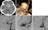
A 63-year-old woman presented with a decreased level of consciousness, nausea, vomiting, and visual discomfort. She was found to have a wide necked, giant, right cavernous aneurysm with a calcified wall that was eventually obliterated with coil embolization. She remained neurologically undisturbed except for the visual discomfort. Her follow-up angiography 6 months later revealed coil compaction. Additionally, worsening of coil compaction was observed on the 1-year follow-up angiography. Eventually, the patient underwent re-embolization with stable occlusion and a patent right internal carotid artery (ICA) was visible on the magnetic resonance angiogram (MRA) more than 2 years after diagnosis. Her diagnostic imaging findings and periprocedural images are presented in Fig. 1.
Patient 5
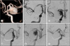
A 63-year-old woman presented with homonymous hemianopsia in her right eye. A computed tomographic scan and lumbar puncture did not produce any evidence of SAH. Angiography identified a giant saccular right cavernous aneurysm and large fusiform petrous aneurysm. The former was treated with stent-assisted coil embolization and the latter was treated only with coil embolization. Angiography performed at 9 months and MRA done at 15 months revealed that the patient was in a stable state. Eventually, the patient's vision improved about 15 months after the procedure. Her diagnostic imaging findings and periprocedural images are presented in Fig. 2.
Patient 8
A 61-year-old woman presented with a severe headache. A computed tomographic scan revealed subarachnoid hemorrhage and an aneurysm with a calcified wall. The presence of a giant aneurysm in the right posterior communicating artery was confirmed by angiography. The aneurysm was subsequently coiled and the patient was discharged without any neurological deficit. However, follow-up angiography done 12 months after the procedure showed recanalization of the aneurysm in the inflow zone. Thus, another endovascular coiling with stenting was performed. Follow-up simple X-ray and MRA images did not capture any change of coiling until 20 months. However, angiography at 24 months revealed subsequent recanalization. Another session of coiling was then performed and was successful. Thirty months follow-up was attained. Her diagnostic imaging findings and periprocedural images are presented in Fig. 3.
DISCUSSION
The prognosis of giant aneurysms is very poor because of hemorrhage, mass effect, or thromboembolism.3)5)21) Drake et al.6) reported the natural history of untreatable giant aneurysms. These lesions are associated with a mortality rate of 66% at 2 years and over 80% at 5 years. Because of this, therapeutic strategies should be carefully selected based on lesion locations, morphological features, sizes, and type of associated symptoms.
The transcranial surgical clipping can be a good treatment option if it is feasible. There are several reports of giant aneurysms treated with surgical clipping that produced good results.9)18) Despite these reports, many neurosurgeons have noted that giant aneurysms are associated with higher surgical morbidity and mortality than small aneurysms, especially when they are located in the posterior circulation.13)19) Due to these characteristic features, surgical clipping of giant aneurysms is a heavy burden to neurosurgeons.
Endovascular embolization with detachable coils was introduced in the early 1990s. As a matter of course, giant aneurysms are also the most complex types of aneurysms to treat because of their size, incorporation of parent and perforator vessels, thrombus, thin wall tissue, and calcification.16) The selection of endovascular candidate is different from the selection of surgical candidates. The aneurysm geometry (dome to neck ratio and neck width) is the most important factor because it will determine the coil stability, primary neck occlusion, later recanalization, and strategies using adjunctive devices such as stents or balloon. Recent technical improvements in coil and stent technology has enabled easier, safer, and more effect packing of the aneurysmal sac. These have also allowed coiling in broad-necked aneurysms that was previously impossible.
When the giant aneurysms are associated with surgical complexity, comorbidity, poor-grade according to the Hunt-Hess grade and Fisher grade, and ideal morphology for coiling, neurosurgeons can choose to perform endovascular coil embolization. Above all, the most important factor is anticipated surgical morbidity. Mass effect is the most common presentation of giant aneurysms which range from 39 to 75% of cases.4)5)22) Isolation of the aneurysm from circulation by endovascular parent occlusion, trapping, or endosaccular obliteration improves mass effect symptoms due to reduction in the pulsation of the aneurysmal mass to the surrounding brain parenchyma.6) In contrast, aggravation of mass effect symptoms has been reported.15) Gruber et al.12) reported 27.2% of their patients had worsening symptoms after embolization and required additional treatment such as trapping and surgical decompression. The development of new devices to occlude the aneurysmal neck without using coils makes it possible to reduce the mass effect.
Our results showed that primary endovascular coil embolization of giant aneurysms with a low rate of procedure-related complications is technically possible. Satisfactory clinical results (occlusion rates of 90% or greater) were achieved in 89% of patients at a median follow-up period of 27.9 months. Our cases met the three objectives for endovascular treatment summarized by Gonzalez et al.10): 1) protection from bleeding, 2) reduction of size for mass effect relief, and 3) prevention of thromboembolic complications. No bleeding, rebleeding, or thromboembolic complications occurred, and symptoms related to the mass effect were relieved in all of the patients. However, recanalizations occurred in two patients that required additional coiling (one case needed one more coiling). These two patients had near-complete occlusion rates at their last follow-ups and these additional procedures did not result in any procedure-related morbidity.
Our series had a few significant limitations.
We performed retrospective chart and angiogram reviews. Therefore, we could not eliminate biases inherent of retrospective reviews.
Because the occlusion rate in our series was measured using post-operative angiograms, this was probably not the absolute occlusion rate or indicative of stability.
Giant aneurysms are not a common aneurysm form. Thus, we did not experience the sufficient cases with surgical clipping comparable to our cases with endovascular coiling.
Because the mean follow-up period in our series was short, there is the possibility that the regrowth rate was underestimated.
Due to the small size of our study, we could not rule out the chance that the favorable results were somewhat exaggerated.
The favorable results of our cases do not necessarily mean that endovascular embolization is the treatment of choice for giant aneurysms. However, this technique is one possible treatment option that neurosurgeons should consider when they encounter this type of lesion.

 XML Download