Journal List > Clin Endosc > v.46(4) > 1151823

Lee, Kim, Kim, Lee, Lee, Keum, Cheung, Yang, and The Stent Study Group of the Korean Society of Gastrointestinal Endoscopy: Evidence-Based Recommendations on Colorectal Stenting: A Report from the Stent Study Group of the Korean Society of Gastrointestinal Endoscopy

Abstract

Recently, placement of self-expandable metallic stents has been used for the treatment of colorectal obstruction. As domestic awareness of colorectal cancer has increased, the number of colorectal stenting procedures performed has also increased. We aimed to provide evidence-based recommendations for colorectal stenting to aid gastroenterologists in making informed decisions regarding the management of patients who present with colorectal obstruction. The working group consisted of eight gastroenterologists who actively practice and conduct research in the field of colorectal stenting and are the members of the Stent Study Group of the Korean Society of Gastrointestinal Endoscopy. A literature search was conducted using the PubMed, Embase, KoreaMed, and the Cochrane Library databases to identify relevant articles published between January 2001 and June 2012. Based on the modified Delphi process, 10 recommendation statements regarding indications, usefulness, methodology and complications of colorectal stenting, and alternative treatments for malignant colorectal obstruction were determined. The contents will be widely distributed, and periodically revised to reflect the latest knowledge. These evidence-based recommendations for colorectal stenting will provide gastroenterologists and patients with appropriate and balanced information, and will improve the quality of care.

INTRODUCTION

This report presents the evidence-based recommendations for colorectal stenting for benign and malignant conditions. It includes the current view of the Korean Society of Gastrointestinal Endoscopy on endoscopic colorectal stenting and describes the associated indications, outcomes, models of available stents, and colorectal stenting techniques. Colorectal obstruction usually requires rapid intervention because patients are not able to ingest a meal and rapidly deteriorated. Several modalities have been employed to restore colorectal patency. The traditional modality included surgical diversion and stoma formation. However, surgery has the disadvantages of surgery-related mortality and morbidity. Emergency surgical treatment has been performed in patients with this condition, but its morbidity and mortality rates are relatively high.1-3 Patients with impending intestinal rupture require immediate decompression, and emergency operations increase the risk of perioperative complications. Recently, placement of self-expandable metallic stents (SEMSs) has been used for the treatment of colorectal obstruction. SEMS insertion is a safe and effective modality that has been accepted as a suitable or preferable alternative to surgery. As domestic awareness of colorectal cancer has increased, the number of colorectal stenting procedures performed has also increased. Colorectal stenting is mainly performed in secondary or tertiary hospitals. However, it is difficult to find international guidelines regarding the treatment of colorectal obstruction with SEMSs. Furthermore, no domestic consensus recommendations or evidence-based guidelines have been reported to date. Therefore, we aimed to provide evidence-based recommendations for colorectal stenting to aid gastroenterologists in making informed decisions regarding the management of patients who present with colorectal obstruction. The development of evidence-based recommendations for colorectal stenting will provide gastroenterologists and patients with appropriate and balanced information, and will improve the quality of care.

Development processes

This work began in June 2011 with the formation of the colorectal stenting working group. The working group consisted of eight gastroenterologists who actively practice and conduct research in the field of colorectal stenting. All these physicians are the members of the Stent Study Group of the Korean Society of Gastrointestinal Endoscopy. No participant declared a conflict of interest. The Korean Society of Gastrointestinal Endoscopy supported this work.

Distribution and revisions of the evidence-based recommendations

This report will be published in the official journal of the Korean Society of Gastrointestinal Endoscopy and will be provided free of charge on the website of the Korean Society of Gastrointestinal Endoscopy. The contents will be widely distributed through a summary book. Moreover, the contents will be periodically revised to reflect the latest knowledge.

METHODS

Literature search

A literature search was conducted using the PubMed, Embase, KoreaMed, and the Cochrane Library databases to identify relevant articles published between January 2001 and June 2012. Keywords used in the literature searches included "colon OR colonic OR colorectal OR rectum OR rectal" AND "obstruction OR stenosis" AND "stent." The articles were included if they met the following criteria: 1) the manuscript is written in English or Korean; 2) the full manuscript is available; and 3) the study participants are older than 18 years of age. Seven working group members performed the searches and summarized the data using standardized report forms. Subsequently, 10 key questions were identified and distributed to each member of the working group. Based on the relevant literature articles, the working group members rated the level of evidence and created the draft statements. The level of evidence and the strength of recommendation were defined and graded (Table 1).4,5 The working group members checked and revised the draft statements and their ratings after further review and discussion. Based on the modified Delphi process, the draft statements were circulated electronically to all participants prior to the meeting. The first round of voting on the statements was conducted by email. Each statement was assessed on a 5-point Likert scale as follows: 1) accept completely; 2) accept with some reservation; 3) accept with major reservation; 4) reject with reservation; and 5) reject completely. Based on the voting results and comments, the statements were modified. The second round of voting was carried out during the face-to-face meeting, followed by the modification of the statement when the proportion of the working group answering 1 plus those answering 2 was <75%. Subsequently, participants voted again on the statement. When the proportion of the working group answering 1 plus those answering 2 was ≥75%, the statement was finally accepted. Funding was provided by the Korean Society of Gastrointestinal Endoscopy, and any industrial influence on the process of consensus development was avoided. The funding source had no role in identifying statements, abstracting data, grading evidence, or preparing the manuscript. This manuscript was reviewed by the external reviewers (J.O.K. from the Department of Gastroenterology, Institute for Digestive Research and Digestive Disease Center, Soonchunhyang University Hospital and S.A.J. from the Department of Gastroenterology, Ehwa Womans University School of Medicine) prior to submission to Clinical Endoscopy and was approved by the Korean Society of Gastrointestinal Endoscopy.

Recommendation statements

Indications and usefulness of colorectal stenting

1) Is colorectal stenting useful for the management of leftsided colon or rectal malignant obstruction as a bridge to surgery?

Colorectal stenting using SEMSs can be performed in the management of left-sided colon or rectal malignant obstruction as a bridge to surgery in order to avoid emergency surgery.
Grade of evidence: moderate.
Level of agreement: 1) accept completely (82%); 2) accept with some reservation (18%); 3) accept with major reservation; 4) reject with reservation; and 5) reject completely.
The use of SEMSs as a bridge to surgery is useful for avoiding emergency surgery and allowing time to improve the patient's medical conditions before elective surgery. A systemic review comparing elective surgery after SEMS placement and emergency surgery demonstrated that elective surgery following SEMS insertion showed twice the rate of primary anastomosis compared to emergency surgery alone. Moreover, colostomy rates were higher in the emergency surgery group. A shorter hospital stay was required in the elective surgery group following SEMS insertion.6 A meta-analysis including 10 studies and 451 patients was performed, comparing SEMS placement and elective surgery with open surgery. The success rate of stent insertion was 88% to 100%. A shorter hospital stay was noted in the SEMS group. Lower rates of stoma formation and fewer posttreatment medical complications were likely in the SEMS group, although there was no difference in long-term outcomes between the groups.7 Recently, several small-sized, randomized, controlled trials (RCTs) comparing SEMS and elective surgery with emergency resection surgery were reported. Cheung et al.8 demonstrated that the clinical success rate of stent insertion was 83%. In the SEMS group, 67% of the patients underwent a 1-stage operation, and the postoperative morbidity was significantly low. In a meta-analysis of RCTs,9,10 SEMS placement as a bridge to surgery was shown to be beneficial, particularly with regard to short-term outcomes. In the SEMS group, higher primary anastomosis and lower overall stoma rates were shown. A shorter hospital stay, less blood loss, and a lower procedure time were noted in the SEMS group. However, there were no significant differences in long-term outcomes, including permanent stoma rates, hospital mortality, and postoperative complication rates. Although there are no proven benefits in long-term outcomes, SEMS placement before surgery improves the quality of the primary surgical procedure as a bridge to surgery.
On the other hand, several controversies related to the safety and efficiency of SEMSs remain. Some authors indicate the risk of potentially life-threatening complications such as perforation and the lack of strong evidence of clinical benefit, which hampered the use of SEMSs for the management of left-sided or rectal malignant obstruction. Colorectal stenting using SEMSs can be performed in the management of leftsided colon or rectal malignant obstruction as a bridge to surgery in order to avoid emergency surgery. However, in order to confirm the potential evidence related to safety and efficiency of SEMS placement before surgery, further RCTs with larger sample sizes and well-designed trials are needed. A few ongoing prospective studies are expected to provide additional assurance for the usefulness of SEMSs in these conditions.11

2) Is the placement of SEMSs necessary for malignant proximal colon obstruction?

SEMS placement for malignant proximal colon obstruction can allow the elective surgery.
Grade of evidence: low.
Level of agreement: 1) accept completely (27%); 2) accept with some reservation (73%); 3) accept with major reservation; 4) reject with reservation; and 5) reject completely.
Literature articles related to the use of SEMSs for malignant obstruction in the proximal colon include case series or retrospective comparative studies. There has been no prospective controlled study. Elsberger et al.12 reported seven procedures using SEMSs for obstruction in the transverse colon (n=4) and splenic flexure (n=3). Technical success was achieved in six patients. There was no SEMS-related morbidity or mortality. This report showed that the placement of colonic stents proximal to the descending colon was feasible, effective, and safe. Repici et al.13 described 21 patients who underwent placement of SEMSs proximal to the midtransverse colon. Obstruction was complete in eight patients and subtotal in 13 patients. Initial technical success was achieved in 20 of 21 patients (95%). Complete relief of obstruction was achieved in 17 of 20 patients who had technical success (85%). There were no procedure-related complications. Among these patients, eight underwent elective surgery after SEMS placement, and no surgical complications were noted. These results suggest that SEMS placement is safe and effective for the treatment of malignant obstruction of the proximal colon as a bridge to surgery as well as palliation.
In a previous study including 16 SEMS placement proximal to the splenic flexure, technical success was achieved in 94% of patients and clinical success was noted in 88%. Stenting was attempted as a bridge to definitive surgery (n=5) and for palliation (n=9). In the patients who received stenting as a bridge to surgery, colectomy was performed within 1 to 12 weeks (mean, 6 weeks). Among these five patients, no major surgical complication occurred, and none required temporary stoma formation.14 These results demonstrate that SEMS placement can allow the elective surgery in acute malignant proximal colon obstruction with high efficacy and safety. Kim et al.15 reported 57 patients who underwent preoperative SEMS insertion for obstruction of resectable colorectal cancers proximal to the sigmoid colon (n=13) and in the sigmoid colon (n=22) and rectum (n=22). There were no significant differences in stent-related complications, clinical improvement rates, and 1-stage resection rates among these three groups. The postoperative complications, the requirement rate of intensive care unit (ICU) care, the period of ICU stay, duration of postoperative hospital stay and hospital mortality rate did not significantly differ among the three groups. These results suggest that clinical improvement rates and postoperative results following successful placement of stents for obstruction of resectable colorectal cancers do not differ according to the location of the obstructing lesion.
Based on these data, SEMSs can be used as a bridge to surgery for patients with malignant obstruction in the proximal colon to avoid emergency operations. However, obstruction complicated by proximal colon cancer is not as severe as distal colon cancer due to the relatively small amount of fecal material retained. Many proximal colon cancers are primarily managed by 1-stage surgery without the need for bowel preparation and stoma formation. Therefore, SEMS placement for proximal colon obstruction as a bridge to surgery should be carefully selected with the consideration of the patient's conditions such as age and underlying comorbidity.

3) Is stent placement useful for unresectable malignant colorectal obstruction?

In patients with unresectable malignant colorectal obstruction, SEMS placement can not only relieve symptoms and improve quality of life but also allow chemotherapy and/or radiotherapy for palliation.
Grade of evidence: low.
Level of agreement: 1) accept completely; 2) accept with some reservation (100%); 3) accept with major reservation; 4) reject with reservation; and 5) reject completely.
SEMSs have provided an alternative for relieving colorectal obstruction. Several studies have demonstrated that SEMSs are an effective palliative measure in treating colorectal obstruction.16-18 There are several case-control reports of stent placement in patients with unresectable malignant colorectal obstruction. Carne et al.19 compared expandable metal stents (n=25) with open surgery (resection, bypass, or stoma; n=19) in patients with left-sided, nonresectable metastatic colon cancer, and reported that patients treated with stents are discharged earlier than those who underwent open surgery and that the survival difference between these two groups was insignificant. In two previous studies that compared stent placement with palliative surgical intervention in patients with unresectable colorectal cancer, palliative treatment of incurable obstructive colorectal carcinoma using stents was shown to be an effective and suitable alternative to palliative surgery, with no negative impact on the survival rate but with less peri-interventional morbidity and mortality and comparable overall morbidity rates.20,21 In one of these case-control studies, the authors suggested that insertion of SEMSs in obstructing colon cancer with nonresectable synchronous metastases may increase the resectability rate of metastases and favorably affect survival.21 However, the risk of tumor perforation during chemotherapy should be considered. Cennamo et al.22 described nine metastatic colon cancer patients that had received chemotherapy (capecitabine monotherapy in two patients, 5-fluorouracil+folinic acid+oxaliplatin in one patient, capecitabine+ oxaliplatin [XELOX] in two patients, XELOX+ panitumumab in two patients, and XELOX+bevacizumab in two patients) after SEMS placement, and delayed bowel perforation occurred in only two XELOX+bevacizumab-treated patients. Further studies are required to clarify whether SEMS placement increases the risk of perforation caused by bevacizumab-based therapies. Unlike the previous reports, another study reported a significant survival difference between palliative surgery (n=53) and SEMS placement (n=45) as a palliative treatment in patients with incurable stage IV colorectal cancer.23 In this study, stent placement offers advantages regarding early morbidity, hospital stay and stoma creation. Surgery offers a benefit in survival in patients who receive chemotherapy but not in noncandidates to chemotherapy. Recently, Lee et al.24 compared long-term outcomes of endoscopic stenting (n=7) with those of palliative surgery (n=73) in patients with unresectable obstructive colorectal cancer. Early success rates between the SEMS group and the surgery group were not different (95.8% vs. 100%; p=0.12). The SEMS group had fewer early complications compared to the surgery group (15.5% vs. 32.9%; p=0.015). Although the patency duration of the first stent in the SEMS group was shorter than that in the surgery group (p<0.001), the median patency duration after a second stenting procedure was comparable to that of the surgery group (p=0.239). There were more late complications in the SEMS group than in the surgery group (p=0.028), although the rates of major complications did not differ between the two groups (p=0.074). The authors suggested that SEMSs could be recommended not only to patients with malignant colorectal obstruction and short life expectancy but also to those with a longer life expectancy.
Consequently, in patients with unresectable malignant colorectal obstruction, SEMS placement can not only relieve symptoms and improve quality of life but also allow chemotherapy and/or radiotherapy for palliation.

4) Is stent placement useful for the management of colorectal obstruction by malignancies other than those of the colon and rectum?

SEMS placement may be useful in the management of colorectal obstruction by malignancies other than those of the colon and rectum, but should be considered as an alternative to surgical treatment with consideration of the surgery-related risks and the benefits of successful stenting.
Grade of evidence: low.
Level of agreement: 1) accept completely (18%); 2) accept with some reservation (82%); 3) accept with major reservation; 4) reject with reservation; and 5) reject completely.
Most malignant colorectal obstructions result from intrinsic factors, i.e., colorectal cancer. However, malignancies of extracolonic origin could also manifest as obstructions of colorectal patency. The stomach is the most frequent site of origin of extracolonic cancer causing colorectal obstruction (Table 2). Yoon et al.25 published a report of 114 patients with colorectal obstruction due to extracolonic cancers. Stomach cancer occurred in 72% of all cases, followed by gynecologic malignancies and pancreatobiliary malignancies. Eleven literature articles that described the origins of extracolonic cancers resulting in colorectal obstruction were included. The most common site of origin was the stomach, followed by the ovary and uterus, pancreatobiliary system, bladder and prostate, lung, neuroendocrine tumor of gastrointestinal tract, breast and head and neck (in order of frequency).
The 11 studies on the stenting for the malignant colorectal obstruction reported 91.5% to 100% technical success rates and 81.8% to 100% clinical success rates. These studies included both colorectal cancer and extracolonic cancers as the cause of malignant colorectal obstruction. The proportion of extracolonic cancers ranged from 2% to 45%.26,27 Luigiano et al.28 analyzed the cases of stent failure and reported that technical failure was related to female sex (p=0.04) and the extracolonic etiology of the stricture (p<0.001). Additionally, the SEMSs placed in patients with an intrinsic colorectal malignancy showed higher patency compared to the patients with extracolonic malignancies (p=0.045).
There have been various reports comparing the success rates of SEMS placement for colorectal obstruction by intrinsic and extrinsic malignancies. The overall outcomes varied, ranging from 42% to 100% for the technical success rate and from 25% to 87.5% for the clinical success rate.29-34 Some reports found little difference in the success or complication rates between these two groups,30,33 whereas others reported lower success rates and higher complication rates for SEMS placement for extrinsic colorectal obstructions.29,31,34 Shin et al.30 reported an 87.2% technical success rate and an 82.1% clinical success rate. Complications occurred in 22 of 57 stents, including stent reobstruction (n=14) and stent migration (n=5). All stent migrations but one developed with covered stents. Trompetas et al.29 reported disappointing results. Among 11 patients with obstruction, only two patients with a clinically successful stenting (18%) survived over 1 month. Five of 11 patients underwent surgical colostomy after failed stenting.
Although there have been no RCTs, six studies compared the outcomes of stenting for malignant colorectal obstruction between patients with colorectal cancer and those with extracolonic cancers (Table 3).25,33-36 Four studies compared the technical success rates between stenting in patients with colorectal cancer and extracolonic cancers. The technical success rates ranged from 84% to 97.1% for colorectal cancer and 65% to 94% for extracolonic cancer. Kim et al.33 reported a technical success rate of 84% for colorectal cancer and 94% for extracolonic cancer (p=0.137) along with a clinical success rate of 82.8% for colorectal cancer and 90% for extracolonic cancer (p=0.533). Perforation rates were 2% and 11% for colorectal cancer and extracolonic cancer patients, respectively. Migration occurred in 10% and 4%, bleeding in 6% and 9%, procedure-related pain in 4% and 13%, and tumor overgrowth in 6% and 2%, respectively. Obstructive symptom-free overall survival rates were 4 months (95% confidential interval [CI], 2.2 to 5.8) and 3 months (95% CI, 2.8 to 4.2; p=0.72) for colorectal and extracolonic cancer patients, respectively. However, in the largest study conducted by Yoon et al.,25 the technical success rates differed between colorectal cancer and extracolonic cancer patients (94.3% vs. 80.7%, respectively; p=0.01). In that study, once the stent was accurately deployed on the site, the clinical success rates were not different (83.8% and 83.7%, respectively). Keswani et al.34 reported worse outcomes in extracolonic cancer patients. Five patients (33.3%) with extracolonic cancer obstruction had at least one complication, including two deaths, while complications occurred in three patients (8.8%) with colorectal cancer (p=0.046). Surgical diversion to relieve persistent obstructive symptoms was required in significantly more patients with extracolonic cancers. Selinger et al.35 reported that SEMS insertion had a significantly higher long-term success rate in patients with intrinsic colorectal cancer (81%) compared to those with extracolonic malignancies (43%; p=0.049).
In conclusion, the clinical outcome of stenting for colorectal obstruction in patients with extracolonic malignancies is likely to be less favorable than that of intrinsic colorectal cancer patients. The outcomes range from 42% to 100% for technical success rates and from 20% to 90% for clinical success rates. More complications tend to occur in patients with extracolonic malignancies. Therefore, although SEMS placement may be useful in the management of colorectal obstruction by malignancies other than those of the colon and rectum, it should be considered as an alternative to surgical treatment with consideration of the surgery-related risks and the benefits of successful stenting.

5) Can stenting be used as a treatment option for benign colorectal strictures?

SEMSs can be used in the management of benign colorectal strictures as a bridge to surgery in order to avoid emergency surgery or as a palliative treatment in patients with high surgical risks or those who are unfit for surgery. However, it should be carefully selected with consideration of the considerable risk of complications.
Grade of evidence: low.
Level of agreement: 1) accept completely (27%); 2) accept with some reservation (73%); 3) accept with major reservation; 4) reject with reservation; and 5) reject completely.
The literature articles regarding the use of SEMSs for benign colorectal strictures primarily include case reports or case series. There have been no controlled studies to date (Table 4). Small et al.37 described 23 patients with benign obstructive disease who underwent endoscopic SEMS placement. The etiologies of the stricture were diverticular/inflammatory (n=16), postsurgical anastomotic (n=3), radiation-induced (n=3), and Crohn disease (n=1). Uncovered Enteral Wallstents or Ultraflex Precision Colonic stents (Boston Scientific, Natick, MA, USA) were used. Clinical success was achieved in 95% of the patients, although major complications occurred in 38% of patients, including migration (n=2), reobstruction (n=4), and perforation (n=2). Eighty-seven percent of these complications occurred 7 days after the placement. Eighty-four percent of the 19 patients who underwent planned surgical resection were successfully decompressed and converted from an emergent operation to an elective operation. In six of these patients, surgery was delayed for longer than 30 days after stent placement. These results demonstrated that SEMSs could decompress high-grade, benign colonic obstructions, thereby enabling elective surgery. However, this approach is associated with a high rate of delayed complications. Thus, data from this small study suggest that SEMSs can be used in the management of benign colorectal strictures as a bridge to surgery in order to avoid emergency surgery.
Keränen et al.38 reported 23 procedures in 21 patients who had an obstruction in the surgical anastomosis (n=8), anastomotic strictures due to Crohn disease (n=2), obstruction caused by diverticular disease (n=10), and a stricture after radiation therapy (n=1). Clinical success was achieved in 76% of these patients. The strictures were resolved in 63% of the eight patients with anastomotic strictures and 30% of the three patients with diverticular strictures. Nine patients (43%) had a complication, most of which occurred in patients with diverticular strictures. The role of endoluminal stenting in benign obstruction, especially for Crohn disease, is controversial, with limited data. A case series in a single center suggests that endoluminal stenting of Crohn disease-induced strictures is an effective alternative to surgery in selected patients.39 However, instead of stenting, endoscopic balloon dilatation can be performed with reliable success for strictures in Crohn disease patients. The relapse rate seems to be higher than after surgery, but a repeated endoscopic treatment can be performed. Therefore, serial balloon dilations can be considered first to avoid or postpone repeated resections.40
Based on the current data, SEMSs can be used in the management of benign colorectal strictures as a bridge to surgery in order to avoid emergency surgery or as a palliative treatment in patients with high surgical risks or those who are unfit for surgery. However, it should be carefully selected with consideration of the considerable risk of complications. Both the benefits and risks must always be considered.

Methodology for colorectal stenting

1) How should colorectal stenting be performed for colorectal malignant obstruction: by endoscopy or fluoroscopy?

Colorectal stenting for colorectal malignant obstruction can be performed using endoscopy, fluoroscopy, or both.
Grade of evidence: low.
Level of agreement: 1) accept completely (55%); 2) accept with some reservation (45%); 3) accept with major reservation; 4) reject with reservation; and 5) reject completely.
Colonic stent insertion for colorectal malignant obstruction is performed by an endoscopist using a combination of endoscopic and fluoroscopic guidance, or by a radiologist under fluoroscopic guidance alone. During radiological stent placement, colon obstruction is located fluoroscopically using a water-soluble contrast medium. Then, a guidewire is inserted from the anus to the malignant stenosis, and it should be passed through the stenotic lesion under fluoroscopic guidance alone. If the guidewire is placed through the lesion, the stent is inserted into the obstruction and released fluoroscopically.41,42 On the other hand, during combined endoscopic and fluoroscopic placement, the distal end of the obstruction is documented endoscopically. If the stenosis is not too tight and the endoscope can be passed through, fluoroscopic guidance is not necessary for stent placement. However, if the stenotic lesion cannot be passed with an endoscope, the length and configuration of the stenosis should be identified fluoroscopically using an endoscopic catheter. The catheter is used to cannulate the stenosis and water-soluble contrast is injected through the stenotic lesion. After identification of the stenosis, a guidewire is inserted through the obstruction, and a through-the-scope (TTS) stent delivery system is placed over the guidewire. Then, the stent is deployed under endoscopic and fluoroscopic guidance.42,43
It was reported that the technical and clinical success rates were not different between these two methods, and that both methods were effective. However, there have been no RCTs comparing endoscopic/fluoroscopic guidance with fluoroscopic guidance only for the placement of colonic stents. The decision of which method should be selected depends on the individual situation, although some advantages of the endoscopic procedure were suggested,42 which include 1) biopsies for histologic confirmation can be taken during the procedure; 2) the endoscope provides easier passage through the sigmoid colon than a guidewire alone, which can reduce the procedure time and deliver the stent to wherever the endoscope can reach, even in proximal parts of the colon; 3) the risk of misplacement can be decreased; and 4) a shorter procedure time can minimize the amount of radiation exposure for the patient, doctor, and assistants.

2) What type of stents should be selected?

The type of SEMSs that are best suited for each situation should be used, with consideration of the features such as the stent material, design, deployed diameter and length, radial force exerted, flexibility, degree of shortening during expansion, recapturability, delivery system, etc.
Grade of evidence: low.
Level of agreement: 1) accept completely (64%); 2) accept with some reservation (27%); 3) accept with major reservation (9%); 4) reject with reservation; and 5) reject completely.
Many types of colorectal stents have unique and common features related to the stent material, design, deployed diameter and length, radial force exerted, flexibility, degree of shortening during expansion, recapturability, and delivery system. These features represent factors to consider when selecting the stent model; however, there is not enough evidence to demonstrate whether these factors could affect the clinical outcomes of patients.44-55
With regard to the stent material, nitinol (a metal alloy composed of nickel and titanium) has been used as a wire due to its shape memory and super-elasticity. Polytetrafluoroethylene or silicone membranes have been utilized as the covered stent membrane material due to their high biocompatibility and strong physical and chemical resistance.
Many stent models have improved flexibility to conform to complex curves, and the deployed diameter and length of the stents vary to meet diverse needs. Although the ideal stent diameter for proper radial force is unknown, the postdeployment diameter of colonic stents is usually approximately 20 to 30 mm and the deployed length of colonic stents ranges from 6 to 16 cm. The end portion of most stents is designed to prevent mucosal damage due to the sharp end and to have a flare to potentially reduce the risk of migration. In addition, most of the stents show various degrees of shortening in length during expansion (20% to 45%), and to facilitate accurate stent placement, highly visible endoscopic and/or fluoroscopic markers have been placed on the delivery system or stent.
Although both uncovered and covered stents show similar technical and clinical success rates, they present unique advantages and disadvantages.56-59 Uncovered stents have the advantage of less stent migration and the disadvantage of a high risk of stent occlusion by tumor ingrowth. In contrast, covered stents are associated with less tumor ingrowth, while they appear to be more prone to stent migration. However, these two stent types did not show significant differences in overall complication rates and stent patency duration.56,57
Stent delivery systems may vary according to the approach method used for SEMS placement, such as TTS and non-TTS stent placement. The delivery system used for the TTS method has a longer total working length and smaller predeployment diameter (10 Fr) to allow the passage of stents directly through the working channel of a therapeutic colonoscope (diameter, ≥3.8 mm). Delivery systems have been developed to have excellent pushability, efficient maneuverability (especially in difficult anatomical locations), and recapturability during deployment. The recapturable ranges of the deployed portion during deployment vary according to the stent models. To improve the clinical outcomes of patients or the ease of maneuverability for its operators, the operators need to understand the features of each stent through experience.
In conclusion, the operators need to select the type of SEMSs that are best suited for each situation, with consideration given to the features of stent material, design, deployed diameter and length, radial force exerted, flexibility, degree of shortening during expansion, recapturability, delivery system, etc.

3) Is the preoperative evaluation of the colon proximal to the obstructive lesion necessary after stent placement?

After SEMS placement for malignant colorectal obstruction, preoperative evaluation of the colon proximal to the obstructive lesion is necessary for detecting synchronous lesions.
Grade of evidence: low.
Level of agreement: 1) accept completely (100%); 2) accept with some reservation; 3) accept with major reservation; 4) reject with reservation; and 5) reject completely.
Synchronous cancers are reported to be present in 1.5% to 9.0% of patients with colorectal cancer.60-64 While a complete preoperative evaluation of the entire colon in patients with colorectal cancer is important, full colonoscopic evaluation of the entire colon is not always feasible, particularly in the setting of malignant colorectal obstruction. There are two single center studies on the subject of preoperative examination of the proximal colon of the obstructive lesion after stent placement, and no controlled studies have been conducted to date. Vitale et al.65 reported the results of preoperative colon evaluation using colonoscopy after SEMS placement in acute malignant colon obstruction. Among 57 patients with acute neoplastic colon obstruction, SEMSs were placed in 50 of 57 patients (87.8%). Thirty-one of 50 patients had resectable cancer (62%), and a complete preoperative colonoscopy was possible in 29 of 31 patients (93.4%). Synchronous cancer and adenomas were detected in three and eight patients (9.6% and 25.8%), respectively. The presence of synchronous cancer lesions led to a change in surgical plan. Seven patients in whom SEMS placement (12.2%) was unsuccessful underwent an urgent surgical intervention. Nineteen of 50 patients who underwent stent placement were ineligible for surgery due to unresectable cancer. No major complication occurred in any patient. The only complication that occurred during the endoscopic procedure was minor bleeding at the stent site, which was noted in five of 31 patients. No mechanical damage of the endoscope due to passage through the stent was detected at the end of the procedure. Cha et al.66 used computed tomography (CT) colonography as a preoperative examination modality. Fifty patients (age, 58.5±11.7 years), who demonstrated no postprocedural complications after successful placement of SEMSs to treat cancer-induced acute colon obstruction, underwent CT colonography 1 to 43 days (median, 5 days) after stent placement. Per-lesion and per-patient sensitivities of CT colonography for lesions 6 mm or larger in diameter in the colon proximal to the stent were 85.7% (12 of 14 lesions; 95% CI, 58.8 to 97.2) and 90% (nine of 10 patients; 95% CI, 57.4 to 99.9), respectively. CT colonography depicted all synchronous cancers (two lesions) and advanced adenomas (five lesions). Per-patient specificity for lesions 6 mm and larger in the proximal colon was 85.7% (18 of 21 patients; 95% CI, 64.5 to 95.9). CT colonography did not generate any false diagnosis of synchronous cancer. False-positive findings on CT colonography did not result in a change in surgical plan for any of the patients. No CT colonography-associated stent dislodgment/migration or colonic perforation occurred in any patient (95% CI, 0% to 6.2%). Both studies demonstrate the importance of preoperative examination of the proximal colon of malignant obstructive lesions after stent placement.
Therefore, after SEMS placement for malignant colorectal obstruction, preoperative evaluation of the colon proximal to the obstructive lesion is necessary for detecting synchronous lesions. However, well-planned randomized trials comparing the efficacy and cost-effectiveness of these approaches are required.

Complications related to colorectal stenting

What complications can occur in SEMS insertion for colorectal obstruction?

SEMS insertion for colorectal obstruction can be associated with complications such as perforation, migration, tumor ingrowth/outgrowth, stool impaction, bleeding, pain, tenesmus, fecal incontinence, or death.
Grade of evidence: low.
Level of agreement: 1) accept completely (82%); 2) accept with some reservation (18%); 3) accept with major reservation; 4) reject with reservation; and 5) reject completely.
Colorectal stenting is a relatively low-risk procedure with a mortality rate below 1%.26,67,68 However, the mortality rate of patients experiencing perforation was 16%.67 Perforation is one of the most serious complications of colorectal stenting. The overall risk of perforation is approximately 5%,24,52,67,69,70 and procedure-related perforation is usually related to wire or catheter misplacement or stricture dilatation.71
The migration rates of uncovered stents range from 3% to 36%, while the migration rates for covered stents are 8% to 50%.52,57,59,70,72-74 It is unclear whether migration is related to chemotherapy.75,76 In patients who have a dilated cecum, the amount of air inflation should be limited during the procedure to avoid cecal perforation.43,76 In up to two thirds of the cases with colorectal stenting-related perforations, an emergency surgical intervention is required, and only cases with minor perforation can be managed with bowel rest and broad spectrum antibiotics.43,70 Stent migration can be managed by the insertion of a second stent if obstruction still exists.71
Stent occlusion can be caused by tumor ingrowth or outgrowth. Since colorectal cancers usually proliferate and invade local tissues, stent occlusion due to tumor growth can occur over time. According to Korean studies, stent occlusion related to tumor ingrowth or outgrowth occurs in 15% to 25% of patients after a mean of 127 to 137 days, which can be managed with successful additional stenting.24,72 Stool impaction is another cause of stent occlusion.24,28,57,70 Enema or endoscopic lavage can be tried to remove impacted stool and laxatives may be helpful to prevent stool impaction.70
Bleeding, pain, tenesmus, and fecal incontinence are minor complications of colorectal stent insertion.52,70,77 Most of bleeding can be managed conservatively.13 Tenesmus, pain, or fecal incontinence can occur by stents placed very distally in the rectum. If the stent is placed within 2 cm proximal to the upper end of the anal canal, it may interfere with anal function.76 More than half of patients who have obstruction within 5 cm of the anal verge complained of severe pain and foreign body sensation after stent insertion and some of them required narcotics for analgesia.78 Retrievable stents can be a good option for patients with lower rectal obstruction since the stent should be removed when the patient complained of severe pain, incontinence or tenesmus.70
In summary, SEMS insertion for colorectal obstruction can be related with perforation, migration, tumor ingrowth/outgrowth, stool impaction, bleeding, pain, tenesmus, or fecal incontinence. Perforation is one of the most serious complications with higher mortality, which usually require surgical intervention. Migration occurs more frequently for covered stents and reinsertion of a second stent may be required. Stent occlusion can be also caused by tumor ingrowth/outgrowth and stool impaction. Additional stenting is a choice of treatment for tumor ingrowth/outgrowth, and stool impaction can be managed by enema or endoscopic lavage. Distally placed stents in the rectum can induce pain, tenesmus or fecal incontinence, which can be managed by conservative treatments or stent retrieval.

Alternative treatments for malignant colorectal obstruction

Which SEMS alternatives can be used for the management of malignant colorectal obstruction?

Endoscopic laser ablation, argon plasma coagulation (APC), and transanal drainage tubes can be alternatives to the use of SEMSs for the management of malignant colon obstruction.
Grade of evidence: low.
Level of agreement: 1) accept completely; 2) accept with some reservation (82%); 3) accept with major reservation (18%); 4) reject with reservation; and 5) reject completely.
Endoscopic laser ablation and APC have been used to reduce the tumor volume in unresectable malignant colorectal obstruction. Rao et al.79 reported 11 patients who were offered palliation with endoscopic Nd-YAG laser ablation for rectal carcinoma (n=8), rectosigmoid tumor (n=2), and recurrent tubulovillous adenoma (n=1). The number of treatment episodes varied from 1 to 12, and the symptom-free interval was 2 to 18 months between treatment episodes. There were three failures. There were no immediate posttreatment complications. Another study described 57 patients who underwent high-powered diode laser therapy for colorectal carcinoma. The median number of treatments received by each patient was 3 (range, 1 to 16), with a median interval between treatments of 9.5 (range, 1 to 25) weeks. Lifelong palliation of symptoms occurred in 89% of patients (n=51). The major complications included two perforations and one hemorrhage (5.3% of patients).80 Endoscopic laser therapy can be an alternative to other palliative treatment modalities for the management of patients with unresectable colon cancer. Eickhoff et al.81 reported 100 patents who underwent APC tumor desobstruction in the esophagus (n=22), gastrointestinal junction (n=8), stomach (n=18), and rectum (n=8). The overall local response rate was 85%. Transanal decompression tubes provide another alternative to SEMSs or palliative surgery. Horiuchi et al.82 reported 54 patients who were treated for acute colorectal obstruction by endoscopic decompression using Deniss colorectal tubes. The site of obstruction was the cecum in four patients, the ascending colon in two, the transverse colon in seven, the descending colon in 11, the sigmoid colon in 18 and the rectum in 12. The technical success rate was 96.3% (n=52). There were no procedure-related complications. Transanal decompression tubes may be considered a primary method for decompression of the obstructed colon before considering surgery or stenting.
Based on the current data, endoscopic laser ablation, APC tumor desobstruction, and transanal decompression tubes can be alternatives to SEMSs for the management of malignant colon obstruction. However, there are no controlled studies comparing SEMS placement with these procedures.

CONCLUSIONS

  1. Colorectal stenting using SEMSs can be performed for the management of left-sided colon or rectal malignant obstruction as a bridge to surgery in order to avoid emergency surgery.

  2. The placement of SEMSs for malignant proximal colon obstruction can allow for elective surgery.

  3. In patients with unresectable malignant colorectal obstruction, SEMS placement can not only relieve symptoms and improve quality of life but also allow chemotherapy and/or radiotherapy for palliation.

  4. SEMS placement may be useful in the management of colorectal obstruction by malignancies other than those of the colon and rectum, but should be considered as an alternative to surgical treatment with consideration of the surgery-related risks and the benefits of successful stenting.

  5. SEMSs can be used in the management of benign colorectal strictures as a bridge to surgery in order to avoid emergency surgery or as a palliative treatment in patients with high surgical risks or those who are unfit for surgery. However, it should be carefully selected with consideration of the considerable risk of complications.

  6. Colorectal stenting for colorectal malignant obstruction can be performed using endoscopy, fluoroscopy, or both.

  7. The type of SEMSs that are best suited for each situation should be used, with consideration of the features such as the stent material, design, deployed diameter and length, radial force exerted, flexibility, degree of shortening during expansion, recapturability, delivery system, etc.

  8. After SEMS placement for malignant colorectal obstruction, preoperative evaluation of the colon proximal to the obstructive lesion is necessary for detection of synchronous lesions.

  9. SEMS insertion for colorectal obstruction can be associated with complications such as perforation, migration, tumor ingrowth/outgrowth, stool impaction, bleeding, pain, tenesmus, fecal incontinence, or death.

  10. Endoscopic laser ablation, APC, and transanal drainage tubes can be alternatives to the use of SEMSs for the management of malignant colon obstruction.

Acknowledgments

We thank Professor Jin-Oh Kim and Professor Sung-Ae Jung for playing the role of external reviewers for this manuscript.

Notes

The authors have no financial conflicts of interest.

References

1. Barillari P, Aurello P, De Angelis R, et al. Management and survival of patients affected with obstructive colorectal cancer. Int Surg. 1992; 77:251–255. PMID: 1335999.
2. Phillips RK, Hittinger R, Fry JS, Fielding LP. Malignant large bowel obstruction. Br J Surg. 1985; 72:296–302. PMID: 3986481.
crossref
3. Deans GT, Krukowski ZH, Irwin ST. Malignant obstruction of the left colon. Br J Surg. 1994; 81:1270–1276. PMID: 7953385.
crossref
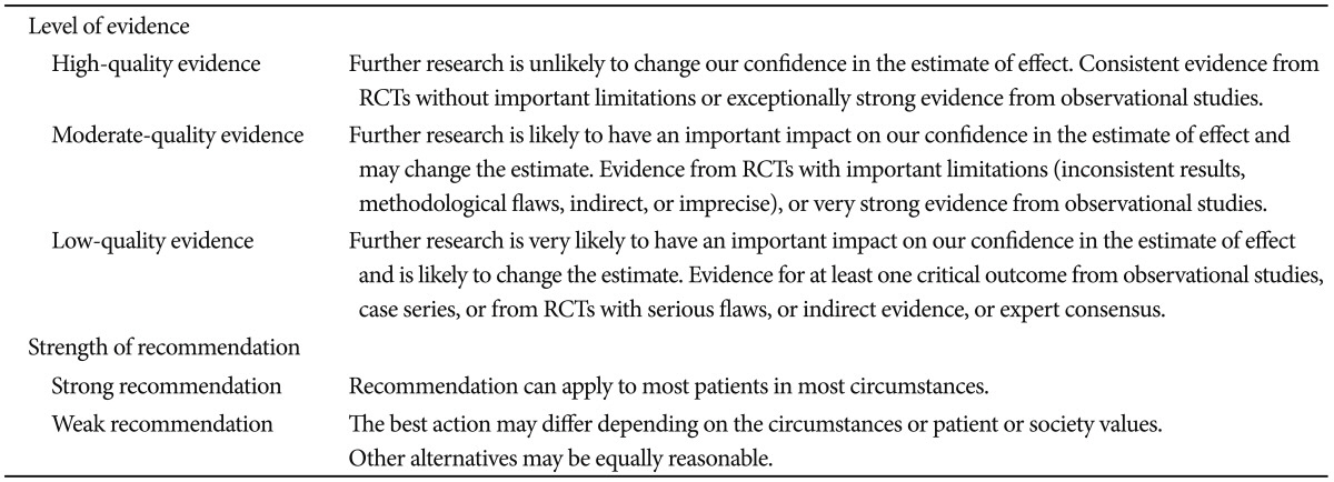
4. Guyatt GH, Cook DJ, Jaeschke R, Pauker SG, Schünemann HJ. Grades of recommendation for antithrombotic agents: American College of Chest Physicians Evidence-Based Clinical Practice Guidelines (8th Edition). Chest. 2008; 133(6 Suppl):123S–131S. PMID: 18574262.
5. Schunemann HJ, Oxman AD, Brozek J, et al. Grading quality of evidence and strength of recommendations for diagnostic tests and strategies. BMJ. 2008; 336:1106–1110. PMID: 18483053.
crossref
6. Watt AM, Faragher IG, Griffin TT, Rieger NA, Maddern GJ. Self-expanding metallic stents for relieving malignant colorectal obstruction: a systematic review. Ann Surg. 2007; 246:24–30. PMID: 17592286.
7. Tilney HS, Lovegrove RE, Purkayastha S, et al. Comparison of colonic stenting and open surgery for malignant large bowel obstruction. Surg Endosc. 2007; 21:225–233. PMID: 17160651.
crossref
8. Cheung HY, Chung CC, Tsang WW, Wong JC, Yau KK, Li MK. Endolaparoscopic approach vs conventional open surgery in the treatment of obstructing left-sided colon cancer: a randomized controlled trial. Arch Surg. 2009; 144:1127–1132. PMID: 20026830.
9. Sagar J. Colorectal stents for the management of malignant colonic obstructions. Cochrane Database Syst Rev. 2011; (11):CD007378. PMID: 22071835.
crossref
10. Tan CJ, Dasari BV, Gardiner K. Systematic review and meta-analysis of randomized clinical trials of self-expanding metallic stents as a bridge to surgery versus emergency surgery for malignant left-sided large bowel obstruction. Br J Surg. 2012; 99:469–476. PMID: 22261931.
11. van Hooft JE, Bemelman WA, Breumelhof R, et al. Colonic stenting as bridge to surgery versus emergency surgery for management of acute left-sided malignant colonic obstruction: a multicenter randomized trial (Stent-in 2 study). BMC Surg. 2007; 7:12. PMID: 17608947.
crossref
12. Elsberger B, Rourke K, Brush J, Glancy S, Collie M. Self-expanding metallic stent insertion in the proximal colon. Colorectal Dis. 2008; 10:194–196. PMID: 17784867.
crossref
13. Repici A, Adler DG, Gibbs CM, Malesci A, Preatoni P, Baron TH. Stenting of the proximal colon in patients with malignant large bowel obstruction: techniques and outcomes. Gastrointest Endosc. 2007; 66:940–944. PMID: 17963881.
crossref
14. Dronamraju SS, Ramamurthy S, Kelly SB, Hayat M. Role of self-expanding metallic stents in the management of malignant obstruction of the proximal colon. Dis Colon Rectum. 2009; 52:1657–1661. PMID: 19690497.
crossref
15. Kim JS, Oh SY, Seo KU, et al. Comparison of effects of preoperative stenting for obstructing colorectal cancers according to the location of the obstructing lesion. Korean J Gastroenterol. 2009; 54:384–389. PMID: 20026893.
crossref
16. de Gregorio MA, Mainar A, Tejero E, et al. Acute colorectal obstruction: stent placement for palliative treatment: results of a multicenter study. Radiology. 1998; 209:117–120. PMID: 9769821.
17. Fernández Lobato R, Pinto I, Paul L, et al. Self-expanding prostheses as a palliative method in treating advanced colorectal cancer. Int Surg. 1999; 84:159–162. PMID: 10408289.
18. Liberman H, Adams DR, Blatchford GJ, Ternent CA, Christensen MA, Thorson AG. Clinical use of the self-expanding metallic stent in the management of colorectal cancer. Am J Surg. 2000; 180:407–411. PMID: 11182388.
crossref
19. Carne PW, Frye JN, Robertson GM, Frizelle FA. Stents or open operation for palliation of colorectal cancer: a retrospective, cohort study of perioperative outcome and long-term survival. Dis Colon Rectum. 2004; 47:1455–1461. PMID: 15486741.
crossref
20. Ptok H, Marusch F, Steinert R, Meyer L, Lippert H, Gastinger I. Incurable stenosing colorectal carcinoma: endoscopic stent implantation or palliative surgery? World J Surg. 2006; 30:1481–1487. PMID: 16850152.
crossref
21. Karoui M, Charachon A, Delbaldo C, et al. Stents for palliation of obstructive metastatic colon cancer: impact on management and chemotherapy administration. Arch Surg. 2007; 142:619–623. PMID: 17638798.
22. Cennamo V, Fuccio L, Mutri V, et al. Does stent placement for advanced colon cancer increase the risk of perforation during bevacizumab-based therapy? Clin Gastroenterol Hepatol. 2009; 7:1174–1176. PMID: 19631290.
crossref
23. Súarez J, Jiménez J, Vera R, et al. Stent or surgery for incurable obstructive colorectal cancer: an individualized decision. Int J Colorectal Dis. 2010; 25:91–96. PMID: 19859722.
24. Lee HJ, Hong SP, Cheon JH, et al. Long-term outcome of palliative therapy for malignant colorectal obstruction in patients with unresectable metastatic colorectal cancers: endoscopic stenting versus surgery. Gastrointest Endosc. 2011; 73:535–542. PMID: 21257165.
crossref
25. Yoon JY, Jung YS, Hong SP, Kim TI, Kim WH, Cheon JH. Clinical outcomes and risk factors for technical and clinical failures of self-expandable metal stent insertion for malignant colorectal obstruction. Gastrointest Endosc. 2011; 74:858–868. PMID: 21862005.
crossref
26. Jiménez-Pérez J, Casellas J, García-Cano J, et al. Colonic stenting as a bridge to surgery in malignant large-bowel obstruction: a report from two large multinational registries. Am J Gastroenterol. 2011; 106:2174–2180. PMID: 22085816.
crossref
27. Inaba Y, Arai Y, Yamaura H, et al. Phase II clinical study on stent therapy for unresectable malignant colorectal obstruction (JIVROSG-0206). Am J Clin Oncol. 2012; 35:73–76. PMID: 21293242.
crossref
28. Luigiano C, Ferrara F, Fabbri C, et al. Through-the-scope large diameter self-expanding metal stent placement as a safe and effective technique for palliation of malignant colorectal obstruction: a single center experience with a long-term follow-up. Scand J Gastroenterol. 2011; 46:591–596. PMID: 21271788.
crossref
29. Trompetas V, Saunders M, Gossage J, Anderson H. Shortcomings in colonic stenting to palliate large bowel obstruction from extracolonic malignancies. Int J Colorectal Dis. 2010; 25:851–854. PMID: 20390286.
crossref
30. Shin SJ, Kim TI, Kim BC, Lee YC, Song SY, Kim WH. Clinical application of self-expandable metallic stent for treatment of colorectal obstruction caused by extrinsic invasive tumors. Dis Colon Rectum. 2008; 51:578–583. PMID: 18259816.
31. Caceres A, Zhou Q, Iasonos A, Gerdes H, Chi DS, Barakat RR. Colorectal stents for palliation of large-bowel obstructions in recurrent gynecologic cancer: an updated series. Gynecol Oncol. 2008; 108:482–485. PMID: 18190953.
crossref
32. Miyayama S, Matsui O, Kifune K, et al. Malignant colonic obstruction due to extrinsic tumor: palliative treatment with a self-expanding nitinol stent. AJR Am J Roentgenol. 2000; 175:1631–1637. PMID: 11090392.
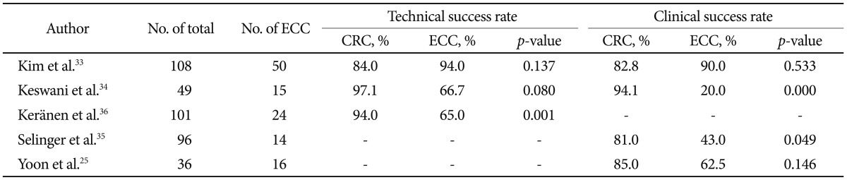
33. Kim JH, Song HY, Park JH, Ye BD, Yoon YS, Kim JC. Metallic stent placement in the palliative treatment of malignant colonic obstructions: primary colonic versus extracolonic malignancies. J Vasc Interv Radiol. 2011; 22:1727–1732. PMID: 21937243.
crossref
34. Keswani RN, Azar RR, Edmundowicz SA, et al. Stenting for malignant colonic obstruction: a comparison of efficacy and complications in colonic versus extracolonic malignancy. Gastrointest Endosc. 2009; 69(3 Pt 2):675–680. PMID: 19251009.
crossref
35. Selinger CP, Ramesh J, Martin DF. Long-term success of colonic stent insertion is influenced by indication but not by length of stent or site of obstruction. Int J Colorectal Dis. 2011; 26:215–218. PMID: 21207043.
crossref
36. Keränen I, Lepistö A, Udd M, Halttunen J, Kylänpää L. Stenting for malignant colorectal obstruction: a single-center experience with 101 patients. Surg Endosc. 2012; 26:423–430. PMID: 21909857.
crossref
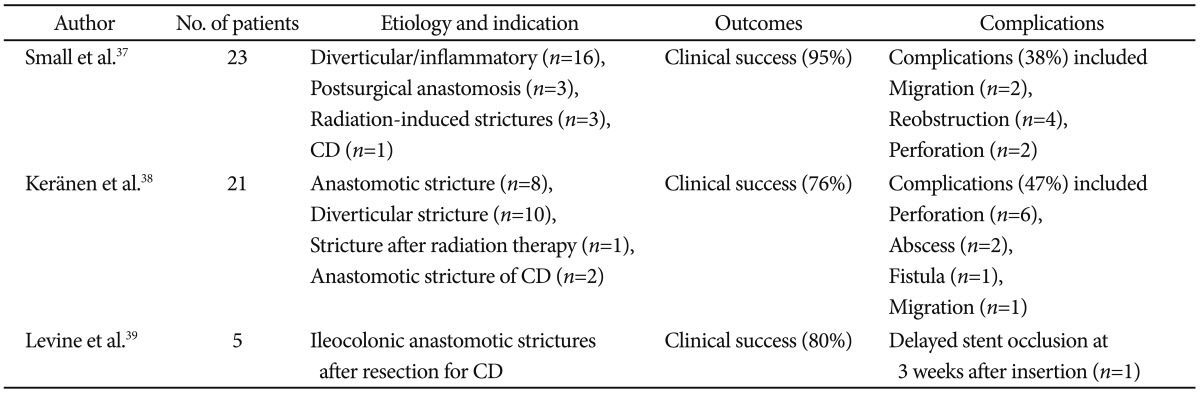
37. Small AJ, Young-Fadok TM, Baron TH. Expandable metal stent placement for benign colorectal obstruction: outcomes for 23 cases. Surg Endosc. 2008; 22:454–462. PMID: 17704890.
crossref
38. Keränen I, Lepistö A, Udd M, Halttunen J, Kylänpää L. Outcome of patients after endoluminal stent placement for benign colorectal obstruction. Scand J Gastroenterol. 2010; 45:725–731. PMID: 20205505.
crossref
39. Levine RA, Wasvary H, Kadro O. Endoprosthetic management of refractory ileocolonic anastomotic strictures after resection for Crohn's disease: report of nine-year follow-up and review of the literature. Inflamm Bowel Dis. 2012; 18:506–512. PMID: 21542067.
crossref
40. Stienecker K, Gleichmann D, Neumayer U, Glaser HJ, Tonus C. Long-term results of endoscopic balloon dilatation of lower gastrointestinal tract strictures in Crohn's disease: a prospective study. World J Gastroenterol. 2009; 15:2623–2627. PMID: 19496192.
crossref
41. Farrell JJ. Preoperative colonic stenting: how, when and why? Curr Opin Gastroenterol. 2007; 23:544–549. PMID: 17762561.
crossref
42. Keymling M. Colorectal stenting. Endoscopy. 2003; 35:234–238. PMID: 12584644.
crossref
43. Repici A, Pagano N, Hervoso CM, et al. Metal stents for malignant colorectal obstruction. Minim Invasive Ther Allied Technol. 2006; 15:331–338. PMID: 17190657.
crossref
44. Domingo S, Puértolas S, Gracia-Villa L, Puértolas JA. Mechanical comparative analysis of stents for colorectal obstruction. Minim Invasive Ther Allied Technol. 2007; 16:126–136. PMID: 17474056.
crossref
45. Repici A, De Caro G, Luigiano C, et al. WallFlex colonic stent placement for management of malignant colonic obstruction: a prospective study at two centers. Gastrointest Endosc. 2008; 67:77–84. PMID: 18155427.
crossref
46. Repici A, Fregonese D, Costamagna G, et al. Ultraflex precision colonic stent placement for palliation of malignant colonic obstruction: a prospective multicenter study. Gastrointest Endosc. 2007; 66:920–927. PMID: 17904133.
crossref
47. Fregonese D, Naspetti R, Ferrer S, et al. Ultraflex precision colonic stent placement as a bridge to surgery in patients with malignant colon obstruction. Gastrointest Endosc. 2008; 67:68–73. PMID: 18028916.
crossref
48. Small AJ, Baron TH. Comparison of Wallstent and Ultraflex stents for palliation of malignant left-sided colon obstruction: a retrospective, case-matched analysis. Gastrointest Endosc. 2008; 67:478–488. PMID: 18294511.
49. Kim JH, Song HY, Li YD, et al. Dual-design expandable colorectal stent for malignant colorectal obstruction: comparison of flared ends and bent ends. AJR Am J Roentgenol. 2009; 193:248–254. PMID: 19542421.
crossref
50. Shim CS, Cho JY, Jung IS, et al. Through-the-scope double colonic stenting in the management of inoperable proximal malignant colonic obstruction: a pilot study. Endoscopy. 2004; 36:426–431. PMID: 15100952.
crossref
51. Li YD, Cheng YS, Li MH, et al. Management of acute malignant colorectal obstruction with a novel self-expanding metallic stent as a bridge to surgery. Eur J Radiol. 2010; 73:566–571. PMID: 19167177.
crossref
52. Small AJ, Coelho-Prabhu N, Baron TH. Endoscopic placement of self-expandable metal stents for malignant colonic obstruction: long-term outcomes and complication factors. Gastrointest Endosc. 2010; 71:560–572. PMID: 20189515.
crossref
53. Song HY, Kim JH, Shin JH, et al. A dual-design expandable colorectal stent for malignant colorectal obstruction: results of a multicenter study. Endoscopy. 2007; 39:448–454. PMID: 17516352.
crossref
54. van Hooft JE, Fockens P, Marinelli AW, et al. Early closure of a multicenter randomized clinical trial of endoscopic stenting versus surgery for stage IV left-sided colorectal cancer. Endoscopy. 2008; 40:184–191. PMID: 18322873.
crossref
55. Jung MK, Park SY, Jeon SW, et al. Factors associated with the long-term outcome of a self-expandable colon stent used for palliation of malignant colorectal obstruction. Surg Endosc. 2010; 24:525–530. PMID: 19597776.
crossref
56. Park S, Cheon JH, Park JJ, et al. Comparison of efficacies between stents for malignant colorectal obstruction: a randomized, prospective study. Gastrointest Endosc. 2010; 72:304–310. PMID: 20561619.
crossref
57. Moon CM, Kim TI, Lee MS, et al. Comparison of a newly designed double-layered combination covered stent and D-weave uncovered stent for decompression of obstructive colorectal cancer: a prospective multicenter study. Dis Colon Rectum. 2010; 53:1190–1196. PMID: 20628284.
crossref
58. Choi JS, Choo SW, Park KB, et al. Interventional management of malignant colorectal obstruction: use of covered and uncovered stents. Korean J Radiol. 2007; 8:57–63. PMID: 17277564.
crossref
59. Lee KM, Shin SJ, Hwang JC, et al. Comparison of uncovered stent with covered stent for treatment of malignant colorectal obstruction. Gastrointest Endosc. 2007; 66:931–936. PMID: 17767930.
crossref
60. Langevin JM, Nivatvongs S. The true incidence of synchronous cancer of the large bowel. A prospective study. Am J Surg. 1984; 147:330–333. PMID: 6703204.
61. Slater G, Aufses AH Jr, Szporn A. Synchronous carcinoma of the colon and rectum. Surg Gynecol Obstet. 1990; 171:283–287. PMID: 2218832.
62. Ekelund GR, Pihl B. Mutiple carcinomas of the colon and rectum. Cancer. 1974; 33:1630–1634. PMID: 4834157.
63. Adloff M, Arnaud JP, Bergamaschi R, Schloegel M. Synchronous carcinoma of the colon and rectum: prognostic and therapeutic implications. Am J Surg. 1989; 157:299–302. PMID: 2537586.
crossref
64. Pagana TJ, Ledesma EJ, Mittelman A, Nava HR. The use of colonoscopy in the study of synchronous colorectal neoplasms. Cancer. 1984; 53:356–359. PMID: 6690019.
crossref
65. Vitale MA, Villotti G, d'Alba L, Frontespezi S, Iacopini F, Iacopini G. Preoperative colonoscopy after self-expandable metallic stent placement in patients with acute neoplastic colon obstruction. Gastrointest Endosc. 2006; 63:814–819. PMID: 16650544.
crossref
66. Cha EY, Park SH, Lee SS, et al. CT colonography after metallic stent placement for acute malignant colonic obstruction. Radiology. 2010; 254:774–782. PMID: 20177092.
crossref
67. Datye A, Hersh J. Colonic perforation after stent placement for malignant colorectal obstruction: causes and contributing factors. Minim Invasive Ther Allied Technol. 2011; 20:133–140. PMID: 20929424.
68. Donnellan F, Cullen G, Cagney D, et al. Efficacy and safety of colonic stenting for malignant disease in the elderly. Int J Colorectal Dis. 2010; 25:747–750. PMID: 20213457.
crossref
69. Sagar J. Colorectal stents for the management of malignant colonic obstructions. Cochrane Database Syst Rev. 2011; (11):CD007378. PMID: 22071835.
crossref
70. Lopera JE, De Gregorio MA. Fluoroscopic management of complications after colorectal stent placement. Gut Liver. 2010; 4(Suppl 1):S9–S18. PMID: 21103302.
crossref
71. Repici A, de Paula Pessoa Ferreira D. Expandable metal stents for malignant colorectal strictures. Gastrointest Endosc Clin N Am. 2011; 21:511–533. PMID: 21684468.
crossref
72. Suh JP, Kim SW, Cho YK, et al. Effectiveness of stent placement for palliative treatment in malignant colorectal obstruction and predictive factors for stent occlusion. Surg Endosc. 2010; 24:400–406. PMID: 19551432.
crossref
73. Park JK, Lee MS, Ko BM, et al. Outcome of palliative self-expanding metal stent placement in malignant colorectal obstruction according to stent type and manufacturer. Surg Endosc. 2011; 25:1293–1299. PMID: 20976501.
crossref
74. Branger F, Thibaudeau E, Mucci-Hennekinne S, et al. Management of acute malignant large-bowel obstruction with self-expanding metal stent. Int J Colorectal Dis. 2010; 25:1481–1485. PMID: 20607252.
crossref
75. Fernández-Esparrach G, Bordas JM, Giráldez MD, et al. Severe complications limit long-term clinical success of self-expanding metal stents in patients with obstructive colorectal cancer. Am J Gastroenterol. 2010; 105:1087–1093. PMID: 19935785.
crossref
76. Baron TH. Colonic stenting: technique, technology, and outcomes for malignant and benign disease. Gastrointest Endosc Clin N Am. 2005; 15:757–771. PMID: 16278137.
crossref
77. Park JH, Oh SH, Lee WY, Choo SW, Do YS, Chun HK. Flexible rectal stent for obstructing colonic neoplasms. J Korean Soc Coloproctol. 2000; 16:267–273.
78. Song HY, Kim JH, Kim KR, et al. Malignant rectal obstruction within 5 cm of the anal verge: is there a role for expandable metallic stent placement? Gastrointest Endosc. 2008; 68:713–720. PMID: 18561924.
crossref
79. Rao VS, Al-Mukhtar A, Rayan F, Stojkovic S, Moore PJ, Ahmad SM. Endoscopic laser ablation of advanced rectal carcinoma: a DGH experience. Colorectal Dis. 2005; 7:58–60. PMID: 15606586.
80. Courtney ED, Raja A, Leicester RJ. Eight years experience of high-powered endoscopic diode laser therapy for palliation of colorectal carcinoma. Dis Colon Rectum. 2005; 48:845–850. PMID: 15747077.
crossref
81. Eickhoff A, Jakobs R, Schilling D, et al. Prospective nonrandomized comparison of two modes of argon beamer (APC) tumor desobstruction: effectiveness of the new pulsed APC versus forced APC. Endoscopy. 2007; 39:637–642. PMID: 17611919.
crossref
82. Horiuchi A, Nakayama Y, Tanaka N, et al. Acute colorectal obstruction treated by means of transanal drainage tube: effectiveness before surgery and stenting. Am J Gastroenterol. 2005; 100:2765–2770. PMID: 16393233.
crossref
Table 1
Level of Evidence and Strength of Recommendation
ce-46-355-i001

RCT, randomized controlled trial.

Table 2
Distribution of Origin of Extracolonic Malignancies Causing Colorectal Obstruction in Articles Cited in the Literature25,28-30,33,35,58,71
ce-46-355-i002
Table 3
Comparative Studies for Colorectal Stents in Patients with Colorectal Cancer and Extracolonic Malignancies
ce-46-355-i003

ECC, extracolonic cancer; CRC, colorectal cancer.

Table 4
Case Series of Self-Expandable Metallic Stents for Benign Colorectal Strictures
ce-46-355-i004

CD, Crohn disease.

TOOLS
Similar articles