

 PDF
PDF ePub
ePub Citation
 Print
Print


Diabetes mellitus (DM) encompasses a heterogeneous group of disorders with the common characteristic of elevated blood glucose, i.e., Hyperglycemia, due to disruption in metabolism of carbohydrates, fat, and protein, resulting from impaired insulin secretion [1,2,3]. Based on etiology, DM can be divided into four groups, including type 1 DM, type 2 DM, other specific types of diabetes, and gestational diabetes [4]. Among them, type 2 DM is characterized by relative moderate insulin deficiency resulting from a decreased effect of insulin in target tissues, e.g., muscles and adipose tissue (insulin resistance), or a secretory defect of insulin with or without insulin resistance [4,5].
Use of appropriate animal models for type 2 DM is important for understanding its pathogenesis and development and testing of therapeutics for treatment of this disease. Rodent models provide a number of advantages over other mammals; however, there are also several drawbacks, which might primarily include the fact that, genetically and physiologically, rodents are not as close to humans, compared to other mammals, e.g., monkey and pig, etc. Among them, pigs share the structure and function of the gastrointestinal tract, the development and morphology of the pancreas, and overall metabolic status with humans [6,7,8], and have also demonstrated comparable pharmacokinetics of the compound via subcutaneous administration [9] with humans; therefore, use of pigs might be a good alternative for replacement of rodents for use in diabetes research. Owing to their small size and ease of handling, even at full maturity, mini-pigs were found to be suitable for use in long-term studies. The pancreas of mini-pigs resembles the human pancreas in size, shape, and position. In addition, the amino acid sequences of insulin between pig and human differ only in a single residue. Based on these various advantages, the mini-pig is of particular interest for studies related to human type 1 DM [10].
High fat and cholesterol (HFC) diets with or without streptozocin (STZ) have been adapted for the establishment of animal models for type 2 DM, such as pig [7,11,12] and rat [13]. STZ is a chemical that is particularly toxic to insulin-producing pancreatic β-cells in mammals. It is used in medical research for the construction of animal models for type 1 DM [14,15]. In addition, treatment with STZ with nicotinamide (NIA), which modulates STZ-mediated β-cell destruction and reduces its severity, has been widely used for induction of type 2 DM in animal models, including mouse [16], rat [17,18], hamster [19], and Göttingen minipig [9]. However, variability in the establishment of STZ-mediated DM, including both type 1 DM and type 2 DM, has restricted development of a general protocol using STZ alone as well as STZ with NIA [20,21]. Therefore, testing and optimization of procedures for induction of type 2 DM for the new line of animals is worthwhile. In this study, in order to establish a useful animal model for diabetes research, we performed the induction of type 2 DM in Micro-pigs, which differ from Göttingen minipigs [22]. The Micro-pig is a breed of laboratory swine developed and maintained until 2005 by the Medi kinetics Co (MK) breeding program for biomedical research and nonclinical trials.
All animals included in the study were adult male Micro-pigs over six months of age with less than 20 kg of body weight. Animals were obtained from the barrier unit at Medi Kinetics Co (Pyeongtaek, Korea). All animals were housed in single pens under controlled conditions (temperature was kept between 18 and 22℃, relative air humidity was 30-70% with 15 air changes/h) with a 12:12-hr light-dark cycle and allowed free access to water. Principles of laboratory animal care were followed in accordance with the Guide for Animal Experimental Protocol (MK-IACUC:2010-0088) and the type of study was approved by the Institutional Animal Care and Use Committee of Medi kinetics Co. The standard feeding regime was applied to the animals used in this study.
Animals were fed a pellet diet sterilized by 2M rad radiation (Purina, Seongnam, Korea) and received sterilized water ad libitum. For induction of type 2 DM, NIA and STZ were administered to male Micro-pigs via intravenous (i.v.) injection. NIA (Cat No. N0636) and STZ (Cat No. S0130) were purchased from Sigma-Aldrich Co. (St. Louis, MO). NIA solution was prepared by dissolving in sterile saline solution at a concentration of 300 mg/mL and administered via i.v. within 15 min prior to administration of STZ. Injection of 67 mg/kg of NIA was administered to each animal. STZ was dissolved in sodium citrate buffer at a concentration of 62.5 mg/mL and injected via i.v. for a period of 1-2 minutes. A total of 125 mg/kg STZ was administered per animal. Blood was taken before feeding, following fasting for 12-16 hr in controls or 12-16 hr in NIA/STZ administration groups. Measurement of blood glucose levels was performed weekly before and after induction of DM, using a blood glucose tester (Accu-Chek® Go) (Roche Diagnostics). At the end of the experiments, all Micro-pigs were scarified and subjected to autopsy. Specifically, pancreatic glands were removed and fixed in 10% neutralized buffered formalin, processed, and embedded in paraffin, followed by sectioning, hematoxylin and eosin (H&E) staining, and immunostaining with insulin antibody (Cat No. ab58977, Abcam).
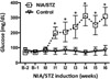
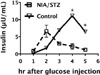
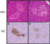
In this study, Micro-pigs were treated with NIA/STZ in order to recapitulate human type 2 DM, which is characterized by moderately elevated levels of glucose and insulin resistance. Compared to controls, Micro-pigs treated with NIA/STZ showed elevated levels of glucose (P<0.05, t-test). Elevation of glucose occurred two weeks after administration of NIA/STZ in type 2 DM pigs and the maximum increase was achieved at four weeks after treatment, with the maintenance of a hyperglycemias trend until the end of the experiment (Figure 1). These data provided indication that the NIA/STZ regimes used in this study might be effective for induction and maintenance of type 2 DM in Micro-pigs, which is in agreement with findings from previous reports using Göttingen minipigs. The dose of NIA/STZ used in this study differs from that used in the previous study, reflecting the intrinsic variability of STZ-induced DM. In addition, lower levels of serum insulin were observed in NIA/STZ-treated Micro-pigs (P<0.05, Figure 2), compared to controls, suggesting impaired production and/or insulin from β-islet cells. An insulin antibody was used to perform immunohistochemistry (IHC) in NIA/STZ-induced type 2 DM pancreas. Results of IHC analysis revealed a significant decrease in the positivity of insulin expressing β-cells from NIA/STZ treated pancreas (Figure 3D). In addition, enlarged islets were observed, indicating an inflammatory response to NIA/STZ treatment (Figure 3B). In conclusion, based on the results described above, we suggest that the chemically induced Micro-pig diabetic model can be used for testing of the efficacy of candidate diabetic drugs and products for use in diagnosis and treatment of patients with diabetes mellitus.
 XML Download