

 PDF
PDF ePub
ePub Citation
 Print
Print


Introduction
The application of cone-beam computed tomography (CBCT) in dentistry has increased rapidly since it was introduced.1 CBCT has been used as a method for creating three-dimensional radiographs,2 which are one of the most sensitive imaging modalities for dental diagnostic purposes, and as a substitute for traditional CT in the assessment of pathology and dysfunction of the craniomaxillofacial complex.3 Furthermore, CBCT has been increasingly applied in endodontics for the diagnosis of periapical lesions, root canal observation, assessing the extent of internal and external resorption, and identifying root fractures.4
Radiographic and clinical examinations are essential for the diagnosis of vertical root fractures, and CBCT is more sensitive than periapical radiography in detecting vertical root fractures.5 Two-dimensional conventional radiographs with bony structure superimposition may hide the root fracture, especially when the X-ray position is not parallel to the plane of the fracture, meaning that the fracture may not be observed in the radiograph.4
One feature of vertical root fractures is a local deep pocket, the presence of a sinus tract, and halo-type lateral radiolucency on a radiograph.6 The clinical features of root fractures can be misleading or present with a delay. They can also be hard to distinguish from pulp and periapical diseases, making it challenging to identify root fractures in teeth with non-displaced parts.4 The prognosis of a tooth with a root fracture depends on various factors, such as the patient's age, the stage of root formation, the amount of displacement of the coronal part of the tooth, the extent of looseness in the coronal part of the tooth, and the distance between the separated parts.7 Therefore, a definitive diagnosis of vertical root fracture is essential in order to avoid unnecessary tooth extraction.
Based on the field of view (FOV), CBCT systems are classified into three types: small-volume (also known as limited-volume) systems, which are usually used for scanning a sample of contiguous teeth or one jaw; medium-volume systems that are used to image both jaws, the maxillary sinus, and part of the nose; and large-volume systems that are applied to image the entire maxillofacial area, and in some systems even the cranial vertex.8
Limited-volume systems have some advantages in terms of price, image resolution (voxel size), and radiation dose in comparison with large-volume systems. In CBCT imaging with a limited volume, the maxillofacial hard tissue structures located outside the imaging volume cause discontinuity of beam information. This influences image fidelity and results in areas with variable density.1
Metal artifacts occur in all CT imaging systems. Many small and large metal items may be present in the human body, especially in the head and neck area. Metal restorations, metal posts located in the canal, crowns, brackets, and implants can influence the quality of the acquired CT image due to effects such as quantum noise, starvation photons, and beam hardening.9 Beam hardening results in two kinds of artifacts: the distortion of metal structures that is known as the cupping artifact because of the pattern of differential absorption that results, and streaks and dark bands that can appear between two dense substances10 or around metal substances. The artifact may even cause a complete loss of gray values between the adjacent metal substances. As a result, the area in question is not accurately imaged, with consequences for the treatment plan.9
In 2012, Costa et al.8 investigated horizontal root fractures in teeth with and without metal posts using large-volume CBCT by designing a cylinder measuring 20 cm×15 cm. The identification of horizontal fractures was more accurate in the group of teeth without a metal post (with or without a fracture), while weak or very weak agreement was found between two observers in the group of teeth with a metal post. In another investigation in 2011, Costa et al.11 studied on horizontal root fractures in teeth with and without a metal post located in the canal using small-volume CBCT. They likewise found that the identification of fractures was more accurate in the group of teeth without a metal post (with or without a fracture) than in the teeth with a metal post.
The presence of prefabricated posts has little effect on the accuracy of CBCT,12 but it influences the treatment plan based on CBCT images. We hypothesized that root fractures may be more accurately observed in images with a small FOV, due to the smaller size of the pixels and the higher image resolution. Thus, the aim of this study was to investigate the effects of metal artifacts in CBCT images on the accurate diagnosis of root fractures in large and small/limited fields of view.
Materials and Methods
In this study, 40 extracted human mandibular molar and premolar teeth without fractures, periapical pathology, root resorption, or any other anomalies were collected. In order to disinfect and remove the soft cellular tissue, the teeth were placed in a 5.25% sodium hypochlorite solution (Golrang, Tehran, Iran) for one hour and then placed in normal saline until the experiment was started. The collected teeth had not undergone any endodontic treatment, and the absence of vertical root fractures was established by examination with a stereomicroscope at 20× (Wild Photomakroskop M400, Heerbrugg, Switzerland).
Access canals in all teeth were prepared using a diamond bur, and the opening of both the molar distal canal and the premolar canal was accomplished with a #15 K-file. The length of the canals was visually determined with this file, and the measurement was decreased by 1 mm in order to reflect functional length of the canals. The canals were prepared according to the manufacturer's instructions using a rotary system (NSK, Fukushima, Japan), with a speed of 300 rpm, controlled torque, and SX, S1, S2, F1, and F2 ProTaper files (Dentsply, Ballaigues, Switzerland). Each canal was frequently washed with a 2% sodium hypochlorite solution. The prepared canals were filled with guttapercha (Pro Taper F2 & F3, Dia Dent, Burnaby, BC, Canada).
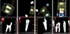
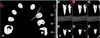
In order to create fractures in the canals, the teeth were first put in acryl (Acropars 200, Marlic Medical Industries Co., Tehran, Iran) to avoid splitting, and a thin layer of wax was added to simulate soft tissue. One screw driver-tape wedge was then placed in the canal. The fractures were created through the application of mild pressure with a hammer in the canals of the premolar teeth and the distal root of the molars. Fractures were created in half of the teeth. The teeth were evaluated again with the stereomicroscope with the same magnification in order to confirm the absence of fractures in teeth in which a vertical root fracture was made. The teeth were then coded and randomly placed in a wax rim made of three to four wax layers on an acryl base designed in the shape of a mandible. The mandibles were fixed in cardboard boxes, so that the central beam ray was perpendicular to the longitudinal axis of the teeth. CBCT scans were obtained using a Newtom 5G apparatus (QR s.r.1., Verona, Italy), with a potential difference of 110 kV, a tube current of 9.6 mA, a FOV of 18 cm×16 cm for the large FOV and 6 cm×6 cm for the small FOV, a voxel size of 0.3 mm, denture scan mode, and 4.8 seconds of exposure. The guttapercha that had been located in the canal was removed in order to place a #3 gold-plated pin (Nordin, Montreux, Switzerland). The fracture line pattern was denoted with 1% methylene blue, metal pins were put in all of the teeth, and CBCT imaging was again performed using the previous specifications. The images were evaluated in the axial plane and in multiplanar image using the NNT viewer software version 3.0 (QR s.r.1., Verona, Italy), with a thickness of 0.3 mm and an interval space of 0.3 mm. All scans were studied by two oral and maxillofacial radiologists in axial and cross-sectional views (Fig. 1) in a dim room using an 18.5-inch LG Flatron monitor (LG, Seoul, Korea), with a resolution of 0.3×0.3 pixel pitch. The images were randomly observed in limited numbers over consecutive days in order for the observers to identify tooth fractures. Specificity, sensitivity, positive predictive value (PPV), and negative predictive value (NPV), and the positive and negative likelihood ratios were calculated.
Results
The small-volume CBCT images of teeth without pins showed 100% sensitivity and specificity.
In the small-volume images, 100% specificity was also observed in the presence of a pin. Although the sensitivity in the small-volume images without a pin was 5% higher than in the small-volume images with a pin, no statistically significant difference was found between these two groups.
The sensitivity in the large-volume images without a pin was 14% lower than in large-volume images with a pin, whereas the specificity was 11% higher in the large-volume images without a pin. Overall, the sensitivity of the diagnoses of vertical root fractures made using small-volume images was higher than that of the diagnoses made using large-volume images. The positive predictive value in all groups was 100%, except in large-volume images with a pin. The negative predictive value in small-volume images without a pin and in large-volume images with a pin was 100%, but was lower in small-volume images with a pin and large-volume images without a pin.
Table 1 shows the sensitivity, specificity, positive predictive value, the negative predictive value, and positive and negative likelihood ratios of the various groups after CBCT was used to identify teeth root fractures, along with 95% confidence intervals.
Discussion
The statistical analysis demonstrated that CBCT images of teeth without pins were highly sensitive in the diagnosis of fracture lines in small-volume mode, in comparison with other modes. The main goal of this ex vivo study was to investigate the diagnostic accuracy of fracture lines in large and small CBCT radiographs in the presence of root canal fillings and metal pins. The sensitivity of small-volume images without pins was 100%, but was reduced to 86% in the large-volume group without pins.
This is a reasonable finding in light of the decreased imaging area, the lower amount of ray dispersion, the higher signal-to-noise ratio, and the higher resolution of the radiographs. Our findings correspond to those of Costa et al.,11 who investigated horizontal fracture lines in small-volume CBCT. They are also in agreement with the findings of Iikubo et al.,13 who compared conventional periapical radiographs to CBCT images. The sensitivity of the large-volume CBCT images was 100% for the group with pins, but a lower value of specificity was reported in this group (89%). This finding is likely explicable through the beam-hardening phenomenon. Beam hardening occurs in connection with the increase of medium- energy X-rays, because lower-energy photons are absorbed by the structures being imaged instead of higher-energy photons. As mentioned above, this phenomenon results in artifacts that obscure the fracture lines in the region of interest. The artifacts that appear in the shape of dark areas or lines around endodontic materials are similar to the lines that reflect root fractures, resulting in false positives (Fig. 2).14
As seen in Figure 2A, the fracture lines and the lines due to metal artifacts are not distinguished easily. A study carried out by Wang et al.15 with the aim of identifying tooth fracture lines using CBCT in comparison with conventional radiography and to determine the effect of root canal fillings found that CBCT was more accurate in identifying root fractures. The sensitivity of CBCT decreased in the presence of root canal fillings, but the specificity was not influenced by any of the factors they studied. In this study, specificity decreased with the presence of pins in the large-volume images, but not in the small-volume images. This effect might be due to the voxel size and issues involving contrast and spatial resolution.16 Large-FOV CBCT provides less contrast resolution and spatial resolution than small-FOV CBCT.6
Negative predictive value is an important functional index, because when a radiologist confirms that no fracture is present in a scan, he/she estimates the possibility that a vertical root fracture is not present.4 The negative predictive values in the small-volume CBCT group without pins and the large-volume group with pins were 100%. The positive predictive value was 100% in all groups except the large-volume CBCT group with pins, where a slightly lower value was observed. A 100% positive predictive value means that the lines displayed as fracture lines were 100% likely to be real fracture lines.
In conclusion, small-volume CBCT scans are very accurate in diagnosing vertical root fractures, and the presence of pins has only a small effect on detecting root fractures.

 XML Download