

 PDF
PDF ePub
ePub Citation
 Print
Print


INTRODUCTION
Hematospermia can be defined as the appearance of blood in ejaculate [123]. While it is usually self-limited in most patients, in some cases, persistent, recurrent symptoms have been reported, causing serious concerns.
We could classify the many suggested etiologies for hematospermia into inflammatory or infectious diseases and anatomical abnormalities or disorders [123]. Urethritis, prostatitis, and epididymitis are a prototype of inflammatory hematospermia, while posterior urethral obstruction, calculi in the ejaculatory duct, and prostate cancer can be a prototype of anatomical hematospermia.
Tests for evaluating lower urinary tract infection such as urine analysis and Gram staining are routine tests for hematospermia [245]. Because transrectal ultrasonography (US) can show the anatomical structures of the prostate, seminal vesicles, and adjacent structures, many urologists have applied this modality for evaluating the anatomical causes of hematospermia [456].
Prostatitis, an inflammatory disorder of the prostate, has been considered an important disease causing bloody ejaculation [12345]. It has been well known that the basic methods for evaluating prostatitis include the National Institutes of Health Chronic Prostatitis Symptom Index (NIH-CPSI) questionnaire, and the white blood cell (WBC) counts and bacterial culture of expressed prostatic secretion (EPS) [78910]. Unfortunately, very few studies on the determination of prostatitis-related etiologies for the origin of hematospermia have been performed using robust prostatitis evaluation methods. Therefore, in this study, we have aimed to evaluate the association between prostatitis and hematospermia by using systematic methods for evaluating prostatitis, and to clarify the role of prostate inflammation in bloody ejaculate.
MATERIALS AND METHODS
1. Study design and participant characteristics
Informed consent was obtained from the study participants. The research protocol was also approved by the Institutional Review Board of Dankook University Hospital. We collected data from 37 patients who visited a single university hospital complaining of hematospermia between April 2009 and November 2014.
We evaluated prostatitis-associated symptoms by using a Korean version of the NIH-CPSI questionnaire, and prostate inflammation by using an EPS wet smear and culture. On the basis of these evaluations, eligible participants were classified into patients without any evidence of prostatitis (negative prostatitis), with chronic bacterial prostatitis (NIH-category II), with chronic nonbacterial prostatitis (NIH-category IIIA), with prostadynia (NIH-category IIIB), and with asymptomatic inflammatory prostatitis [810]. In particular, we defined the group of negative prostatitis patients as hematospermia patients who did not have a specific prostatitis symptom according to the NIH-CPSI questionnaire or display any relevant signs in the EPS studies. We defined chronic prostatitis as a condition of pelvic pain or discomfort that lasted for at least three months; we further divided the chronic prostatitis group into chronic bacterial prostatitis, prostadynia (<16 WBC per high-power field [hpf] in EPS), and chronic nonbacterial prostatitis (≥16 WBC per hpf in EPS) groups [11]. Finally, we defined the asymptomatic prostatitis patients as people who had high WBC counts in EPS (≥16 WBC per hpf in EPS) without any specific symptom of chronic prostatitis.
The exclusion criteria were the presence of genitourinary cancer, urinary stone disease, gonococcal or chlamydial urethritis, acute epididymitis, acute cystitis, acute prostatitis, traumatic urethral stricture, or seminal vesicle or ejaculatory duct calculi, as well as experience of prostate surgery or biopsy within the past 6 months.
2. Laboratory tests
We strongly recommended serum prostate-specific antigen (PSA), which was determined with a PSA kit (PSA-RIACT; CIS Bio International, Gif-sur-Yvette, France). We also evaluated the structures of the seminal vesicles, prostate, and adjacent organs with transrectal US (HD7 Ultrasound System; Philips, Shenyang, China). The prostate size was automatically determined, and US-guided biopsy was mandatorily performed on the prostate nodule to differentiate prostate cancer. Patients with a serum PSA level of more than 4 ng/mL underwent transrectal biopsy to exclude prostate cancer.
Culture specimens for diagnosing prostatitis were obtained using the modified Meares-Stamey method [101112]. In brief, after periurethral cleansing with an alcohol sponge, the patient provided a VB1 specimen, followed by a VB2 urine specimen. After the production of EPS by a digital prostatic massage, the patient provided 5 to 10 mL of voided urine for the VB3 specimen. We divided the VB1 urine sample for the VB1 culture, automatic urine analysis with Sysmex UF-1000i (TOA Medical Electronics, Kobe, Japan), and multiplex polymerase chain reaction (PCR) tests for sexually transmitted infections. The results of the WBC count in the first-voided urine were classified into two categories: WBC counts of 0 to 1 and ≥2. EPS was collected by a digital rectal massage into a sterile 1.5-mL tube. Using a micropipette, we placed 5 µL of EPS on a glass slide and covered it with a 22-mm2 No. 1 cover glass. The slide was examined with Olympus BX40F (Olympus, Tokyo, Japan). At least 25 fields were examined. The results of the WBC count in EPS were classified into two categories: <16 and ≥16 WBC per hpf [11]. Each VB1 specimen was cultured within 4 hours of collection by spreading 100 µL onto plates containing 5% sheep blood agar, incubated aerobically, and examined for bacterial growth after 48 hours. Similarly, 100 µL of EPS was cultured onto the same culture plates for 2 days. The criterion for chronic bacterial prostatitis was that the bacterial colony concentration of the EPS specimens increased by at least 10-fold (one log) compared with the colony numbers of the VB1 specimen. Only one VB1 and one EPS specimen were recorded per person.
Using the stored VB1 and EPS samples, DNA extraction was performed with a genomic DNA purification kit (Qiagen, Hilden, Germany) according to the manufacturer's instructions. Multiplex PCR was performed using KL1-KL2 primers for Chlamydia trachomatis infection and HO1-HO3 primers for Neisseria gonorrhoeae as previously described [11].
3. Statistical methods
All data are expressed as mean±standard deviation (SD). A Mann-Whitney U test was performed to test for the differences in the mean ages, serum PSA level, prostate volume, and the items of the NIH-CPSI questionnaire between the patients. Two-sided null hypotheses of no difference were rejected if p values were less than 0.05. All analyses were performed using the SPSS software for Windows ver. 11 (SPSS Inc., Chicago, IL, USA).
RESULTS
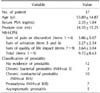
The mean age±SD, mean serum PSA level, and mean prostate size of the patients were 55.89±14.87 years, 2.35±1.84 ng/mL, and 34.35±15.25 mL, respectively. From the items of the NIH-CPSI questionnaire, we found that the sum scores of the pain or discomfort, urination, quality of life impact, and total items were 3.86±5.07, 2.27±2.54, 3.64±3.04, and 9.72±8.63, respectively. Thirty-seven patients were classified into the following groups: no evidence of prostatitis (12 patients; 32.43%), chronic bacterial prostatitis (3 patients; 8.1%), chronic nonbacterial prostatitis (10 patients; 27.02%), prostadynia (7 patients; 18.91%), and asymptomatic prostatitis (5 patients; 13.51%) (Table 1).
We found Enterococcus faecalis in three chronic bacterial prostatitis patients.
To differentiate patients with prostatitis-associated hematospermia (PAH) from those without any evidence of prostatitis (HWP), we divided the 37 hematospermia patients into 12 patients for HWP and 25 patients for PAH. We could not find any statistically significant differences between the two groups in terms of the age interval, serum PSA level, and prostate volume (Table 2). Even though no statistically significant difference was found in the sum items of urination (items 5 and 6) between the two groups, we were able to find a statistically significant difference in the sum of the quality of life impact (items 7~9) (Table 2).
DISCUSSION
Even though the prevalence of hematospermia is not high in the general population and its clinical course is usually self-limited, it often causes considerable anxiety to patients [1231314]. Some patients presuppose hematospermia to be linked to sexual behaviors such as prolonged sexual abstinence or overindulgence, while others worry that genitourinary malignancies and sexually transmitted diseases are the key causes of hematospermia [1234561516]. Theoretically, it may be derived from anatomical conditions along the sperm passageway, such as in the lesions of testis, epididymis, vas deferens, seminal vesicles, prostate, and posterior urethra. Urologists have also categorized hematospermia on the basis of pathophysiologic mechanisms such as inflammation and infections, ductal obstruction and cysts of accessory sexual glands, tumors, vascular abnormalities, and systemic and iatrogenic factors [3].
Inflammatory or infectious conditions in the genitourinary tract have appeared to be the most common causal factors of hematospermia [12345]. An irritation of the mucosa, mucosal edema, and hyperemia of the accessory sexual glands and their corresponding ducts may lead to bleeding and hematospermia [3]. The suggested infectious etiologies include viral, bacterial, mycobacterial, and parasitic infections such as bilharziasis [3]. Bamberger et al [16] investigated sexually transmitted diseases from infected patients who demonstrated hematospermia and found infections of Herpes simplex virus (42%), C. trachomatis (33%), E. faecalis (17%), and Ureaplasma urealyticum (8%). We also evaluated the C. trachomatis and gonococcal infection with a multiplex PCR method in this study. However, we could not find any trace of the two pathogens in the specimens of urine or EPS. This discrepancy may be attributed to the difference in the age distributions between the two studies. The age range of Bamberger et al's [16] study was 17 to 66 years (median: 33 years), with 4 patients over the age of 40 years. In contrast, the mean age of the patients in this study was 55.89±14.87 years. It has been well known that young age is an important factor associated with chlamydial infection [17].
Total ejaculate contains from 60% to 80% biological materials of prostate origin [18]. Therefore, we easily presuppose the association between hematospermia and prostatitis. We found E. faecalis infection in the prostate of the three hematospermia patients. E. faecalis is a gram-positive, commensal bacterium inhabiting the human gastrointestinal tract [19]. These bacteria frequently colonize the urinary tract and cause infection [11]. We have previously reported that only 41 samples from 1,021 patients with chronic prostatitis/chronic pelvic pain syndrome (CP/CPPS) revealed a significant E. faecalis manifestation in their prostate for defining chronic bacterial prostatitis [11]. When we compare the incidence of E. faecalis between the 8.1% (3 E. faecalis-infected patients/37 hematospermia patients) found in this study with the 4% (41 E. faecalis-infected patients/1,021 CP/CPPS patients) in the previous study, we can infer that a bacterial prostatitis infection may lead to hematospermia in certain patients. Furthermore, the hematospermia symptoms of all E. faecalis patients improved with an appropriate antibiotic (fluoroquinolone) treatment.
Prostatitis, an inflammatory or infectious disorder of the prostate, can be classified into acute, chronic bacterial, chronic nonbacterial, prostadynia, and asymptomatic forms [820]. In this study, we could not find any acute prostatitis patients. Interestingly, 25 patients with hematospermia revealed prostatitis-like symptoms or signs. Among them, three patients revealed an E. faecalis infection in the prostate, defined as chronic bacterial prostatitis.
With a strict criterion (≥16 WBC per hpf in the EPS specimen) for defining inflammatory prostatitis, we were able to diagnose 10 patients with chronic nonbacterial prostatitis.
We strongly recommend robust studies for CP/CPPS for patients with hematospermia for determining specific prostatitis etiologies because 25 of the 37 enrolled hematospermia patients were associated with CP/CPPS, as well as a higher incidence of chronic nonbacterial prostatitis than prostadynia.
From the meta-analysis studies, the mean age of hematospermia is 37 years, showing prevalence in young males [215]. However, the mean age in our study was 55.89 years. From the data of the mean serum PSA level (2.35± 1.84 ng/mL), prostate size (34.35±15.25 mL), and mean International Prostate Symptom Score (IPSS) score (9.64±8.27), our patients may include patients with benign prostatic hypertrophy. The mean age in our study was very similar to the mean age reported by Ng et al [1] (54 years; age range: 16~82 years). Around this age, the major concern for many patients is to know whether they have prostate cancer. In general, the incidence of prostate cancer is not high in patients with hematospermia.
We could not find any statistically significant differences between the PAH patients and the patients without hematospermia in terms of the age interval, serum PSA level, prostate volume, and sum of items about urination (items 5 and 6 of the NIH-CPSI questionnaire). Interestingly, the PAH patients revealed a relatively poor quality of life compared with the patients without prostatitis. One of the treatment targets of prostatitis is to improve the quality of life in patients with chronic prostatitis [21]. Therefore, we must consider more specific treatments for hematospermia patients with prostatitis symptoms or signs.
We recognize two limitations to this study. First, even though we ardently collected samples from hematospermia patients for five years, we found very few well-characterized samples for evaluating CP/CPPS. Second, this study can be classified as a correlation study. While our results showed that the hematospermia patients were associated with a higher prevalence of prostatitis than the normal population, as well as for inflammatory prostatitis than prostadynia, we could not determine whether this association is a true etiologic factor in the manifestation of hematospermia.
CONCLUSIONS
Two-thirds of the hematospermia patients were associated with evidence of prostatitis. We must carefully evaluate the association of prostatitis in hematospermia patients. Further, hematospermia patients with prostatitis revealed a relatively poor quality of life compared with those without prostatitis. To improve the quality of life of the patients, we must control the symptoms of CP/CPPS in hematospermia patients with prostatitis.

 XML Download