

 PDF
PDF ePub
ePub Citation
 Print
Print


Introduction
Creating a manual or mechanical glide path was shown to be the first step for safer use of nickel titanium (Ni-Ti) rotary instrumentation because this procedure prevents fracture, shaping aberrations, and torsion of instruments.1,2 Additionally, creating a glide path is recommended to reduce the risk of taper lock and frictional forces to the canal walls especially in curved canals.3,4
Several path-finding rotary systems are available for use in creating a glide path. The new ProGlider (PG) Ni-Ti rotary instrument for mechanical pre-flaring was recently introduced by Dentsply Maillefer. It is manufactured from M-Wire Ni-Ti alloy to enhance flexibility and cyclic fatigue resistance as claimed by the manufacturer. The system consists of a single instrument, with variable progressive taper. The PG instrument is available with 21, 25, and 31 mm length and tip size 16 with a taper of 0.02 at the tip of the file.
Numerous studies have investigated the effects of different path-finding systems on root canal anatomy preservation, remaining dentin thickness, and separation incidence of instruments.35678 It was reported that glide path preparation reduced root canal modifications, canal aberrations, excessive dentin removal, and separation incidence.3578 However, in some previous studies it was found that creation of a glide path did not influence the apical canal transportation in curved root canals.67
Furthermore, several studies have investigated the dentinal damage associated with different Ni-Ti rotary instruments.9101112 Thus far, there is no research dealing with the effect of glide path on creating dentinal damage during root canal preparation with rotary Ni-Ti instruments. Therefore, the aim of this study was to investigate the dentinal crack formation after using ProTaper Next (PTN) system with and without the newly introduced path-finding system (PG).
Materials and Methods
Mandibular first molars with similar lengths and curvatures of 25 - 35° were selected from the collection of teeth that had been extracted for reasons unrelated to this study and kept in distilled water until use. Canal curvature angles of the teeth were measured according to the method described by Schäfer et al.13 All the external root surfaces were inspected with transmitted light and a stereomicroscope to exclude those with any pre-existing craze lines or cracks. According to these criteria, 45 mandibular first molar teeth were selected. The distal roots with the respective part of the crown were sectioned at the furcation level by using a low-speed saw (Isomet, Buehler Ltd, Lake Bluff, IL, USA). For each sample, a size 10 K-type file was progressed through the root canal until it was visible at the apex, and the working length of the canal was determined to be 1 mm short from this point. During root canal preparation, a reference point was determined and care was taken to provide the same position of rubber stops to ensure stable working length on each sample. A silicon impression material was used to coat the external root surface to simulate the periodontal ligament space. All the roots then were embedded in autopolymerizing acrylic blocks. Fifteen teeth were left unprepared as the control group and remaining 30 teeth were assigned into two experimental groups.
Group PG/PTN, a glide path was prepared with a size 16 ProGlider instrument. Then root canals were instrumented with ProTaper Next files (Dentsply Maillefer, Ballaigues, Switzerland) in the sequence of X1 and X2 at a rotational speed of 300 rpm and 200 g/cm torque according to the manufacturer's instructions. The instruments were used up to the working length.
Group PTN, glide path was not prepared. The root canals were instrumented with ProTaper Next files up to size 25 (X2) using the instrument order specified by the manufacturer as described in PG/PTN group.
The rotary file systems were used with an electrical motor (X-Smart, Dentsply Maillefer) and a 16:1 reduction handpiece. Each instrument was used in 3 canals. The root canals were irrigated with 2 mL of 1% NaOCl solution after each instrument change. After preparation, the specimens were rinsed with 5 mL of distilled water.
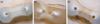
The roots were horizontally sectioned at 1, 2, 3, 4, 6, and 8 mm from the apex using a low-speed saw (Isomet, Buehler Ltd) under water-cooling. To avoid any artifacts by dehydration, the teeth were kept moist in distilled water during all the experimental procedures. All slices were observed under a stereomicroscope (Olympus SZ-CTV, Olympus, Tokyo, Japan) at ×20 magnification and pictures were taken. All root canal preparations were performed by one operator and the assessment of each cross section was performed by another examiner who was blinded in respect to all experimental groups. In each group, a total of 90 slices were examined for cracks. To define crack formation, two categories were made as 'no crack' and 'crack'. 'No crack' was defined as root dentine without cracks either at internal or external surface of the root. 'Crack' was defined as all lines observed on the slice that either extended from the root canal lumen to the dentin or from the outer root surface into the dentin (Figure 1).14
The results were expressed as the number and percentage of cracked roots in each group. The Chi-square test with Yates correction was used to determine for difference between groups. P value of less than 0.05 was considered statistically significant for all tests. Statistical analyses were performed using SPSS 19.0 software (SPSS, Inc., Chicago, IL, USA).
Results
The numbers of roots with cracks for all groups are shown in Table 1. No cracks were observed in the control group. When considering the crack formation in total sections, crack formation was found to be 17.8% in PG/PTN group, while 28.9% was observed in PTN group. However, there were no significant differences between the two experimental groups (p = 0.134). Regarding the different section levels (1, 2, 3, 4, 6, and 8 mm), no significant difference was found between the experimental groups at any level.
Discussion
Creating a glide path provides several advantages such as preserving original canal anatomy, less canal aberrations, less postoperative pain, less separation rates of Ni-Ti rotary instruments and avoiding excessive instrument binding in the canal.1234151617 The possibility of dentinal defects may increase due to the excessive instrument binding and the maximum contact between the file and dentin. Thus, the present study aimed to evaluate the crack formation of the PTN system both with and without a glide path by using the PG system.
In the present study, a newly introduced pathfinding system ProGlider was used to create glide path. It has a size 16 with a taper of 0.02 at the tip of the file. It consists of one file with a variable progressive taper. It is manufactured by using M-Wire Ni-Ti alloy to enhance its flexibility and its cyclic fatigue resistance of the files. It is recommended by the manufacturer to use PG for creating a glide path before instrumentation with the PTN system. The methodology used was based on previous researches.141819 The biological width was simulated and the crowns corresponding to mesial roots were kept intact to simulate clinical conditions where the interference of cervical dentin could induce unwanted tension or resistance.5
This study revealed that dentinal cracks occurred independent of creating the glide path. The incidence of cracks was observed in 17.8% and 28.9% of root in PG/PTN and PTN groups, respectively. Regarding the crack formation, there was no significant difference between glide path versus no glide path groups. This might be attributed to the cross-sectional design and innovative M-Wire Ni-Ti technology for PTN system. ProTaper Next files exhibit a rectangular cross-section design for superior strength and an exceptional asymmetric rotary motion that improves the file's canal shaping effectiveness according to the manufacturer. This manufacturing process provides greater flexibility and greater resistance to cyclic fatigue for the system.5 Moreover, PTN system has off-centered rectangular design that generates a swaggering motion, which decreases the screw effect, dangerous taper lock, and torque on any given file by minimizing the contact between the file and the dentin.20 It can be speculated that as a result of the reduction in the contact area between the file and canal system, lower defects may occur because of lower frictional forces to canal walls.
In the literature, there were limited studies available regarding the crack formation of PTN system.202122 Capar et al. reported cracks in 28%, Karataş et al. reported cracks in 33.3%, whereas Çiçek et al. reported in 64.44% of roots instrumented with the PTN system.202122 These contradictory results may be attributed to a number of reasons, and the most likely one might be related to the type of teeth included. Unlike the present study, single rooted teeth were selected in those studies.2021 Moreover, Capar et al. performed a glide path via a size 15 K type file before instrumentation with ProTaper Next files in mandibular premolar teeth, whereas Karataş et al. and Çiçek et al. did not mention any glide path preparation before instrumentation in mandibular central incisors and mandibular molar, respectively.202122
Conclusions
Within the limitations of this in vitro study, the creation of the glide path before ProTaper Next rotary system did not influence dentinal crack formation in root canals. Further studies should be conducted to evaluate the effect of glide path created with different type of path-finding systems on crack formation.

 XML Download