

 PDF
PDF ePub
ePub Citation
 Print
Print


Introduction
Gastric cancer is the third most common cause of cancer-related death worldwide.1 The highest incidence rates are in East Asia, Eastern Europe, and South America.2,3,4,5 Studies have shown that the clinicopathological presentation of gastric cancer, including histology, location, environmental exposure, and dietary factors, varies widely between Eastern and Western countries.6,7,8 Moreover, the 5-year survival rate after curative gastrectomy for gastric cancer is lower in the West than in the East.9,10,11 It is well known that many diseases in Korea and Japan share similar clinicopathological characteristics; these countries also have similar treatment policies and screening programs for gastric cancer, and achieve comparable results.12,13,14 In contrast, there are little data on the differences in the clinicopathological characteristics of gastric cancer between China and other Asian countries.15
The aim of this study was to compare the clinicopathological variables and outcomes between 2 high-volume gastric cancer centers in China and Korea. The goal of this work was to identify critical clinicopathological differences between Chinese and Korean patients and consequently improve treatment for gastric cancer patients.
Materials and Methods
Patients diagnosed with gastric cancer and eligible for curative resection (R0) at either Peking University People's Hospital, China or Seoul St. Mary's Hospital, Korea between 1998 and 2009 were included in this study. We analyzed patient demographics, tumor factors, surgical factors, and survival. Patients who underwent R0 resection but who had no other history of cancer were included. Patients were excluded if they underwent neoadjuvant chemotherapy, wedge resection, or endoscopic mucosal resection. tumor-node-metastasis (TNM) classification was based on the 7th edition of the American Joint Committee on Cancer staging system. D1 or D1+ lymphadenectomy was performed for early gastric cancer. D2 or D2+ (D2+14v, or +12p, or +8p, or +16a) lymphadenectomy was performed for advanced cancer. The criteria for follow-up and recurrence were similar for both Korean and Chinese patients. Follow-up evaluation was repeated every 3 months for 2 years, every 6 months from the third to fifth post-operative year, and every year thereafter. The follow-up rates were 92.5% and 94.8% for Chinese patients and Korean patients, respectively.
1. Statistical analysis
The Statistical Package for the Social Sciences (SPSS) version 17.0 (SPSS Inc., Chicago, IL, USA) was used. The chi-square test was employed to assess differences in the categorical clinicopathological variables. The independent t-test was used to evaluate differences in continuous variables. Overall survival (OS) was calculated from the time of surgery to the last follow-up or date of death. For patients who experienced recurrence, progression-free survival (PFS) was calculated as the time from surgery to the time of first recurrence; for those with no recurrence, PFS was defined as the time from surgery to the last follow-up or death. Univariate survival analysis of OS and PFS was estimated using the Kaplan-Meier method. The Cox proportional hazards model was used for multivariate analysis. Variables in the model included patient group, gender, age, body mass index (BMI), family history of cancer, operation type, digestive tract reconstruction methods, lymphadenectomy type, tumor location, tumor size, tumor differentiation, TNM stage, adjuvant chemotherapy, and number of harvested lymph nodes. P-values<0.05 were considered statistically significant.
Results
1. Demographic data of gastric cancer patients
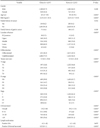
A total of 1,637 Chinese and 2,231 Korean gastric cancer patients were identified and compared. The age of the Chinese patients was significantly higher than that of the Korean patients (P<0.001). The BMI of the Chinese patients was significantly lower than that of the Korean patients (P<0.001). There was no significant difference in family history of any type of cancer, including gastric cancer, between the 2 institutions (Table 1).
2. Surgical characteristics of gastric cancer patients
Korean surgeons performed significantly more total gastrectomies, multivisceral resections, and Billroth II digestive tract reconstructions compared with their Chinese counterparts, and less postoperative chemotherapy was administered in Korea (P<0.001; Supplementary Table 1). D2+ lymphadenectomy was performed in 79% of advanced cases in Korea (797/1,009), but was not performed in any of the Chinese cases in this study (0/1,440).
There were no significant differences in the incidence of major surgery-related complications between the 2 institutions (P=0.42). These complications included anastomotic leakage, anastomotic stenosis, intra-abdominal bleeding, postoperative ileus, and post-operative intra-abdominal infection (Supplementary Table 2).
3. Pathological characteristics of gastric cancer patients
Chinese gastric cancer patients had more tumors located in the esophagogastric (EG) junction and whole stomach (P<0.001), and a greater number of undifferentiated tumors (P<0.001) compared with Korean patients. Tumors in Chinese patients were generally larger than those in Korean patients (P<0.001).
There were more gastric cancer patients with relatively early-stage disease including T1, T2, N0, and stage I, in Korea than in China (P<0.001).
More lymph nodes were harvested from Korean patients than from Chinese patients (P<0.001). The ratio of positive lymph nodes to total nodes examined was significantly higher in China than in Korea (P<0.001; Table 1).
4. Survival analysis
1) Overall survival
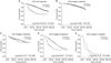
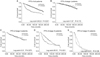
The OS of Korean patients was longer than that of Chinese patients (P<0.001), especially for stage III disease (P<0.001). Analysis based on T and N stages showed that the OS for Korean patients with stages T3 (P=0.011) and T4 (P=0.036) or N2 (P=0.002) and N3 (P<0.001), but not T1 (P=0.299) and T2 (P=0.085) or N0 (P=0.062) and N1 (P=0.090), was significantly longer than that of Chinese patients with an equivalent disease stage (Fig. 1, 2).
2) Progression-free survival
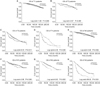
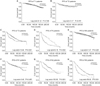
Korean patients with either stage III (P<0.001) or stage IV (P=0.106), but not stage I or II disease, had a longer PFS than Chinese patients with the same disease stage. Korean patients with stages T3 (P=0.001), T4 (P=0.002), N2 (P<0.001), or N3 (P<0.001), but not T1 (P=0.726), T2 (P=0.075), N0 (P=0.226), or N1 (P=0.485), had a significantly longer PFS than Chinese patients (Fig. 3, 4).
3) Independent factors affecting overall survival and progression-free survival
The Cox proportional hazards model showed that patient group, age, and TNM stage were independent risk factors affecting both OS and PFS, whereas BMI was an independent risk factor for OS but not PFS (Supplementary Table 3).
Discussion
To date, there has been little research comparing the different gastric cancer characteristics in Korea and China. Our study showed that Chinese patients were older than Korean patients and had lower BMI values. These findings can be partly explained by the nationwide screening system that was introduced in Korea in 1999 as part of the National Cancer Screening Program,16,17,18 which ensured early cancer detection.19,20,21
Korean and Chinese institutions have different surgical policies.22 Our data showed that Korean surgeons performed more total gastrectomies for tumors located in the proximal part of the stomach than their Chinese counterparts. They also preferred extensive surgery for T4 gastric cancer, in contrast to most Chinese surgeons who follow the National Comprehensive Cancer Network guidelines and prefer pre-operative chemotherapy followed by radical resection of the tumor for these patients.
Interestingly, our results showed that the rate of EG junction cancer was significantly higher in Chinese than in Korean patients. Patients in Western countries have more EG junction tumors than those in Asian countries.23 Data from the Memorial Sloan-Kettering Cancer Center8 showed that 18% of all gastric cancers were located at the EG Junction. In this study, we found that Chinese patients had significantly more EG junction tumors than Korean patients, but fewer EG junction tumors than those in the US.24 Although both China and Korea are in Asia, the countries have very different diets, which may contribute to clinicopathological differences. Chinese food is typically oily and resembles a Western-style diet.
Korean patients with advanced-stage cancer had longer OS and PFS compared to Chinese patients. We hypothesized that this difference might be due to different treatment policies, while the longer OS of Korean patients might be partially related to their younger age. Korean surgeons traditionally prefer extended surgery, especially D2+ lymphadenectomy, for advanced gastric cancer. D2+ lymphadenectomy was performed in 79% of advanced cases in Korea, but was not performed in any of the Chinese cases in this study. However, the overall percentage of D2+ lymphadenectomies was lower in Korean patients due to the lower ratio of advanced gastric cancer in this group. In fact, the benefit of extended D2 lymphadenectomy for gastric cancer remains unclear, although some studies have proposed its use.25,26,27 However, other researchers have contrasting opinions.28,29,30 While the number of lymph nodes harvested in Korean patients was significantly greater than that in Chinese patients, multivariate analysis showed that this was not an independent risk factor affecting OS and PFS. Therefore, we speculated that the number of lymph nodes harvested and extent of lymphadenectomy might be important for the survival of gastric cancer patients, especially those with non-metastatic advanced gastric cancers such as stage III; however, it is probably irrelevant for early stage (stage I, II) and metastatic cancers (stage IV). Moreover, the poorer survival of Chinese patients, particularly those with stage III disease, might also be partially attributed to downstaging31 because of the insufficient number of lymph nodes harvested (approximately 20% of patients had <15 lymph nodes harvested).
The number of lymph nodes harvested is dependent on both surgical technique and the pathologist's experience. A multidisciplinary team (MDT) could improve communication between surgeons and pathologists, and ensure that the lymph nodes are checked by pathologists.32 In this study, a MDT was established in the Chinese institution in 2010. For patients with fewer than 15 lymph nodes harvested, accurate staging might be accomplished by dividing by the metastatic lymph node ratio,33 referred to in the TNM staging system, but more evidence is needed to support this strategy.
In summary, we find that some clinicopathological variables are different between Korean and Chinese gastric cancer patients. Korean gastric cancer patients have longer OS and PFS compared to Chinese patients with advanced disease stages. This study may guide the future direction of gastric cancer research in both China and Korea, and may provide evidence to influence surgical treatment policies in both countries.
 XML Download