

 PDF
PDF ePub
ePub Citation
 Print
Print


INTRODUCTION
Spontaneous bowel perforations not associated with disease or blunt trauma, particularly a colon perforation, are uncommon beyond the neonatal period. Spontaneous colon perforation (SCP) has been reported infrequently in the literature. It could be defined as a sudden perforation of normal colon in the absence of disease or injury, and is difficult to diagnose preoperatively. These conditions usually occur sporadically without preceding events and could often result in delayed management.
In adults and premature neonates, it has been associated with ischemic insult or necrotizing enterocolitis. However, it is limited to study and guessing in infants and children without health problems. Because of its rarity, it is also challenging to assess with clinical manifestations and other physical findings. In addition, when neglected, a colon perforation may cause fecal peritonitis with life-threatening conditions and result in sepsis and high mortality.
In this study, we reviewed cases of SCP beyond the neonatal period and attempted to suggest its clinical implication in management by identifying the clinicopathological characteristics.
MATERIALS AND METHODS
Patient selection
There were 127 cases of gastrointestinal perforation in pediatric patients which were confirmed after operation in Pusan National University Children's Hospital between January 2009 and June 2015. We reviewed cases of colon perforation in pediatric patients (under age of 18 years), which were identified after the operation. Excluding cases of neonates, perforations associated with inflammatory disease and other traumatic injury, 11 patients with spontaneous perforation were selected as the subject group.
Evaluation of clinical characteristics and comparison
A retrospective study was conducted using patients' clinical data. We analyzed basic demographic characteristics including sex, age at operation, and body weight, as well as clinical characteristics including presented symptoms, laboratory and radiologic findings, accompanied conditions, operative methods, pathologic and postoperative results. Because a pneumoperitoneum is usually a pathognomonic sign and the results of gastrointestinal perforation, it is presumed to affect delayed diagnosis and management in absence of this sign. Then laboratory finding and clinical course after the operation were compared according to the presence of pneumoperitoneum as initial findings.
RESULTS
Demographic findings
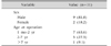
There were 9 boys and 2 girls. According to age at operation, seven patients (63.6%) were under 2-year-old and one was over 7-year-old. Because of the small sample size, it was difficult to describe the relation to sex and age (Table 1).
Clinical characteristics, preoperative and postoperative findings
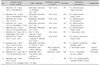
All patients were transferred to emergency department. They had a history of hospitalization with mean 6.7±3.0 days and most were associated with management for fever (9/11, 81.8%), the others were viral gastroenteritis and respiratory tract infection. When they visited the emergency department, the chief complaint of seven patients was abdominal distention and others were abdominal pain, diarrhea. Simple radiologic examination was performed in all patients before and after transfer, but only seven patients showed a sign of pneumoperitoneum (7/11, 63.6%). Therefore, abdominal computed tomography (CT) performed additionally in four patients who did not show a pneumoperitoneum showed positive findings in two patients. In addition, radiologic study showed no suspicious finding of perforation in one patient and it was detected during the operation (Table 2). Perforation was found evenly in all segments of colon, most commonly at the sigmoid colon in 5 cases. Its pattern was single or two perforations with pin-point or round shape in 8 cases, the others were multiple or necrotic shallow shape. Especially in cases with a single perforation, most of them were identified at antimesenteric surface of involved colon. Only a simple primary closure was sufficient in the case of a single perforation with minimal inflammatory change, otherwise a resection with primary anastomosis was performed. Postoperative complication developed in three patients; wound infection, immediate septic condition, and pancreatitis. However there were no cases of mortality (Table 2).
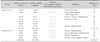
Laboratory findings and pathologic results according to the presence of pneumoperitoneum (Table 3)
At the initial radiologic examination, a pneumoperitoneum was detected in 7 patients (group A), and not in 4 patients (group B).
Blood test of complete blood cell showed an elevated white-blood cell (WBC) count, mean 19.7±9.2×10³/µL in group A and 24.5±6.8×10³/µL in group B. Serum level of C-reactive protein (CRP) was also highly increased, mean 22.2±7.7 mg/dL in group A and 21.7±5.8 mg/dL. However review of a serologic marker for bacteria (Salmonella and Clostridium) and virus (Cytomegalovirus and Rotavirus) showed no positive findings in both groups. Histology of perforated colon showed only inflammatory change without definite pathologic condition. Considering the hospital stay after the operation, it was longer in group B (mean 23.3±8.7 days vs. 11.1±3.0 days).
DISCUSSION
SCP in pediatric populations are usually encountered as necrotizing enterocolitis in the neonatal period, but are rare in infants and children without preceding conditions including Hirschsprung's disease, inflammatory bowel disease, connective tissue disorder, lymphoma, and infective colitis [123]. It is more frequent in elderly with history of constipation at age older than 60 years [456].
SCP can be classified according to 2 different types, stercoral and idiopathic. Stercoral type is associated with chronic constipation, thus it is rare in infants and children, however the idiopathic type is sporadic and could occur at any age [5678]. In our study, none of the patients had preceding condition, fecal impaction and revealed no histopathology reports of perforated lesion affecting perforation. This idiopathic type is less common and shows better prognosis possibly because of minimal fecal contamination [910]. Interestingly, all patients in this study had a history of hospitalization before transferring to our institution. There were variable reasons for admission with staying for a mean 6.7±3.0 days. Fever was the most common cause (9/11, 81.8%), followed by upper respiratory infection, bronchiolitis, and acute gastroenteritis. In situations requiring a hospital stay over several days for fever or associated symptoms, there may be many chances of prescribing non-steroidal anti-inflammatory drug (NSAID). Even a short period, it could cause gastrointestinal problems including perforation. There were a few reports about the case of NSAID-induced colon perforation in pediatric patient [111213]. This phenomenon seems to be related to factors induced by NSAID: increased intestinal permeability, bacterial-mediated production of toxic-free acids, and drug synergism with bowel ischemia [14151617].
Common symptoms were abdominal pain and diarrhea during the febrile period, but these were non-specific gastrointestinal symptoms. Sudden onset of abdominal distention presuming pneumoperitoneum was the most common sign (7/11, 63.6%). Pneumoperitoneum was considered as a suggestive finding of perforation of hollow viscus including a gastrointestinal tract. Thus, whether it was found at initial clinical course could be assumed that it could affect early diagnosis and management. Although most patients showed pneumoperitoneum at their visit to the emergency department or before transfer, four patients did not. Therefore it was necessary to perform an additional CT scan. In two cases, it was not identified by all radiologic examinations and diagnosed during the operation itself. Patients who did not show pneumoperitoneum on an initial simple radiologic examination underwent a segmental resection and had a long hospital stay. They also showed higher WBC count and lower CRP level. However, when comparing patients who showed a pneumoperitoneum at initial radiologic examination, there were no significant differences in laboratory findings and clinical course after the operation.
In this study, most patients (90.9%) were infants and preschool children. But, it is difficult to guess an age-relation to occurrence. Considering the possibility of colon perforation, it should be to paid particular attention to management of this age group for fever with a long hospital stay over average of 6.7 days and associated with a non-specific gastrointestinal symptoms.
Colon perforation requires prompt surgical intervention for diagnosis and treatment because a good outcome can be expected when detected early with less fecal contamination. Surgical management for colon perforation depends on the time of onset, degree of peritonitis, and general condition of the patient. In the past, it consisted of fecal diversion enterostomy after removal of the involved segment, but a recent trend moved toward a conservative operation including a minimal invasive surgery when possible [18192021]. Our experience showed good outcomes by primary repair only, and primary repair (6 cases) was slightly more common than resection (5 cases). Although this procedure could usually be attempted in colon perforation during or after colonoscopy because of good bowel preparation, we experienced good results even if it was not similar. Therefore we could carefully suggest that a simple procedure like a primary repair may be attempted in cases of SCP with a stable condition in pediatric populations.
In previous reports, colon perforation in infants occurred more commonly at the proximal colon and appendix [2223]. Nevertheless, in our study, perforation occurred evenly through the segment of the colon; sigmoid colon (5 cases), cecum (2 cases), ascending colon (2 cases), and transverse colon (2 cases). Moreover, the lesion of perforation was located at antimesenteric surface of colon in most single perforation. Idiopathic perforation can occur at any part of the colon, but more commonly in the sigmoid colon, which is a vulnerable anatomical part in the vasculature, it has been proposed to explain the colon perforation [1024]. However, a histologic study of perforated colon did not reveal ischemia, instead chronic inflammation or with ulcerative lesion was usually confirmed. And serologic makers in all patients showed no association with significant pathogen. Postoperative stay was mean 15.5±8.1 days, which is shorter compared with previous reports [2225]. Morbidity was found in two patients without pneumoperitoneum on the initial examination, overall morbidity 20%, but there was no case of mortality. Previous studies reported some mortality related to infectious agents despite of surgical management [10242627]. However, we could encounter a favorable outcome without mortality because there were no other associated gastrointestinal conditions and infectious agents, a prompt surgical management as well.
In conclusion, when previously healthy infants and children present with a sustained fever and a sudden onset of abdominal distention during management for fever associated with respiratory or gastrointestinal problems, there is a great likelihood of colon perforation with no pathological condition in involved site. Thus, a prompt surgical management as a timely decision-making is required in order to achieve a good progress with least surgical procedure. Additionally, this study was a single center experience and has a limitation of data collection, so it is necessary to undertake further studies about SCP.
 XML Download