

 PDF
PDF ePub
ePub Citation
 Print
Print


Abstract
Microscopic examination of peripheral blood smear (PBS) for detection of microorganisms is simple method that can be used for doctors to confirm the septicemia more swiftly and to select more specific therapy. But it is unusual to find microorganisms in PBS. We report a case of gram negative bacteremia diagnosed by PBS in a severe thrombocytopenic pediatric surgical patient. A 6-month and 2 week old baby with cyanosis was diagnosed congenital heart diseases such as transposition of great arteries, at-rial septal defect, and patent ductus arteriosus. The infant underwent surgical operations and the post-operative platelet count progressively decreased in spite of transfusion of multiple platelet concentrates. We performed routine examination of a PBS for evaluation of severe thrombocytopenia. The PBS revealed severe thrombocytopenia, leukopenia with left shifted and some extracellular bacilli. Toxic granulations, toxic vacuoles and some bacilli were observed in the neutrophils. The bacilli were identified as Pseudomonas aeruginosa and Serratia marcescens in blood culture. To our knowledge, this is the second case of bacteremia diagnosed by PBS before the positive blood culture in Korea. We suggest that a PBS is useful for the rapid detection of organisms in cases of septicemia with severe thrombocytopenic pediatric surgical patient.
REFERENCES
1. Fife A, Hill D, Barton C, Burden P. Gram negative septicaemia diagnosed on peripheral blood smear appearances. J Clin Pathol. 1994; 47:82–4.

2. Mirza I, Wolk J, Toth L, Rostenberg P, Kranwinkel R, Sieber SC. Waterhouse-Friderichsen syndrome secondary to Capnocytophaga canimorsus septicemia and demonstration of bacteremia by peripheral blood smear. Arch Pathol Lab Med. 2000; 124:859–63.
3. Nakamura H, Saitou M, Kinjo S, Kaneshima H, Higa F, Tateyama M, et al. Overwhelming pneumococcal bacteremia revealed by a peripheral blood smear in a 74-year-old healthy woman. Intern Med. 2007; 46:303–6.
4. Sohn HE and Chung HR. Bacteremia diagnosed on peripheral blood smear before blood cultures become positive: a case report. Korean J Clin Pathol. 1999; 19:27–30.
5. Ristuccia PA, Hoeffner RA, Digamon-Beltran M, Cunha BA. Detection of bacteremia by buffy coat smears. Scand J Infect Dis. 1987; 19:215–7.
6. Rodwell RL, Leslie AL, Tudehope DI. Evaluation of direct and buffy coat films of peripheral blood for the early detection of bacteraemia. Aust Paediatr J. 1989; 25:83–5.
7. Shanholtzer CJ, Schaper PJ, Peterson LR. Concentrated gram stain smears prepared with a cytospin centrifuge. J Clin Microbiol. 1982; 16:1052–6.
8. Branda JA, Ferraro MJ, Kratz A. Sensitivity of peripheral blood smear review for the diagnosis of Candida fungemia. Arch Pathol Lab Med. 2007; 131:97–101.
9. Misawa S. Rapid diagnosis of infectious diseases; features and limitations of the microscopic examination of clinical specimens. Rinsho Biseibutshu Jinsoku Shindan Kenkyukai Shi. 1999; 10:121–31.
10. Graham BS. Detection of bacteremia and fungemia: microscopic examination of peripheral blood smears. Infect Control. 1984; 5:448–52.
11. Kroft SH. Infectious diseases manifested in the peripheral blood. Clin Lab Med. 2002; 22:253–77.
12. van der Meer W, Verwiel JM, Gidding CE, de Metz M, de Keijzer MH. Bacteria in blood smears: overwhelming sepsis or trivial contamination. Acta Haematol. 2002; 107:220–3.
13. Rowe MI, Buckner DM, Newmark S. The early diagnosis of gram negative septicemia in the pediatric surgical patient. Ann Surg. 1975; 182:280–6.
14. François B, Trimoreau F, Vignon P, Fixe P, Praloran V, Gastinne H. Thrombocytopenia in the sepsis syndrome: role of hemopha-gocytosis and macrophage colony-stimulating factor. Am J Med. 1997; 103:114–20.
15. Chang JC. Review: Postoperative thrombocytopenia: with etiologic, diagnostic, and therapeutic consideration. Am J Med Sci. 1996; 311:96–105.
Fig. 1.
The complete blood counts (including segmented neutrophils) results of patient during admission days. ∗after 1st operation; † after 2nd operation; ‡ after 3rd operation; § on presentation. Abbreviations: AD, admission day; HD, hospital day.
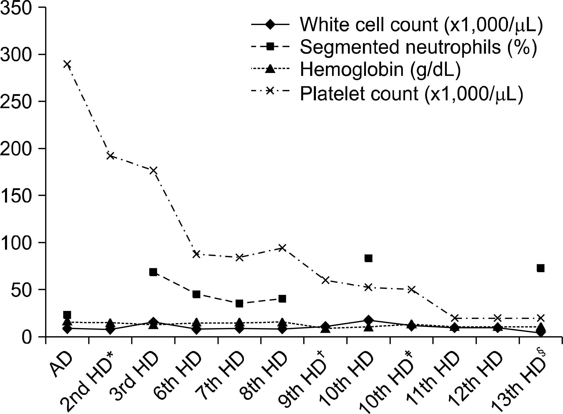
Fig. 2.
Microorganisms shown on peripheral blood smears. (A) Extracellular clustering bacilli and anisocytosis and poikilocytosis of RBCs on peripheral blood smear (Wright stain, ×1,000). (B) Bacilli, vacuoles and toxic granules in cytoplasm of neutrophils (Wright stain, ×1,000). (C) Gram negative bacilli in cytoplasmic and extracellular area on peripheral blood smear (Gram stain, ×1,000). (D) Vacuolation and Gram negative bacilli in neutrophils (Gram stain, ×1,000).

Table 1.
Data of laboratory findings on presentation date
 XML Download