

 PDF
PDF ePub
ePub Citation
 Print
Print


I. Introduction
Oral squamous cell carcinoma (OSCC) is the most common type of head and neck cancer1. It results from diverse genetic mutations of oral epithelial cells. The initiated and promoted cancer cells generally have a monoclonal feature. In the progression stage, however, it mostly has heterogeneity due to the further genetic modifications of the cancer cells2,3. Thus, each of OSCC appears to have biologically diverse patterns and clinically various behaviors4.
There are many kinds of treatment strategies depending on the predicted prognosis of OSCC5. The prediction of its prognosis can be helpful to clinicians in establishing a proper treatment plan. If some cancers are predicted to be aggressive behaviors, clinicians must consider a more aggressive treatment plan6. The TNM staging system has been widely used for the classification of OSCC according to its expected survival rate. This system is a simple method using the tumor size including resectability (T), lymph node involvement (N), and distant metastasis (M). It helps clinicians predict effectively the cancer prognosis and plan the treat ment strategy. This system is generally accepted as a good prognostic indicator for head and neck cancer7.
As a different staging system, specific molecular markers derived from cancer tissue have been applied in recent years for the diagnosis and prediction of OSCC prognosis. Many studies have been conducted to discover useful molecular markers for clinical applications6. Some molecular markers are practically used as tools for adjunctive purposes to evaluate the behavior of OSCC. For instance, epidermal growth factor receptor, cyclin D1, P53, and Matrix metalloproteinase are well-known molecular markers in OSCC. These are related to either cell proliferation or tumor suppression or matrix degradation4,8.
Micro-vessels are an essential part of the growth and progression of a tumor in terms of providing nutrients9,10. In a similar fashion, lymphatic vessels are also an important factor in the lymphatic metastasis of tumor cells11,12. In this study, endoglin and podoplanin as molecular markers were evaluated in tissue specimens from OSCC. Endoglin is one of the representative vascular endothelial cell markers; podoplanin is also a specific marker of lymphatic endothelial cells13,14. Micro-vessels and lymphatic-vessels can be identified by using the primary antibodies for endoglin and podoplanin, respectively. This study was performed to confirm that the formation of micro-vessels and lymphatic vessels is actually correlated with tumor growth and metastasis. If the expressions of endoglin and podoplanin are closely correlated with the clinical stage of OSCC, these markers could be used to evaluate the progression of OSCC. Therefore, this study sought to determine the correlation between the expression of endoglin/podoplanin and the clinical variables of OSCC.
II. Materials and Methods
1. Patient samples
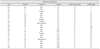
Paraffin specimens from 21 patients diagnosed with OSCC were included in this study. Ten patients were in the early clinical stage (I or II), and eleven patients were in the advanced clinical stage (III or IV). Five patients had positive lymph node involvement, and sixteen patients had negative lymph node involvement by cancer.(Table 1) The exclusion criteria for this study were as follows: (1) recurrent cases of OSCC, and (2) patients with history of adjuvant or curative chemo/radiotherapy before the surgical resection of the main mass. Four surgeons were involved in the surgery of these patients. The patients' charts were reviewed to obtain information such as age, sex, treatment modalities, tumor location, tumor size, lymph node involvement, and distant metastasis.
2. Immunohistochemical staining
Hematoxylin and eosin (H&E) staining and immunohistochemical staining were done for the histological examination. Paraffin-embedded tissue blocks were sliced to thickness of 4 µm. The sections of each tissue were carefully located on the silane-coated slides. These slides were incubated at 60℃ for 1 hour. After cooling to room temperature, the tissue slides were soaked in 100% xylene for 5 minutes in triplicate. At the final step of the xylene treatment, these slides were soaked for a longer time to remove the paraffin completely. The tissue sections were then hydrated by the consecutive application of high- to low-grade ethyl alcohol. Fully hydrated tissue sections were washed with distilled water, and then stored in phosphate-buffered saline solution (PBS, pH 7.5). PBS solutions were applied to wash and preserve the tissue sections in between steps. A universal LSAB+kit (Dako, Glotstrup, Denmark) was used for the immunohistochemical staining. For the antigen, the retrieval procedure was not done. Hydrated tissue sections were treated with 3% hydrogen peroxide for 15 minutes at room temperature to remove endogenous peroxidase and avidins. After that, serum-blocking solutions were applied for 20 minutes to prevent the binding of unspecific proteins. The following antibodies were used as primary antibodies against endoglin and podoplanin: (1) rabbit polyclonal to endoglin (prediluted) (ab27422; Abcam, Cambridge, MA, USA), and (2) mouse monoclonal to podoplanin (ab77854; Abcam). Endoglin was used with no dilution, and podoplanin was used with 1 : 40 dilution. Negative control specimens were not treated with primary antibodies. Tissue sections treated with primary antibodies were incubated overnight at 100% humidity in temperature chambers at 4℃. After adequate binding of the primary antibodies, biotinylated link solution as a secondary antibody was applied to the tissue sections for 30 minutes. After washing to remove any and all unbound secondary antibodies, streptavidin peroxidase solution was applied for 30 minutes. For colorization, diaminobenzidine solution was applied for less than 5 minutes. Mayer's hematoxylin was used for counter-staining. After gradational hydration with ethyl alcohol and clearing with xylene, the tissue sections were fixed by paramount solutions.
3. Quantitative immunohistochemical analysis
Tissue slides were observed with a microscope camera (Olympus DP70 digital microscope camera; Olympus Co., Tokyo, Japan). Positive control of endoglin and that of podoplanin were the human tonsil and the muscularis propria of human bowel, respectively. Images were taken from 3 most highly expressed regions (hot spot)15,16 per specimen under ×100 magnification. All images were blindly assessed to prevent bias of the region. To analyze the expression of endoglin and podoplanin, an image analysis program (SigmaScan Pro 5.0; SPSS Science, Chicago, IL, USA) was used17. The expression of endoglin or podoplanin was measured by the number of pixels, which appeared as red by this program. The average ratio of red pixels to the total pixels was used as variable for each specimen in the analysis.
4. Statistical analysis
Nonparametric tests were used to evaluate the expression of endoglin and podoplanin due to the small sample size. Mann-Whitney U tests were performed to analyze the correlation between the expression of endoglin or podoplanin and clinico-pathological parameters (age, sex, early or advanced stage of OSCC, and lymph node involvement). In addition, Spearman's rank correlation coefficient test was used to evaluate the relationship between the expression of endoglin and podoplanin in each of the OSCC tissue samples. Statistical analysis was performed with IBM SPSS statistics 19.0 (SPSS Inc., Chicago, IL, USA). The significant level of all statistics was set at P<0.05.
III. Results
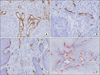
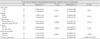
Endoglin and podoplanin, for the most part, were not co-localized in the same region of the serial sections.(Figs. 1. A, 1. B) The average expression of endoglin was 1.691±0.920 in the advanced stages (III, IV) and 0.797±0.583 in the early stages (I, II) (P=0.020). The average expression of podoplanin was 0.286±0.228 in the advanced stages (III, IV) and 0.374±0.157 in the early stages (I, II) (P=0.193). The average expression of endoglin was 1.677±1.246 in the positive lymph node involvement group and 1.137±0.748 in the negative lymph node involvement group (P=0.495). The average expression of podoplanin was 0.345±0.287 in the positive lymph node group and 0.319±0.173 in the negative lymph node group (P=1.000). There was no significant correlation between the expression of endoglin and podoplanin and age or gender. The average expression of endoglin and podoplanin of the entire specimen was 1.266±0.886 and 0.326±0.199, respectively. The correlation coefficient between the expression of endoglin and podoplanin was -0.304 (P=0.193).(Table 2)
IV. Discussion
Many studies have demonstrated that angiogenesis and lym phan giogenesis are correlated with tumor growth and lymphatic invasion in various malignancies including OSCC9-12. Thus, angiogenesis and lymphangiogensis in malignant tissues can be targeted for cancer treatment18-20. In the case of beva cizumab, the vascular endothelial growth factor inhibitor is clinically effective in inhibiting angiogenesis. It has been used as adjuvant to the main chemo-agent in the treatment of breast, lung, and colorectal cancers21-24.
In this study, the primary antibodies for endoglin and podoplanin were used to detect vascular and lymphatic endothelial cells, respectively, in OSCC. Endoglin is a glycoprotein mostly located in the membrane of vascular endothelial cells25. As a component of the transforming growth factor-beta (TGF-β) receptor, it is involved in the TGF-β signaling pathway26. The precise mechanism of its action is still unclear. Note, however, that endoglin is generally known to be linked closely to the formation of the vascular system27. A genetic defect of endoglin causes the underdevelopment of vascular systems28. In several malignancies, high levels of endoglin expression are frequently observed and correlated with poor prognosis13. In our study, the expression of endoglin was significantly higher in the advanced stage group than in the early stage group.(Table 2, Figs. 2. A, 2. B) This suggests that a well-developed micro-vessel is a prerequisite for OSCC growth. These results are consistent with those of other studies. Schimming and colleagues29,30 reported that the expression of endoglin was significantly correlated with the TNM and T stages in OSCC patients. In terms of N stage, our studies showed no correlation between the expression of endoglin and lymph node metastasis. Nonetheless, other studies reported that the expression of endoglin is closely correlated with lymph node metastasis in OSCC15,31,32. These conflicting results might be due to the small number of samples in our study.
Podoplanin is a transmembrane glycoprotein that is mostly observed in lymphatic endothelial cells and not in vascular endothelial cells. Thus, recently, podoplanin antibodies have been widely used to detect lymphatic vessels in several malignancies14,33. For OSCC, Funayama et al.34 reported that podoplanin was expressed less in normal tissues than in malignant tissues. The closer the pre-malignant lesion to the malignant lesion is, the higher the expression of podoplanin34,35. Even though there is some controversy36, the expression of podoplanin is closely correlated with regional lymph node metastasis and poor prognosis in OSCC37-43. In our results, there was no significant correlation between the expression of podoplanin and either the tumor stage or lymph node metastasis. Note, however, that the average expression level of podoplanin in the lymph node positive group was higher than that of the lymph node negative group.(Table 2)
In this study, micro-vessels and lymphatic vessels were successfully distinguished by endoglin and podoplanin antibodies. In most serial sectioned images, the endothelial cell, which is positive to both antibodies, could not be found.(Figs. 1. A, 1. B) These findings are similar to the results of another study that used double immunohistochemical staining to detect micro and lymphatic vessels15. Endoglin was only positive for vascular endothelial cells (Fig. 1. A), whereas podoplanin was positive not only for endothelial cells in the lymphatic vessels but also for basal cells in the basal layer of the mucosa.(Fig. 1. C) Podoplanin-positive basal cells can affect the computer-assisted detection of lymphatic endothelial cells. Kanner et al.44 reported that podoplanin is a novel marker for detecting lymphatic endothelial cells. Note, however, that the density of the lymphatic vessels measured by podoplanin could be misinterpreted because the basal cells of the skin or mucosa also have reactivity to podoplanin. Thus, in this study, for the analysis of only lymphatic endothelial cells, areas that included the podoplanin-positive basal cells were deleted by an image editing process prior to the analysis.
Micro-vessels and lymphatic vessels detected by endoglin and podoplanin antibodies were observed as an oval-shaped ring.(Fig. 1) Various sizes and shapes of vessels were observed in these images. This is because each vessel has originally various shapes and sizes and multidirectional cross sections due to the different planes of the slice. In our study, computer-assisted image analysis was done to evaluate the expression of vascular or lymphatic endothelial cells with several clinical parameters11,45,46. This method might be more accurate than counting the numbers of vascular or lymphatic vessels in specific regions.
This study had a small sample size compared with those of other studies15,29,31. In our results, though, a high expression of endoglin was significantly correlated with advanced-stage OSCC. For clinical application, further studies on the correlation between the expression of endoglin and prognosis-related factors such as survival rate and metastasis in a larger sample population are needed. If our results could be supported by these further studies, analysis of the endoglin expression in the biopsied tissues could be helpful to clinicians and patients in evaluating the prognosis of OSCC.
 XML Download