Abstract
Introduction
Maxillary posterior region, compared to the mandible or maxillary anterior region, has a thin cortical bone layer and is largely composed of cancellous bone, and therefore, it is often difficult to achieve primary stability. In such cases, sinus elevation with bone graft is necessary.
Materials and Methods
In this research, 121 patients who had implant placement after bone graft were subjected to a follow-up study of 5 years from the moment of the initial surgery. The total survival rate, 5-year cumulative survival rate and the influence of the following factors on implant survival were evaluated; the condition of the patient (sex, age, general body condition), the site of implant placement, diameter and length of the implant, sinus elevation technique, closure method for osseous window, type of prosthesis and opposing teeth.
Results
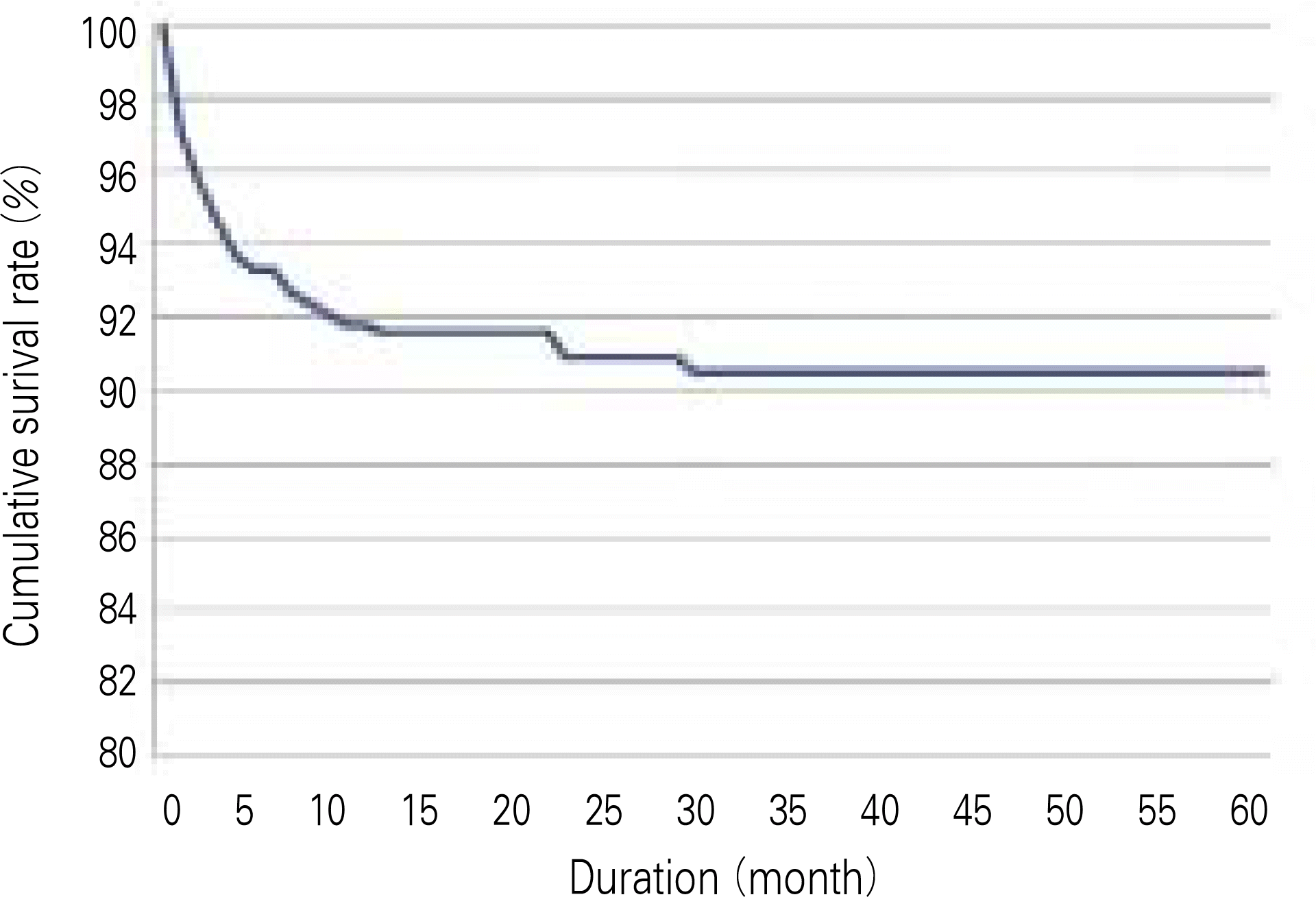
1. The 5-year cumulative survival rate of total implants was 90.5%, there was no significant difference between sex, age, the site of implant placement, diameter and length of the implant, sinus elevation technique, and the type of opposing teeth. 2. Patients with diabetes mellitus & osteoporosis and smooth-surfaced machined group & hydroxyapatite (HA)-treated group and homogenous demineralized freeze dried allogenic bone (DFDB) bone graft only group had significantly lower survival rate. 3. With less than 4 mm of residual alveolar ridge height, lateral approach without closing the osseous window resulted in a significantly lower survival rate. 4. Restoration of a single implant showed a significantly lower survival rate, compared to cases where the superstructure was joined with several implants in the area.
Conclusion
Patients with diabetes or osteoporosis need longer period of time for osseointegration compared to the normal, and the dentists must be prudent when choosing a surface treatment type and the bone graft material. Also, as the vertical dimension of the residual alveolar ridge can influence the result, staged implant placement should be considered when it seems difficult for the implant to gain primary stability from the residual bone with less than 4 mm of vertical dimension. It is recommended to obdurate the bone window and that the superstructure be connected with several impants in the peripheral area.
References
1. Noack N, Willer J, Hoffmann J. Long-term results after placement of dental implants: longitudinal study of 1,964 implants over 16 years. Int J Oral Maxillofac Implants. 1999; 14:748–55.
2. Adell R, Lekholm U, Rockler B, Bra � nemark PI. A 15-year study of osseointegrated implants in the treatment of edentulous jaw. Int J Oral Surg. 1981; 10:387–416.
3. Albrektsson T, Dahl E, Enbom L, Engevall S, Engquist B, Eriksson AR, et al. Osseointegrated oral implants. A Swedish multicenter study of 8,139 consecutively inserted Nobelpharma implants. J Periodontol. 1988; 59:287–96.
4. Adell R, Eriksson B, Lekholm U, Bra � nemark PI, Jemt T. Longterm follow-up study of osseointegrated implants in the treatment of the totally edentulous jaw. Int J Oral Maxillofac Impalnts. 1990; 5:347–59.
5. Chanavaz M. Maxillary sinus: anatomy, physiology, surgery and bone grafting related to implantology-eleven years surgical experience (1979–1990). J Oral Implantol. 1990; 16:199–209.
6. Mish CE. Bone character: second vital implant criterion. Dent Today. 1998; 7:39–40.
7. Friberg B, Sennerby L, Roos J, Lekholm U. Identification of bone quality in conjunction with insertion of titanium implants. A pilot study in jaw autopsy specimens. Clin Oral Impants Res. 1995; 6:213–9.

8. Boyne PJ, James RA. Grafting of the maxillary sinus floor with autogenous marrow and bone. J Oral Surg. 1980; 38:613–6.
9. Tatum H Jr. Maxillary and sinus implant reconstructions. Dent Clinics of North America. 1986; 30:207–29.
10. Summers RB. The osteotome technique: part 3-less invasive methods of elevating the sinus floor. Compendium. 1994; 15:698. 700, 702–4 passim; quiz 710.
11. Kahnberg KE, Ekestubbe A, Gro ¨ndahl K, Nilsson P, Hirsch JM. Sinus lifting procedure. I. One-stage surgery with bone transplant and implants. Clin Oral Implants Res. 2001; 12:479–87.
12. del Fabbro M, Testori T, Francetti L, Weinstein R. Systematic review of survival rates for implants placed in the grafted maxillary sinus. Int J Periodontics Restorative Dent. 2004; 24:565–77.

13. Wennerberg A, Albrektsson T, Andersson B, Krol J. A histomorphometric and removal torque study of screw-shaped titaninum implants with three different surface topographies. Clin Oral Implants Res. 1995; 6:24–30.
14. Buser D, Nydegger T, Oxland T, Cochran DL, Schenk RK, Hirt HP, Snetivy D, Nolte LP. Interface shear strength of titanium implants with a sandblasted and acid-etched surface: a biomechanical study in the maxilla of miniature pigs. J Biomed Mater Res. 1999; 45:75–83.

15. Cochran DL. A comparison of endosseous dental implant surfaces. J Periodontol. 1999; 70:1523–39.

16. Jensen OT, Shulman LB, Block MS, Iacono VJ. Report of the sinus consensus conference of 1996. Int J Oral Maxillofac Impalnts. 1998; 13(S):11–45.
17. Tidwell JK, Blijdorp PA, Stoelinga PJ, Brouns JB, Hinderks F. Composite grafting of the maxillary sinus for placement of endosteal implants. A preliminary report of 48 patients. Int J Oral Maxillofac Surg. 1992; 21:204–9.

18. Yildirim M, Spiekermann H, Biesterfeld S, Edelhoff D. Maxillary sinus augmentation using xenogenic bone substitute material Bio-Oss in combination with venous blood. A histologic and histomorphometric study in humans. Clin Oral Implants Res. 2000; 11:217–9.
19. Del Fabbro M, Testori T, Francetti L, Weinstein R. Systematic review of survival rates for implants placed in the grafted maxillary sinus. Int J Periodontics Restorative Dent. 2004; 24:565–77.

20. Kent JN, Block MS. Maxillary sinus floor bone grafting and placement of hydroxylapatite-coated implants. J Oral Maxillofac Surg. 1989; 47:238–42.
21. Block MS, Kent JN. Sinus augmentation for dental implants: the use of autogenous bone. J Oral Maxillofac Surg. 1997; 55:1281–6.

22. van Steenberge D, Quirynen M, Naert I. Survival and success rates with oral endosseous implants. In: Lang NP, Karring T, Lindhe J, eds. Proceedings of the 3rd European Workshop on Periodontology: implant dentistry; 1999 Jany 30-Feb 3; Charter House at Ittingen Thurgau, Switzerland: Berlin Quintessence Publishing Co;. 1999. 246–54.
23. Albrektsson T, Zarb G, Worthington P, Eriksson AR. The longterm efficacy of currently used dental implants: a review and proposed criteria of success. Int J Oral Maxillofac Implants. 1986; 1:11–25.
24. Misch CE. Contemporary Implant Dentistry. 2nd ed.St. Louis: Mosby;1998. p. 91–123.
25. Rosenberg ES, Cho SC, Elian N, Jalbout ZN, Froum S, Evian CI. A comparison of characteristics of implant failure and survival in periodontally compromised and periodontally healthy patients: a clinical report. Int J Oral Maxillofac Implants. 2004; 19:873–9.
26. Bra � nemark PI, Hansson BO, Adell R, Breine U, Lindstro ¨m J, Halle ′ n O, et al. Osseointegrated implants in the treatment of the edentulous jaw. Experience from a 10-year period. Scand J Plast Reconstr Surg Suppl. 1977; 16:1–132.
27. Zarb GA, Schmitt . The longitudinal clinical effectiveness of osseointegrated dental implants: the Toronto study. Part I: surgical results. J Prosthet Dent. 1990; 63:451–7.

28. Albrektsson T, Bra � nemark PI, Hansson HA, Lindstro ¨m J. Osseointegrated titanium implants. Requirements for ensuring a long lasting, direct bone-to-implant anchorage in man. Acta Orthop Scand. 1981; 52:155–70.
29. Wallace SS, Froum SJ. Efect of maxillary sinus augmentation on the survival of endosseous dental implants. A systematic review. Ann Periodontol. 2003; 8:328–43.
30. Goodacre CJ, Kan JY, Rungcharassaeng K. Clinical complications of osseointegrated implants. J Prosthet Dent. 1999; 81:537–52.

31. Higuchi KW, Folmer T, Kultje C. Implant survival rates in partially edentulous patients: a 3-year prospective multicenter study. J Oral Maxillofac Implants. 1995; 53:264–8.
32. Shin SH, Kim JR, Park BS. Bone formation around titanium impalnts in the tibiae of streptozotocin induced diabetic rats. J Korean Assoc Maxillofac Plast Reconstr Surg. 2000; 22:522–41.
33. Moy PK, Medina D, Shetty V, Aghaloo TL. Dental implant failure rates and associated risk factors. Int J Oral Maxillofac Implants. 2005; 20:569–77.
34. Hu ¨rzeler MB, Kirsch A, Ackermann KL, Quinones CR. Reconstruction of the severely resorbed maxilla with dental implants in the augmented maxillary sinus: a 5-year clinical investigation. Int J Oral Maxillofac Implants. 1996; 11:466–75.
35. Block MS, Kent JN. Maxillary sinus bone grafting. Block MS, Kent JN, editors. Endosseous implants for maxillofacial reconstruction. Philadelphia, PA: WB Saunders Co.;1995. p. 478–503.

36. Wood RM, Moore DL. Grafting of the maxillary sinus with intraorally harvested autogenous bone prior to implant placement. Int J Oral Maxillofac Implants. 1988; 3:209–14.
38. Valentini P, Abensur D. Maxillary sinus floor elevation for implant placement with demineralized freeze-dried bone and bovine bone (Bio-Oss): a clinical study of 20 patients. Int J Periodontics Restorative Dent. 1997; 17:232–41.
39. van den Bergh JP, ten Bruggenkate CM, Krekeler G, Tuinzing DB. Maxillary sinusfloor elevation and grafting with human demineralized freeze dried bone. Clin Oral Implants Res. 2000; 11:487–93.

40. Hallman M, Cederlund A, Lindskog S, Lundgren S, Sennerby L. A clinical histologic study of bovine hydroxyapatite in combination with autogenous bone and fibrin glue for maxillary sinus floor augmentation. Results after 6 to 8 months of healing. Clin Oral Implants Res. 2001; 12:135–43.
41. Hallman M, Sennerby L, Lundgren S. A clinical and histologic evaluation of implant integration in the posterior maxilla after sinus floor augmentation with autogerous bone, bovine hydroxyapatite, or a 20: 80 mixture. Int J Oral Maxillofac Implants. 2002; 17:635–43.
42. Loukota RA, Isaksson SG, Linne′r EL, Blomqvist JE. A technique for inserting endosseous implants in the atrophic maxilla in a single stage procedure. Br J Oral Maxillofac Surg. 1992; 30:46–9.

43. Ioannidou E, Dean JW. Osteotome sinus floor elevation and simultaneous, non-submerged implant placement: Case report and literature review. J Periodontol. 2000; 71:1613–9.

44. Toffler M. Minimally invasive sinus floor elevation procedures for simultaneous and staged implant placement. N Y State Dent J. 2004; 70:38–44.
45. Zitzmann NU, Scha ¨rer P. Sinus elevation procedures in the resorbed posterior maxilla. Comparison of the crestal and lateral approaches. Oral Surg Oral Med Oral Pathol Oral Radiol Endod. 1998; 85:8–17.
46. Tarnow DP, Wallace SS, Froum SJ, Rohrer MD, Cho SC. Histologic and clinical comparision of bilateral sinus floor elevations with and without barrier membrane placement in 12 patients: part 3 of an ongoing prospective study. Int J Periodontics Restorative Dent. 2000; 20:117–25.
47. Froum SJ, Tarnow DP, Wallace SS, Rohrer MD, Cho SC. Sinus floor elevation using anorganic bovine bone matrix (OsteoGraf/N) with and without autogenous bone: a clinical, histologic, radiographic, and histomorphometric analysis–Part 2 of an ongoing prospective study. Int J Periodontics Restorative Dent. 1998; 18:528–43.
48. Bidez MW, Misch CE. Force transfer in implant dentistry: basic concepts and prinsples. J Oral Implantol. 1992; 18:264–74.
49. Enkling N, Nicolay C, Utz KH, Jo ¨hren P, Wahl G, Mericske-Stern R. Tactile sensibility of single-tooth implants and natural teeth. Clin Oral Implants Res. 2007; 18:231–6.

50. Chung DM, Oh TJ, Lee J, Misch CE, Wang HL. Factors affecting late implant bone loss: a retrospective analysis. Int J Oral Maxillofac Implants. 2007; 22:117–26.
Table 1.
Patient distribution according to gender
| Gender | Number of patient | Number of implant | % |
|---|---|---|---|
| Male | 68 | 172 | 60.4 |
| Female | 53 | 113 | 39.6 |
| Total | 121 | 285 | 100 |
Table 2.
Patient distribution according to age
| Age | Number of implant | % |
|---|---|---|
| 21–30 | 5 | 2 |
| 31–40 | 31 | 11 |
| 41–50 | 85 | 30 |
| 51–60 | 96 | 34 |
| 61–70 | 68 | 23 |
| Total | 285 | 100 |
Table 3.
Cumulative survival rate of implants placed in sinus elevated maxilla
Table 4.
Survival rate of implant according to gender
| Gender | State | Survival rate (%) | P value | |
|---|---|---|---|---|
| Placed | Failed | |||
| Male | 154 | 18 | 89.5 | 0.663 |
| Female | 104 | 9 | 92.0 | |
Table 5.
Survival rate of implant according to age
| Age | State | Survival rate (%) | P value | |
|---|---|---|---|---|
| Placed | Failed | |||
| 21–30 | 5 | 0 | 100 | |
| 31–40 | 26 | 2 | 92.9 | |
| 41–50 | 78 | 7 | 91.8 | 0.706 |
| 51–60 | 88 | 11 | 88.9 | |
| 61–70 | 61 | 7 | 89.7 | |
Table 6.
Survival rate of implant according to general disease
| General disease | State S | Survival rate (%) | P value | |
|---|---|---|---|---|
| Placed | Failed | |||
| Normal | 123 | 9 | 93.2 | |
| Diabetes mellitus | 47 | 8 | 85.5 | 0.002 |
| Osteoporosis | 30 | 6 | 83.3 | |
| Others | 58 | 4 | 93.5 | |
Table 7.
Survival rate of implant according to implant location
| Site | State | Survival rate (%) | P value | |
|---|---|---|---|---|
| Placed | Failed | |||
| 1 premolar | 18 | 1 | 94.7 | |
| 2 premolar | 52 | 4 | 92.9 | 0.687 |
| 1 molar | 102 | 11 | 90.3 | |
| 2 molar | 86 | 11 | 88.7 | |
Table 8.
Survival rate of implant according to fixture diameter
| Fixture diameter (mm) | State S | Survival rate (%) | P value | |
|---|---|---|---|---|
| Palced | Failed | |||
| 3.0–3.5 | 49 | 5 | 90.7 | 0.139 |
| 3.75–4.5 | 159 | 14 | 91.9 | |
| 4.75–5.0 | 42 | 5 | 89.4 | |
| 5.75- | 8 | 3 | 72.7 | |
Table 9.
Survival rate of implant according to fixture length
| Fixture length (mm) | State | Survival rate (%) | P value | |
|---|---|---|---|---|
| Placed | Failed | |||
| 8–10 | 78 | 8 | 90.7 | |
| 11–13 | 128 | 14 | 90.1 | 0.562 |
| >14 | 52 | 5 | 91.2 | |
Table 10.
Survival rate of implant according to implant surface texture
| Surface texture | State | Survival rate % | P value | |
|---|---|---|---|---|
| Placed | Failed | |||
| Smooth Machined | 49 | 12 | 80.3 | |
| HA | 52 | 10 | 83.9 | |
| Acid etching | 46 | 2 | 95.8 | 0.002 |
| Rough TPS | 37 | 1 | 97.4 | |
| RBM | 33 | 1 | 97.1 | |
| SLA | 41 | 1 | 97.6 | |
Table 11.
Survival rate of implant according to graft material
Table 12.
Survival rate of implant according to residual bone height
| Residual bone height | State | Survival rate (%) | P value | |
|---|---|---|---|---|
| Placed | Failed | |||
| <4 mm | 30 | 13 | 69.8 | |
| 4–5 mm | 61 | 7 | 89.7 | |
| 5–6 mm | 51 | 5 | 91.0 | 0.000 |
| 6–7 mm | 50 | 1 | 98.0 | |
| >7 mm | 66 | 1 | 98.5 | |
Table 13.
Survival rate of implant according to surgery
| Surgery method | State | Survival rate (%) | P value | ||
|---|---|---|---|---|---|
| Placed | Failed | ||||
| Lateral | Simultaneous | 84 | 10 | 89.4 | |
| approach | Staged | 93 | 8 | 92.1 | 0.957 |
| Crestal approach | 81 | 9 | 90.0 | ||
Table 14.
Survival rate of implant according to window management
| Window management | State | Survival rate (%) | P value | |
|---|---|---|---|---|
| Placed | Failed | |||
| No closure | 87 | 15 | 85.3 | |
| Cortex repositioning | 31 | 1 | 96.9 | 0.022 |
| Resorbable membrane | 59 | 2 | 96.7 | |


 PDF
PDF ePub
ePub Citation
 Print
Print


 XML Download