

 PDF
PDF ePub
ePub Citation
 Print
Print


Abstract
There has been a recent increase in attention focused on the potential risk of radiation-induced carcinogenesis from diagnostic radiology, with a particular emphasis on computed tomography (CT). After the rapid adoption of multidetector CT (MDCT), radiation doses from CT are now the single largest source of diagnostic radiation exposure to patients, and the carcinogenesis risk from diagnostic CT radiation dose exposure can no longer be ignored by physicians. To understand the exposure risk and monitor radiation dose exposure, an understanding and interest in CT dose reports is necessary. Almost all MDCTs now show and allow storage of the volume CT dose index (CTDIvol), dose length product (DLP), and effective dose estimations on dose reports, which are essential to assess patient radiation exposure and risks. To decrease these radiation exposure risks, the principles of justification and optimization should be followed. Justification means that the examination must be medically indicated and useful. Optimization means that the imaging should be performed using doses that are as low as reasonably achievable (ALARA), consistent with the diagnostic task. Optimization includes understanding and changing CT protocols to perform the same diagnostic task with the minimal amount of radiation exposure while maintaining diagnostic accuracy. Physicians and radiologists must be aware of the radiation risks associated with CT exams, and understand and implement the principles for patient radiation dose reduction.
Figures and Tables
Figure 1
Radiation dose difference between general X-ray (A) and computed tomography (CT) (B). General X-ray radiography is projectional passing from one side of the body to the other in a single direction, which results in the highest radiation dose at the entry site of the beam and the least at the exit site of the beam (A). On the other hand, due to the 360 degree rotational nature of CT, radiation enters the body uniformly causing the highest radiation dose near the skin and the least radiation dose at the center of the body (B).
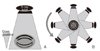
Figure 2
(A) The typical bell curve appearance of computed tomography (CT) radiation dose along the Z axis in a single CT scan slice is depicted. (B) The combined CT radiation dose and resultant multiple scan average dose (MSAD) along the Z axis for multiple CT scan slices is depicted.
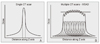
Figure 3
This dose report was generated on a LightSpeed VCT scanner (GE Healthcare, Milwaukee, WI, USA) during a 4 phase dynamic liver computed tomography (CT) in a 49-year-old man. Note the volume CT dose index (CTDIvol) and dose length product (DLP). Dose reports from this GE scanner include scanning type, scan range, CTDIvol, and DLP. Series 200 shows the radiation dose occurring during contrast bolus tracking. In this exam, a scout image was taken, followed by nonenhanced imaging, bolus tracking, arterial, portal, and delayed phase imaging with a total 4 phases. In this case, the portal phase scan range included the pelvis, which explains the relatively larger scan range and higher DLP. The total DLP for this patient is estimated as 1,033.89 mGy×cm.
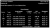
Figure 4
This dose report was generated on a SOMATOM Sensation 64 (Siemens Healthcare, Forchheim, Germany) computed tomography (CT) scanner during a 4 phase dynamic liver CT. Note the volume CT dose index (CTDIvol) and dose length product (DLP). Dose reports from this scanner include kV, mAs/reference mAs, CTDIvol, DLP, and tube rotation time. This dose report shows separate premonitoring and monitoring radiation doses in regard to bolus tracking. In this exam, a scout image was taken, followed by nonenhanced imaging, bolus tracking, arterial, portal, and delayed phase imaging with a total 4 phases. For nonenhanced liver CT, tube current modulation using CareDose 4D was used at 100 kVP with a reference mAs of 180. 120 kVp was used on arterial to delayed phase enhanced images. In this case, the portal phase scan range included the pelvis, which explains the relatively larger scan range and higher DLP. Portal phase CTDIvol was 14.15 mGy, DLP was 787 mGy×cm, and total DLP was 2,096 mGy×cm. TI, time per rotation; cSL, collimated slice.

References
1. United Nations Scientific Committee on the Effects of Atomic Radiation. Sources and effects of ionizing radiation. 2010. New York: United Nations.
2. Mettler FA Jr, Wiest PW, Locken JA, Kelsey CA. CT scanning: patterns of use and dose. J Radiol Prot. 2000. 20:353–359.

3. Health Insurance Review & Assessment Service. 2005 National health insurance statistical yearbook. 2006. Seoul: Health Insurance Review & Assessment Service.
4. You JJ, Levinson W, Laupacis A. Attitudes of family physicians, specialists and radiologists about the use of computed tomography and magnetic resonance imaging in Ontario. Healthc Policy. 2009. 5:54–65.
5. Brenner DJ, Hall EJ. Computed tomography: an increasing source of radiation exposure. N Engl J Med. 2007. 357:2277–2284.
6. Safety investigation of CT brain perfusion scans: update 11/9/2010 [Internet]. 2010. cited 2011 Nov 18. Silver Spring (MD): U.S. Food and Drug Administration;Available from: http://www.fda.gov/MedicalDevices/Safety/AlertsandNotices/ucm185898.htm.
7. Pierce DA, Preston DL. Radiation-related cancer risks at low doses among atomic bomb survivors. Radiat Res. 2000. 154:178–186.
8. Brenner D, Elliston C, Hall E, Berdon W. Estimated risks of radiation-induced fatal cancer from pediatric CT. AJR Am J Roentgenol. 2001. 176:289–296.
9. National Research Council (US). Committee to Assess Health Risks from Exposure to Low Levels of Ionizing Radiation. Health risks from exposure to low levels of ionizing radiation: BEIR VII phase 2. 2006. Washington DC: National Academies Press.
10. McNitt-Gray MF. AAPM/RSNA physics tutorial for residents: topics in CT: radiation dose in CT. Radiographics. 2002. 22:1541–1553.
11. The measurement, reporting, and management of radiation dose in CT: AAPM report no. 96 [Internet]. 2008. cited 2011 Nov 18. College Park (MD): American Association of Physicists Medicine;Available from: http://www.aapm.org/pubs/reports/rpt_96.pdf.
12. Jung SE. Korean Institute for Accreditation of Medical Image. National survey of radiation dose of computed tomography in Korea. 2008. Seoul: Korea Food and Drug Administration.
13. Mettler FA Jr, Huda W, Yoshizumi TT, Mahesh M. Effective doses in radiology and diagnostic nuclear medicine: a catalog. Radiology. 2008. 248:254–263.
14. Shrimptom PC, Jones DG, Hillier MC, Wall BF, Le Heron JC, Faulkner K. National Radiological Protection Board. Survey of CT practice in the UK. 1991. Chilton: National Radiological Protection Board.
15. Kim DS. Guideline for diagnostic reference level of the radiation exposure of CT examination. 2009. Seoul: National Institute of Food and Drug Safety Evaluation.
16. Borgen L, Stranden E, Espeland A. Clinicians' justification of imaging: do radiation issues play a role? Insights Imaging. 2010. 1:193–200.
17. Dougeni E, Faulkner K, Panayiotakis G. A review of patient dose and optimisation methods in adult and paediatric CT scanning. Eur J Radiol. 2011. 06. 16. [Epub]. DOI: 10.1016/j.ejrad.2011.05.025.
18. Payne JT. CT radiation dose and image quality. Radiol Clin North Am. 2005. 43:953–962. vii
19. Curtis JR. Computed tomography shielding methods: a literature review. Radiol Technol. 2010. 81:428–436.
20. Kalra MK, Maher MM, Toth TL, Hamberg LM, Blake MA, Shepard JA, Saini S. Strategies for CT radiation dose optimization. Radiology. 2004. 230:619–628.
21. McCollough CH, Bruesewitz MR, Kofler JM Jr. CT dose reduction and dose management tools: overview of available options. Radiographics. 2006. 26:503–512.
22. Lee CH, Goo JM, Ye HJ, Ye SJ, Park CM, Chun EJ, Im JG. Radiation dose modulation techniques in the multidetector CT era: from basics to practice. Radiographics. 2008. 28:1451–1459.
 XML Download