

 PDF
PDF ePub
ePub Citation
 Print
Print


Introduction
Aging is a set of changes that happen over time in the body. It is primarily affected by sexual and reproductive hormones and is the most important risk factor for several diseases [12]. As the age increases, the ability of women for childbearing decreases. The importance of this issue is determined by the fact that the age of childbearing has been postponed to the fourth decade of life in the modern societies [34]. As a result of declined fertility, such women will have more need to assisted reproductive techniques (ART) to have children [5]. ART, in turn, has high cost and more complications in advanced maternal age which is undesired [67].
Ovarian aging is characterized by declined ovarian reserve [89], low oocyte quality [10], diminished anti-Müllerian hormone [1112], and finally menopause [3]. Ovaries are more susceptible to the complications of natural aging than other tissues for some of the known and unclear reasons [13]. Oxidative stress is one of the disturbing mechanisms involving in aged ovary. During ovarian aging, decreased antioxidant gene expression and increased reactive oxygen species (ROS) resulted in more oxidative damage [1415]. Carbonyl stress due to dysregulation of energetic metabolism in the aging follicles is another distressing mechanism [1617]. It is reported that mitochondrial dysfunction also has a role in ovarian damages in aging [6]. Oxidative stress, carbonyl stress, and mitochondrial dysfunction are related together and affect each other [616].
Application of free radicals scavengers can protect ovary from the damage of oxidative stress [181920]. Vitamin C (L-ascorbic acid) is a natural antioxidant scavenging ROS effectively [2122]. In addition, useful effects of vitamin C on metabolism, collagen synthesis, vasculogenesis, aging, cell proliferation, and differentiation has been reported previously [21222324]. Although, so far, several studies have investigated the role of vitamin C in combination with other antioxidants on female infertility [51925], there are scarcely data regarding to the effect of vitamin C alone on ovarian aging. In the present study, we aimed to evaluate the effects of vitamin C on NMRI mice ovarian aging according to the stereological study.
Materials and Methods
Animals and treatments
In this experimental study, 36 adult female NMRI mice weight of 25–30 g were obtained from Iran Pasteur Institute. The animals were kept in animal house under standard conditions (22±2℃ and 12-hour light/dark) and provided with food and water ad libitum. The mice were then divided into two groups: control and experimental groups. Vitamin C (L-ascorbic acid; Sigma, St. Louis, MO, USA) was prepared by diluting in warm water. The experimental groups were given vitamin C (150 mg/kg) with a 24-hour interval by oral gavage (3.75 mg per animal) for 33 weeks. Control animals were treated with water. On weeks 8, 12, and 33, six animals of each group and right ovary samples were extracted for stereology analysis.
Tissue preparation
The ovaries were placed in 10% formalin fixative for 48 hours. After tissue processing, the samples were placed in paraffin blocks. Following sectioning, hematoxylin and eosin staining was performed.
Stereological study
Volume of ovary, cortex, medulla, and corpus luteum
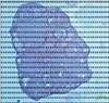
The total volume of the ovary, cortex, medulla and corpus luteum was estimated using the Cavalieri methods applying the following formula [1726]:
In this formula, Σp is the total number of points superimposed on the image, (t) is the thickness of the section and a/p is the area associated with each point (Fig. 1).
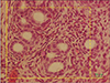
Total number of primordial, primary, secondary, antral follicles, and granulosa cells
The total number of primordial, primary, secondary, and antral follicles were estimated using the optical dissector method (Fig. 2) [1726]. The numerical density (Nv) of primordial, primary, secondary, antral follicles and granulosa cells were calculated with the following formula:
In this formula, “ΣQ” is the number of the nuclei, “ΣP” is the total number of the unbiased counting frame in all fields, “h” is the height of the dissector, “a/f” is the frame area, “t” is the real section thickness measured in every field using the microcator, and “BA” is the block advance of the microtome which was set at 10 µm. The total number of primordial, primary, secondary, antral follicles and granulosa cells was estimated by multiplying the numerical density (Nv) by the total V.
Results
Volume of ovary, cortex, medulla and corpus luteum
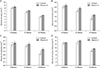
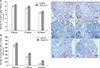
Total volume of ovary at 8, 12, and 33 weeks, total volume of cortex at 12 and 33 weeks and total volume of medulla and corpus luteum at 33 weeks were higher significantly in vitamin C group compared to control group (P≤0.05) (Fig. 4).
Total number of follicles
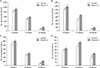
We found a significantly increased the total number of primordial, primary, and antral follicles at 12 and 33 weeks, and also secondary follicle at 33 week in vitamin C group when compared to control group (P≤0.05) (Fig. 6).
Discussion
The scope of the current study was to evaluate the possible beneficial or adverse effects of vitamin C on NMRI mice ovarian aging according to the stereological study. We found that vitamin C could significantly prevent the reduction of ovarian volume, number of ovarian follicles and granulosa cells during a mouse model of ovarian aging, although we did not observe any significant difference in total mean oocyte volume between groups. According to our knowledge, this is the first study evaluating the impact of vitamin C alone on ovarian aging based on stereological parameters.
Following ovarian aging, extensive changes occurs at the level of molecules and genes. Some of these changes are down-regulation of germ line specific genes, oocyte specific genes, mitochondrial electron transport genes and intraovarian signaling pathways as well as up-regulation of genes related to complement activation and membrane receptors. Most of these alterations are specific to ovary and don't happen in somatic organs [2728]. Free radical imbalance is an important part of changes during ovarian aging [29]. Lim and Luderer [15] reported decreased expression of mitochondrial (Prdx3 and Txn2) as well as cytosolic (sGlrx1 and Gstm2) antioxidants genes in ovary with increased age. The main source of free radicals is the oxidative phosphorylation and ATP generation during aerobic metabolism in the mitochondria. Mitochondrial dysfunction is one of causes of increased ROS in aged ovary [1630]. Considering antioxidant properties of estrogen, its deficiency following menopause is one of other causes of oxidative stress in aging [31].
Antioxidant system in ovary is consist of non-enzymatic antioxidants (vitamins A, C, and E) [32] and enzymatic antioxidants (for example antioxidant tripeptide glutathione, glutathione peroxidase (GPX), superoxide dismutase (SOD), and catalase [3334353637]. Based on published studies, ROS scavenging efficiency in ovary decreases during aging including reduced expression and enzymatic activity of SOD in cumulus oophorous cells [38], decreased enzymatic activity of SOD and GPX in postmenopausal women [37] and lower expression of SOD and catalase in cultured granulosa cells collected from old women subjected to in vitro fertilization [36].
In broad terms it seems that decreased antioxidative efficiency on the one hand and increased ROS production on the other hand during aging caused damages in the ovary [29]. Importantly, dysregulation of glucose and energetic metabolism in the aging follicles can produce reactive carbonyl species (RCS) and carbonyl stress. RCS, similar to ROS, contribute to DNA, protein and lipids damages causing deleterious effects in the cells. Carbonyl stress, in turn, strengthens oxidative stress and vice versa. These factors along with mitochondrial dysfunction can cause more age related damages in the ovary [616]. It is reported that AKT and mammalian target of rapamycin (mTOR) signaling pathways are associated with ovarian diseases including ovarian aging [7]. Interestingly, AKT/mTOR signaling is related to oxidative stress and interact on each other [39]. The abnormal perifollicular vascularity also causes abnormal microenvironment in the aged ovary and in turn, may produce oxidative stress [40]. In addition, with increasing the age, the rate of inflammation in the mouse ovaries increases resulting from the function of multinucleated macrophage giant cells and increased expression of inflammatory genes [26].
According to prior published studies, some characteristic of aged ovary are lower follicular quality and quantity [41], increased level of apoptosis and accumulation of lipofuscin pigments in insterstitium [15], fibrosis in the stroma [26], increased DNA fragmentation [42], and chromosomal disturbance [43]. Via stereological analysis, we observed that the total volume of ovary, cortex, medulla and corpus luteum decreased as the age increased (at 8, 12, and 33 weeks) (Fig. 4). Moreover, the total number of follicles, the total mean oocyte volume and number of granulosa cells in antral follicles has declined progressively over time (Figs. 5, 6).
Vitamin C is a natural important water-soluble micronutrient and coenzyme which its deficiency is related to aging of different cells and tissues [44]. It can attenuate vascular dysfunction in several diseases in both in vivo and in vitro studies [22]. Adding vitamin C to culture medium can improve mesenchymal stem cells (MSCs) proliferation and metabolism via mitochondrial activation [23]. It is reported that vitamin C can impact on glucose metabolism through alteration of glucose metabolites [24]. Additionally, anti-inflammatory properties of vitamin C on animal models of ischemia and sepsis has been reported previously [22].
Along with the effects of vitamin C on vascularization, metabolism, and inflammation mentioned above, it has also antioxidant effects. In this regard, vitamin C can postpone aging in MSCs via prevention of the ROS production and AKT/mTOR signaling [21]. Arab et al. [20] found that antioxidant effect of ascorbic acid could ameliorate increased oxidative stress induced by malathion in the rats ovary. In the other study, it has been shown that vitamin c amended Bisphenol A oxidative toxicity in rat ovarian tissue. In that study, total volume of ovaries and oocytes, and also the mean number of antral follicles increased following vitamin C administration [18].
Supplementation of culture medium with vitamin C stimulated the activation and growth of cattle primordial follicles and increased viability of early-stage follicles [45]. Tarin et al. [43] reported that early and late onset administration of vitamins C and E caused improving oocyte quality and quantity in aged mice. In the other study, it has been reported that vitamin C supplementation could improve development and viability of preantral follicles after six days of in vitro culture [40]. In accordance with that studies, we evaluated effects of vitamin C administration on ovarian aging and found that vitamin C enhances the survival of the total number of follicles in different stages. It also could prevent the reduction of the volume of ovary and corpus luteum as well as the number of granulosa cells in antral follicles during aging. It seems that beneficial effects of vitamin C on ovarian aging observed in the present study might be due to its antioxidant effects as well as its impact on vascularization, metabolism, inflammation, and AKT/mTOR signaling pathway as mentioned before.
However, there are contradictions about the benefits of vitamin C on reproduction. The effects of vitamin C in combination with other supplements has been evaluated on cases with female factor infertility such as polycystic ovarian syndrome and unexplained infertility but there was insufficient evidence to support supplemental oral antioxidants prescribing in those women [25]. Similar results have been obtained in pregnant women. Administration of vitamin C alone or in combination with other supplements had no effects on pregnancy results although there were controversial results regarding premature rupture of membranes and placental abruption [46]. In a study, Camarena and Wang [44] reported that adding ascorbic acid to culture medium of hen granulosa cells did not induce antioxidant effects and interestingly the activity of SOD in granulosa decreased in higher doses of ascorbic acid. They concluded that vitamin C might have regulatory role in biochemical and physiological processes in granulosa cells [44].
Collectively, according to our study, vitamin C compensated undesirable effects of aging on ovarian tissue. Our study supports role of vitamin C supplementation for reduce and prevention of ovarian aging complications especially in women delaying childbearing for the several reasons.
 XML Download