

 PDF
PDF ePub
ePub Citation
 Print
Print


INTRODUCTION
The level of periodontal support is the most critical factor for predicting the prognosis of healing after periodontal treatment [1,2]. In general, a tooth with periodontal destruction extending beyond the root apex is considered hopeless, and extraction is the only possible treatment. Neither nonsurgical nor surgical treatment is effective in such cases because of limited accessibility to instrumentation and unfavorable tooth stability after treatment. In the process of periodontal treatment, thorough instrumentation and antibacterial cleansing are performed to remove microbial infection sources. Nevertheless, there are limitations to accessing the periapical area and several studies have reported that microbial etiologic factors were found around the root apex area in recurrent infectious diseases [3,4]. Moreover, the increased mobility of a treated tooth can affect the outcome of periodontal treatment negatively. Tooth mobility may inhibit bone gain and periodontal regeneration, consequently forming a deep pocket and inducing apical migration of epithelial attachment [5].
Intentional replantation may be useful in these situations because this technique seems to resolve the limitations of conventional periodontal treatment. The tooth surfaces, including inaccessible areas, can be visualized and instrumented completely without damaging adjacent periodontal tissue. Some investigators have attempted intentional replantation with periodontally involved teeth. Lu [6] reported successful treatment results with periodontally involved teeth in which intentional replantation was performed first. The researcher intentionally replanted an endodontically mistreated and periodontally involved mandibular first molar and maintained the tooth for 32 months in a functional and asymptomatic condition. Demiralp et al. [7] intentionally replanted fifteen periodontally involved hopeless teeth and followed them for 6 months. They suggested that intentional replantation could be an alternative approach to extraction in cases where advanced periodontal destruction was present and no other treatment could be considered.
However, in spite of the potential of this technique, very few investigators have performed intentional replantation with periodontally involved teeth. This might be due to the questionable prognosis of replanted teeth. Although some studies have shown favorable results, intentional replantation of a periodontally involved tooth seems to carry the risk of reinfection and unstable tooth stability, which results in tooth loss.
Previously, Lee et al. extracted periodontally hopeless teeth and replanted them after a delay to relieve inflammation and provide a scaffold with woven bone formation (unpublished data). They reported successful clinical and radiographic results by delaying the replant procedure, which they named "delayed intentional replantation" and concluded that the procedure could be an alternative treatment option for periodontally involved hopeless teeth. However, in the study, the number of cases involved was limited and statistical analysis was not conducted. Therefore, additional research that evaluates the survival rate of the delayed intentional replantation procedure would be useful in determining the outcomes of the treatment.
The purpose of this study is to retrospectively evaluate the survival of periodontally hopeless teeth that were intentionally replanted after a delay and to compare the radiographic characteristics of the survival group with those of the failure group.
MATERIALS AND METHODS
Study design and criteria
The clinical and radiographic data from patients who underwent delayed intentional replantation in the Department of Conservative Dentistry at the Yonsei University College of Dentistry, Seoul, Korea between March 2000 and July 2010 were reviewed in this study. The patients who were included in the treatment of replantation fulfilled the following requirements:
(1) No systemic disease and no contraindication for periodontal surgery.
(2) The presence of at least one tooth meeting the indications for tooth extraction due to severe periodontal destruction: At least a 6-mm probing depth, minimum 60% radiographic periodontal bone loss, and grade III mobility according to Miller's classification [8].
(3) Patient preference to retain the tooth rather than undergo extraction.
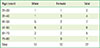
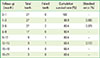
Consequently, 27 patients (17 males, 10 females) ranging in age from 24 to 72 years (mean age, 49.78 years) were included in this study. The patient distribution according to age and intraoral tooth location are as shown in Tables 1 and 2. This study was approved by the Institutional Review Board (IRB) of the Dental Hospital of Yonsei University of College of Dentistry (IRB number: 2-2012-0035).
Treatment procedure
Surgical technique
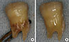
Under local anesthesia, each hopeless tooth was extracted as atraumatically as possible. If necessary, lingual and buccal flap incisions were made and small flap elevation was performed. Pathologic factors existing on the tooth surface were removed gently using an ultrasonic scaler, and then the tooth surface was planed with a fine diamond bur (Fig. 1). Granulation tissues in the extraction socket were thoroughly removed by curettage, and then saline irrigation was performed for disinfection. The extracted tooth was preserved in medium supplemented with antibiotics (1,000-nm dexamethasone solution) at 4℃ for 10 to 14 days. The average time of extraoral tooth storage was 11.3 days. After that, retrograde root canal treatment was carried out using an ultrasonic scaler and super EBA (Harry J. Bosworth Co., Skokie, IL, USA). The tooth was repositioned in the partially healed extraction socket under local anesthesia. The tooth was positioned approximately in line with the adjacent teeth and a resin wire splint was performed in case the stability of the replanted tooth was unstable. When the stability of the replanted tooth was ensured, the patient was instructed to bite down on gauze. Consequently, 18 teeth in total were splinted after delayed replantation. Occlusal adjustment was performed to eliminate occlusal interference in centric and eccentric movements.
Postoperative care
After the tooth extraction and tooth replantation procedures, the patients were prescribed oral antibiotic therapy with amoxicillin (500-mg thrice per day) for three days. All of the patients were recommended to maintain their routine tooth brushing and to use a 0.12% chlorhexidine solution for 2 weeks. Seven days after tooth replantation, the patients were examined and saline irrigation was performed to clean the replantation sites. One month after replantation, the replanted teeth were polished and periapical radiographs were taken. Then patients were recalled every three months for clinical and radiographic examination and maintenance. When there was evidence of the presence of severe inflammation, increasing tooth mobility, or radiographic alveolar bone loss, the replanted tooth was considered a failure and was extracted.
Assessment method
Survival criteria
The replanted teeth that were not extracted during the follow-up periods were deemed to have survived. When severe inflammatory signs or increasing mobility were found during the follow-up period, the replanted teeth were defined as failures and extracted.
Radiographic analysis
Periapical radiographs were taken at baseline, one month postoperatively, and at every recall visit. The tooth distribution according to follow-up period is as shown in Table 3.
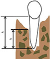
The Heliodent MD (Siemens, Fort Madison, IA, USA) was operated as an x-ray source under 60 kVp, 0.16 mAs conditions, and radiographic images were obtained using a charged coupled device sensor (SIGMA, GE Medical System Instrumentarium Co., Tuusula, Finland). Measuring bone loss was performed using the PiView STAR (Infinitt, Seoul, Korea) program and the method described by Schulte et al. [9]. To determine the amount of alveolar bone loss, two types of measurements were used: the total root length (ht) and the intra-alveolar root length (hi) (Fig. 2). The total root length (ht) was defined as the distance from the proximal cementoenamel junction to the apex parallel to the long axis of the tooth. The intra-alveolar root length (hi) was defined as the distance from the apex to the highest point on the alveolar margin. In the calculation of bone loss, 1.5 mm was subtracted from the total root length because the distance between the crest of the alveolar bone and the cementoenamel junction ranges between 0.75 and 1.49 mm [10]. The average measurement at the mesial and distal sites was determined, and bone loss (BL) and bone gain (BG) were calculated using the following equations:
RESULTS
Cumulative survival rate
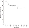
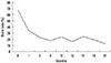
A total of 27 teeth replanted after a delay were followed for 3-21 months. Seven replanted teeth failed, and the overall cumulative survival rate was 66.4%. Five of these teeth were extracted within six months of replantation. The cumulative survival rates for the entire group of replanted teeth by period of time postreplantation are shown in Table 3 and Fig. 3.
Subcategorial descriptive observation: survival group
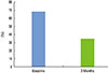
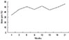
Twenty teeth were not extracted during the follow-up periods and were defined as the survival cases. In this group, all of the patients tolerated the intentional replantation procedure without any discomfort and were satisfied with the result of the replantation treatment. Clinically, all of the teeth were functioning without any signs or symptoms. Radiographs taken at every recall check-up showed remarkable bone gain around the replanted tooth (Fig. 4). The amount of BL was significantly reduced from 68.45% to 34.66% three months after replantation (P<0.05) (Fig. 5). After that, a consistent level of alveolar bone was maintained and the remodeling of new bone was proceeded (Figs. 4 and 6). The amount of BG was 45.02% three months after replantation and maintained a consistent level (Fig. 7). There was radiologic and clinical evidence of ankylosis with 5 teeth. However, root resorption was not found in the entire follow-up period.
Subcategorial descriptive observation: failure group
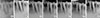
During the follow-up, 7 teeth were extracted and defined as failure. In the failure group, radiographic measurements (BL, BG) were not investigated because the bone formation adjacent to the root of a replanted tooth was rarely seen in radiographs. After replantation, bone formation occurred from the bottom of the socket. However, a remarkable radiolucent line existed along the root of each replanted tooth. The line prolonged and expanded as time passed by. Finally, each tooth that had failed was extracted due to inflammatory signs and increased mobility. As a result, 71% of the teeth that had failed were extracted within 6 months after replantation (Fig. 8).
DISCUSSION
This study describes the clinical and radiological outcomes of delayed intentional replantation of teeth deemed hopeless due to severe periodontal destruction. We assumed that waiting for improved conditions of the extraction socket by delaying the replantation procedure could produce more favorable outcomes from intentional replantation.
Although it is commonly known that intentional replantation is contraindicated in a periodontally involved tooth [11], several studies have reported successful clinical results and have suggested intentional replantation as an alternative treatment technique of last resort for a periodontally involved tooth [6,7]. However, little research has addressed this subject. This may be due to the possible risks mentioned below. Intentional replantation includes the following processes: atraumatic tooth extraction, removal of local factors on both the tooth surface and extraction socket, and reinsertion of the tooth. Through this process, local factors on the tooth surface and extraction socket causing periodontal disease can be eliminated completely. However, forming substantial space between the tooth and socket wall is inevitable as a result of this process. This space can negatively affect the result of the treatment. First, it is likely to increase the mobility of the replanted tooth. Generally, in case of tooth replantation, if the teeth are fixed in the coronal area, the teeth might be mobile in the apical area. Ferencz [5] reported that reduced periodontal regeneration could occur if the treated teeth were not fixed firmly. Mobility can decrease the amount of regenerated periodontal tissue after treatment. Secondly, reinfection and delayed periodontal regeneration might occur because several microorganisms that can invade through the gap between a tooth root and extraction socket wall. In 2003, Demiralp et al. [7] performed intentional replantation using teeth that needed to be extracted due to severe periodontal destruction. After 6 months, they found that alveolar bone loss had diminished somewhat, but was still more than 55% of the total tooth length.
In order to overcome the drawbacks of intentional replantation such as increased tooth mobility and the possibility of reinfection, we separated hopeless teeth from their extraction sockets for 10 days and then performed delayed replantation. Woven bone formed in the extraction socket one to two weeks after tooth extraction [12,13], and this bone not only helped each replanted tooth to be fixed firmly but also acted as a scaffold for periodontal tissue regeneration. Furthermore, 10 days of separation was rarely long enough to permit a gap between the replanted tooth and socket wall and thus decreased the risk of reinfection and reduced regeneration. Additionally, the teeth could be replanted in the healing phase since the inflammatory phase of the extraction socket was converted to the healing phase during that separation period. In a previous immunochemical study [14] regarding the socket from which a periodontally involved tooth was extracted, inflammation persisted in the socket for 3 to 7 days, and was followed by formation of woven bone and angiogenesis. In the same way, we assumed that we could expect a favorable outcome by performing replantation to the socket in the healing phase rather than during the inflammatory phase.
In our study, 27 teeth with severe periodontal destruction were intentionally extracted, replanted after a delay, and followed up for 3 to 21 months. Among these teeth, 7 teeth were removed and the cumulative survival rate was 66.4%. The radiologic examination was performed to all of the replanted teeth during the follow-up period, and there were remarkably different characteristics of the survival and failure groups.
In the survival group, the amount of BL was reduced from 68.4% to 34.77% three months after replantation. After that, a consistent level of alveolar bone was maintained. Similarly, the amount of BG was 45.02% three months after replantation and remained steady after that time. The regeneration of alveolar bone occurred rapidly from the bottom of the socket, and the amount of BG was sufficient to support the tooth stably. The advantages of delaying the replantation mentioned above might contribute to the rapid and sufficient bone formation we observed in our successful cases.
The healing process after replantation of the failure group produced different radiological findings from that of the survival group. Similar to the survival group, new bone rapidly formed from the bottom of socket in the failure group. However, an obvious radiolucent line was commonly exhibited along the replanted root surface in the failure group. The radiolucent line prolonged and expanded as time passed. Finally, the teeth had to be extracted due to signs of inflammation and loss of stability. Based on the radiologic findings, it was thought that the failure to maintain and construct vital periodontal structures on the root surface suitable for bone formation was the cause of extraction. Previously, Nasjleti et al. [15] reported that, as along with other factors, maintaining the periodontal structures on the root surface during the delay period was important for periodontal healing after replantation. In our study, the surface of the extracted tooth was thoroughly debrided to remove pathologic factors completely and stored in dexamethasone solution. Kum et al. [16] studied the effect of dexamethasone solution after delayed tooth replantation and concluded that the use of this solution might reduce the degree or rate of root resorption. However, the extracted teeth were stored for ten days, and periodontal structures on the tooth surface could be compromised by these procedures. According to a previous study, the necrotic periodontal membrane remaining on the tooth surface could cause ankylosis and root resorption [17,18]. Based on these reports, we debrided and polished the tooth surface by mechanical methods with an ultrasonic scaler and fine diamond bur. However, these could produce harmful effects on the periodontal structures needed for periodontal regeneration. In addition, the media used for storing the extracted teeth had no bioactive effect for maintaining and regenerating the periodontal structures. Baltacioglu et al. [19] performed intentional replantation with regenerative techniques using enamel matrix derivative and demineralized freeze-dried bone allograft and reported successful results. Thus alternative methods for treating the tooth surface like using chemical agents and optimal storage media that can maintain regenerative periodontal structures should be developed for improved results of delayed intentional replantation.
In conclusion, delayed intentional replantation has many advantages and could serve as an alternative treatment for periodontally involved hopeless teeth. However, techniques and methods for maintaining the vitality of the periodontal structures on the tooth surface should be developed for improved and more predictable results.


 XML Download