

 PDF
PDF ePub
ePub Citation
 Print
Print


INTRODUCTION
The goal modern dentistry should be to restore the patient to normal contour, function, comfort, esthetics, speech, and health. A dentist provides this restoration for a living, whether removing caries from a tooth or replacing several teeth. What makes implant dentistry unique is the ability to achieve this ideal goal regardless of the atrophy, disease, or injury of the stomatognathic system. That is why the demands for dental implants have recently been increasing.
Long-term success in implant dentistry requires the evaluation of more than 50 dental criteria, many of which are unique to this discipline. However, the amount and density of available bone in the edentulous site of the patient are arguably the primary determining factors in predicting individual patient success [1,2]. The amount of available bone for implant, however, is difficult to evaluate exactly since the bone resorption process occurs soon after tooth extraction, particularly in the posterior maxilla region. In fact, the posterior maxilla bone loses volume faster than any other region. Tooth loss in the posterior maxilla results in a rapid resorption of both horizontal and vertical alveolar bone due to lack of intraosseous stimulation by periodontal ligament fibers. The absence of upper molars leads to increased osteoclast activity in Schneider's membrane, causing pneumatization of the sinus by resorbing bone within a few months [3-5]. Moreover, periodontal disease causes aggressive alveolar bone resorption. The base of the maxillary sinus tends to expand inferiorly to make the alveolar bone height shorter in patients who are edentulous for long periods [6]. According to Lekholom and Zarb's index [7], severely atrophic maxillas usually have unfavorable bone conditions such as type IV bone quality and D or E bone quantity.
These problems have been solved by various surgical methods performed on the maxillary sinus. Maxillary grafting always involves an intraoral maxillary sinusotomy, elevating the mucoperiosteum lining of the maxillary sinus and the Schneiderian membrane upward into the maxillary sinus, and placing a graft - consisting of autogenous bone, allograft, alloplast, or a combination of these materials - into the surgically created submucoperiosteal pocket to augment the alveolar bone and implant the recipient site. Boyne and James [8] carried out maxillary sinus lift for the first time and Tatum [5] first developed two methods which approach from the alveolar crest and lateral wall. Jensen et al. [9] and Adell et al. [10] introduced an onlay graft technique for alveolar ridge augmentation, while Isaksson [11] and Kahnberg et al. [12] introduced a bone graft technique with LeFort I.
Summers first performed a sinus elevation technique using an osteotome to recover the vertically insufficient bone mass for implantation on the posterior region of the maxilla [13] As a modified form of this technique, bone added osteotome sinus floor elevation (BAOSFE), which adds bone graft material to the site of the osteotome procedure was introduced [14].
In the window opening procedure, a primary incision is made in the crestal bone and the flap is reflected bucally to reveal the lateral bony wall of the sinus. In the region of implant placement, the bony wall is carefully perforated, taking care not to damage the sinus mucosa. The caudal border of the window should be about 5 mm above the crest of the alveolar ridge. The sinus mucosa is slowly separated from the bone, creating room for the bone graft. The created space is now filled with the augmentation material, and then covered completely by the mucoperiosteal flap, which must be securely sutured. High success rates have been shown to be achieved using this procedure. Therefore, it is a highly recommended procedure among the optional treatments for the posterior region of the maxilla where the alveolar bone is severely reduced [15]. There are four kinds of bone graft materials which could be used for sinus lift: autogenous bone, allogenic bone, xenogenous bone, and alloplastic bone.
Autogenous bone, as a bone graft material, was first used by Nabers and O'Leary [16]. Dragoo and Sullivan [17] reported that autogenous bone had the highest regeneration ability and Nishibori et al. [18] said that autogenous bone provided appropriate bone quality when used for sinus elevation. However, it is difficult to apply in common clinical situations because of the necessity for additional surgeries, the risk of complications, limitations of bone quantity and additional costs. Allogenous bone also has some issues, including immune problems, potential for infection, and high cost.
Xenogenous bone that originates from bovine bone is being developed to provide solutions to those problems. The xenograft material has osteoconductive abilities, and it is more resistant to resorption than autogenous bone [19]. Both Bio-Oss® (Geistlich Sons Ltd., Wolhusen, Switzerland) and OCS-B® (Nibec, Seoul, Korea) bovine xenograft materials are sold in Korea now.
Stability of graft material grafted into the sinus and changes in the height of graft material over time have been important issues. Wanschitz et al. [20] found a 10-13.9% resorption rate of graft material after a bone graft in the sinus. Hatano et al. [21] tracked vertical changes of mixed bone graft material (autogenous bone : xenogenous bone = 2 : 1) for up to 10 years. Until 2-3 years after grafting, it showed a statistically significant resorption rate with time, but after that, the bone resorption rate was not considerable. Cho and Kim [22] reported that a significant decrease in bone height occurred when either autogenous bone or alloplastic bone was used in a study on the changes in sinus base after graft. In contrast, Keller et al. [23], Blomqvist et al. [24] and Hallman et al. [25] found that the height and volume of bone graft material remained steady.
The present study radiographically compared and evaluated the changes in height of the grafting materials after carrying out maxillary sinus elevation with a window opening procedure. This study also evaluated the difference between two xenogenous bone materials, Bio-Oss® and OCS-B®, when being used for the sinus lifting procedure.
MATERIALS AND METHODS
Selection of experimental groups
The study population was comprised of patients who had been treated with implant surgery using the sinus elevation technique on the maxillary posterior of the edentulous region.
Cases were limited to patients whose charts and radiographic records were trackable. Therefore, only 69 implants in 29 patients met the study's criteria for inclusion. Patients' ages varied from 19 to 61 years (average, 51.7 years). This study was exempted from the approval of the institutional review board because the time zone is not applicable to the process.
Methods
Patients received detailed explanations of the difficulties and complications that could take place during the surgery and all patients agreed before the surgery. All the consenting patients were examined to determine whether there were any signs or symptoms of oral disease before conducting sinus elevation. Patients who had absolute contraindications for implant surgery, such as uncontrolled diabetes, cardiovascular disease, and blood-related disease, were not selected. Two stages of surgery were performed (delayed implantation) for all the patients.
This study restricted patients only to those who had at least 3 panoramic radiographs during the study. All the patients included underwent panorama radiographs at 7 to 12 months and 13 to 24 months after implantation, but only 26 implants in 13 patients had panorama radiography at 25 months after implantation.
For the sinus lift surgery, Bio-Oss® bone graft was used for 40 implants in 15 patients and OCS-B® bone graft was used for 29 implants in 11 patients.
For the implant surgery, two types of implant system were chosen equally: Paragon® (Zimmer Dental Inc, Carlsbad, USA) and Spider II® (BioTIS, Seoul, Korea).
Sinus lifting technique
Local anesthesia was conducted with 2% lidocaine containing 1:100,000 epinephrine. A horizontal incision was made along on the crestal bone in the edentulous area and then buccal vertical incisions were made to elevate the muco-periosteal flap.
After elevation of a full-thickness mucoperiosteal flap, access was gained to the anterior bony wall of the sinus. The lateral bony wall of the sinus was cut by using a small diamond bur under a high speed handpiece. All the cortical bone was removed up to the sinus membrane. Once the membrane was exposed, it was elevated with instruments. The sinus was never lifted more than 2 cm to avoid occluding the sinusal ostium and was never lifted less than 12 mm to allow placement of implants of sufficient size to guarantee adequate long-term stability of the implant-supported prosthesis. The sinus cavity was then packed with mixtures of bovine xenograft material and PRP. An absorbable collagen membrane (Bio-Gide®, Geistlich Pharma AG, Wolhusen, Switzerland) was then placed on the vestibular wall of the sinus to avoid migration of the graft and its invasion by soft tissues. A complete wound closure was performed.
The bovine xenograft materials Bio-Oss® and OCS-B® were used for sinus elevation for the comparison. After the surgery, patients were prescribed 875 mg of augmentins twice a day for a week, and advised to rinse their mouths daily with chlorhexidine (0.2%) for 10 days. The patients were examined 1 week post-surgery when the sutures were removed. All patients were checked regularly to verify healing. After a period of 6 to 8 months, the implants were placed by the traditional method. The choice of the implant length was based on the postpanorama after the sinus lift surgery. Two types of implant system were chosen equally: the Paragon Implant from Zimmer Dental Inc, United State and the Spider II Implant from BioTIS, Korea.
Measurement of graft material's height
Height of graft material was measured at the following points.
Three to four panoramic radiographs were taken between right after the implantation to the last observation period. Not all patients were tested for a 4th time. If the border of the graft material was not clear, then it was not measured. The implant length, alveolar crest, the original base line of the sinus and the base line of the sinus were traced on tracing paper. Two measurement points were measured with a digital caliper to the nearest 1/10 mm according to the method suggested by Hatano et al. [21] (Fig. 1).
To evaluate changes in the height of graft material, these values were measured:
Implant length (IL): the distance from implant platform to the apex.
Bone length (BL): the distance from implant platform to the base of the maxillary sinus, which was elevated with graft material.
BL ratio (BL/IL ratio): this value used to evaluate changes in mass of the graft material under the implant.
Statistical analysis
All the data were classified according to those values measured from the radiographs and treatment records. Their means and standard deviations were calculated. Statistical program (SPSS™, SPSS Inc., Chicago, USA) was used to evaluate the resorption rate of graft material with time and the differences between graft materials. Changes in the BL/IL ratio with time were evaluated through ANOVA and simple linear regression analysis, while comparisons between graft materials were assessed through repeated measures of the general linear model. We considered it statistically significant when P < 0.05.
RESULTS
Changes of BL/IL with time
Sixty-nine dental implants with sinus grafts were placed in 26 patients. There were 26 fixtures with radiographs acquired after 25 months from implant placements. The mean BL was 23.50 mm right after placement, 21.97 mm after 7 to 12 months, 20.63 mm after 13 to 24 months and 20 mm after at least 25 months. The value of BL/IL was 1.54 right after placements, 1.44 after 7 to 12 months, 1.35 after 13 to 24 months, and 1.31 over 25 months (Table 1).
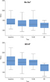
Changes of BL/IL in the Bio-Oss® group with time
There were 21 fixtures with radiographs acquired at least 25 months after implant placement. In patients with Bio-Oss®, the mean BL right after placement was 22.70 mm, 21.23 mm after 7 to 12 months, 21.21 mm after 13 to 24 months, and 20.41 mm after 25 months. The mean IL was consistently 15.37 mm. The mean value of BL/IL was 1.47 directly after the placement, 1.38 after 7 to 12 months, 1.38 after 13 to 24 months, and 1.33 after 25 months (Table 2).
Changes in BL/IL in the OCS-B® group with time
There were 5 fixtures with radiographs acquired at least 25 months after implant placement. The mean BL was 24.50 mm right after placement in patients with OCS-B®, 22.90 mm after 7 to 12 months, 22.68 mm after 13 to 24 months, and 18.53 mm after 25 months. The mean IL was consistently 15.21 mm. The mean value of BL/IL was 1.61 right after placement, 1.50 after 7 to 12 months, 1.49 after 13 to 24 months, and 1.22 at 25 months or more (Table 3).
Results with statistical analysis
Changes in BL/IL showed a statistically significant decreasing tendency over time (P < 0.05) (Fig. 2). There was no significant change in the Bio-Oss® group (P > 0.05) (Fig. 1). In contrast, the OCS-B® group showed a significant decrease with time (P < 0.05) (Fig. 3). However, no significant difference was observed between the 2 groups (P > 0.05) (Fig. 4).
DISCUSSION
It is difficult to place dental implants where maxillary posterior teeth are lost due to insufficient bone quantity and compromised bone quality caused by pneumatization [26-30]. It is difficult to gain a large enough amount of bone for implant placement when there are alveolar bone loss after extractions of maxillary posterior teeth and severe pneumatization. In these situations where alveolar bone has poor quality and short height, the sinus lift technique could be the first option for treatment.
Air pressure from respiration may bring about pneumatization in the maxillary sinus [3], and this could accelerate resorption of graft material in the maxillary sinus [31]. It is known that the stability of the graft material is one of the major factors influencing further implant stability, and many studies have been done on this issue.
One in vivo experiment used autogenous bone graft materials. Graft height reduced continuously and finally the apex of the implant was exposed to the maxillary sinus [32]. Another study reported that it was impossible to place dental implants after the bone graft technique was usedbecause of rapid bone resorption [33].
Jensen et al. [34] reported on different resorption patterns with various kinds of graft material. Every case showed a resorption tendency regardless of the type of graft materials used. Block et al. [35] performed sinus lift procedures with different types of graft material and evaluated the changes in height of the graft materials by computed tomography. At observations 5 to 10 years after the procedure, all the kinds of graft materials showed height reduction. Furthermore, in 90% of cases, the graft materials were positioned superior to the apex of the implant.
Hatano et al. [21] reported that graft materials were reduced a statistically significant amount during 2 to 3 years after a sinus lift. On the other hand, Nystrom et al. [36], Listrom and Symington [37] observed that the force loading to dental implants caused graft height to be sustained at a consistent level. Jensen et al. [34] reported that the resorption rate of bone is influenced by the types of graft materials, and that the amount of resorption was 1.8 mm in an autograft, 2.1 mm in an demineralized allograft, 0.9 mm in an alloplast and 0.8 mm in an autograft mixed with alloplast.
In the present study, as in most other studies, graft materials were resorbed over time with statistical significance. The value of BL/IL was 1.54 at the time of placement, 1.44 after 7 to 12 months, 1.35 after 13 to 24 months and 1.31 after at least 25 months. There was a significant decrease in comparison with data directly after placement, after 7 to 12 months, after 13 to 24 months and after at least 25 months; however, reduction rates from one period to the next were not statistically significant. However, bone grafts were resorbed with time, as mean BL/IL was observed to be more than 1 in every period. Grafted bones remained in the superior position of the implant apex.
In the Bio-Oss® group, the mean value of BL/IL was 1.47 right after the placement, 1.38 after 7 to 12 months, 1.38 after 13 to 24 months and 1.33 after at least 25 months, but there was no statistical significance.
In the OCS-B® group, the mean value of BL/IL was 1.61 right after placement, 1.50 after 7 to 12 months, 1.49 after 13 to 24 months, and 1.22 over at least 25 months, and it showed a reduction with statistical significance. However, no significant changes occurred until 24 months. Therefore, it is thought that a significant decrease occurred since only 5 out of 29 fixtures that had been observed for more than 25 months were investigated. also In addition, there was no significant difference in resorption between the two graft materials.
In conclusion, graft materials are resorbed with time, but without resorption to the apex of the implant, the graft supported the implant structure consistently. Furthermore, no significant difference in height change was observed between the two graft materials.
In the current study, radiographs taken directly after placement, after 7 to 12 months, and 13 to 24 months were from the same patients. However, the number of radiographs taken after at least 25 months was insufficient; there were limitations in achieving accurate follow-up on the changes in grafts with time. We faced limitations in analysis because the number of cases was not large enough and only 2 types of graft material were investigated.
In the current study, we selected 2D panoramic images. However, if we took radiographs through 3D images like computed tomography or MRI, we could observe changes in the graft height and volume in maxillary sinus more accurately. Since the present study has been done retrospectively without consideration of the type of prosthesis, implant surface modifications, diameter and length of implant, and length of available bone, further studies with controlled variables should be done. It is thought that prospective studies with 3D images are needed.


 XML Download