

 PDF
PDF ePub
ePub Citation
 Print
Print


Dear Editor:
Sebaceous trichofolliculoma (ST) is a rare benign tumor and represents a variant of trichofolliculoma (TF). ST presents as a centrally depressed lesion up to 1 cm in diameter, usually occurring in sebaceous lobules rich areas1. A similar lesion, folliculosebaceous cystic hamartoma (FSCH), is a rare cutaneous hamartoma characterized by follicular, sebaceous, and mesenchymal elements. FSCH typically presents as a small solitary symmetrical papule, usually occurring on the head and neck2.
The relationship between ST and FSCH is controversial. According to Schulz and Hartschuh3, early stage TF is characterized by several curved vellus hair follicles, whereas late stage shows changes in catagen and telogen hair follicles and prominent sebaceous differentiation. However, late stage ST has been postulated to be identical to FSCH3. Here, we report a rare case exhibiting the common histopathologic findings of ST and FSCH, and we discuss the association between these two diseases.
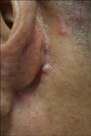
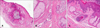
A 56-year-old male presented with an asymptomatic papule on the left postauricular area for several years. Physical examination revealed a single, skin-colored, firm, hairless papule that was 4×5 mm in diameter (Fig. 1). The tumor was completely removed by excision. Histopathology revealed a markedly dilated follicular cystic structure lined by a stratified squamous epithelium that was connected to the epidermis. Abundant mature sebaceous lobules radiated outward from the cystic structure, with secondary follicles in the dermis. The epithelial component was surrounded by dense, laminated, collagenous fibroplasia and the fibroepithelial unit was separated by a cleft-like space. There are mature adipocytes and perivascular mucin deposition in variable collagenous mesenchymal stroma (Fig. 2).
The features of ST are similar to those of FSCH. On histopathology, FSCH involves the upper segment, particularly around the isthmus, and is characterized by an infundibular cyst, hyperplasia of the sebaceous lobules, an abundant fibrous stroma, and mesenchymal components such as collagen, adipose, vascular tissue. FSCH tumors are separated by a cleft. Although, initially thought to be uncommon, epidermal communication may be present. ST has somewhat similar lesions, but it is also characterized by secondary follicles with features of lower segment differentiation4. ST also lacks the mesenchymal proliferation seen in FSCH. This case exhibited secondary follicles, which are the main finding in ST, and a fibroepitheilal unit with clefting and bundle of collagen, adipocytes, and increased number of small venules in mesenchymal stroma, as is typically observed in FSCH. Therefore, the findings in this case suggest that very late stage ST is identical to FSCH, as proposed previously4.
Alternately, FSCH and TF may be considered distinct entities. They may, however, follow similar patterns of pathogenesis. The activation of abnormal CK15-positive hair follicle stem cells from a primary infundibulofollicular primordium may occur in both conditions, but FSCH and TF differ from each other in the direction of fundamental differentiation. Bidirectional differentiation is rare5.
In this case, histopathology showed characteristics of two diseases simultaneously, seemingly caused by bidirectional differentiation in a single lesion. Therefore, with the exception of congenital FSCH, we propose that ST and FSCH are on the same disease spectrum.
 XML Download