

 PDF
PDF ePub
ePub Citation
 Print
Print


Dear Editor:
Juvenile xanthogranuloma (JXG) is histologically characterized by dense mononuclear cells with foamy histiocytes and Touton giant cells in a background of lymphocytes and eosinophils123. An unusual variant of non-lipidized JXG has been described in approximately 28 cases, in which Touton giant cells and foam cells are absent or very scant. These cases are described as non-lipidized JXG, early JXG, or a mononuclear JXG variant1345. This variant is typically found in infants or children younger than three years and presents most commonly as a solitary lesion14. There have been few reports of mononuclear JXG variants occurring in adults34. We report an adult female presenting with multiple lesions, consisting of both classic JXG and a mononuclear JXG variant.
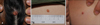
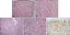
A 20-year-old Korean female presented with a red-to-yellow, dome-shaped asymptomatic scalp nodule that had developed within the last 4 months (Fig. 1A). Subsequently, two additional lesions appeared on her back and thigh, presenting as two yellow papules (Fig. 2B, C). The back lesion was excised, and histologic analysis revealed a dense infiltrate of epithelioid mononuclear cells, with a few containing cytoplasmic vacuoles. However, neither Touton giant cells nor well-developed, foamy histiocytes were detected (Fig. 2A, B). Based on the clinical and histological findings, a mononuclear JXG variant was suspected.
Two weeks after the excision, a punch biopsy of the scalp lesion was performed. The specimen showed mixed proliferation of histiocytes, lymphocytes, and giant cells. Cells were frequently accompanied by a well-developed foamy cytoplasm, and multinucleated giant cells were easily detected, most of which were of the Touton type (Fig. 2C~E). Based on typical clinical and histological JXG features, the second biopsy specimen of the back lesion, for which the histological features were ambiguous, was diagnosed as a mononuclear JXG variant. The thigh lesion, which was clinically similar, was diagnosed as the same entity.
As the initially excised back lesion developed after the scalp lesion, the lack of typical JXG features could be due to insufficient development. This theory is supported by the histologic changes present during JXG development15. Kubota et al.5 suggested that the JXG developmental course, inflammatory cell components, and persistent duration of a few years support the idea that JXG may be a reactive disease.
In summary, several atypical findings that confounded the diagnosis were observed in the current case. First, most cases of mononuclear JXG variants involving multiple lesions have been reported in younger children, with a male preference2. However, our patient was an adult female with multifocal cutaneous lesions. Second, multiple lesions appeared within a short period (4 months), resulting in coexistence of a fully developed lesion and immature lesions in early evolutionary stages. The simultaneous presence of these uncommon features in a single patient is unique, and this supports the hypothesis that JXG may develop through a reactive rather than neoplastic process5.
 XML Download