

 PDF
PDF ePub
ePub Citation
 Print
Print


Dear Editor:
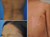
A 10-year-old girl presented with a round, brown to whitish, atrophic plaque on the right side of her back, which she has had for 5 years. Histologic findings showed thick collagen bundles with lymphocytic infiltrations around the vessel in the dermo-subcutaneous tissue, which are consistent with morphea. Only topical calcipotriol was used due to the limited localization of the plaque and the young age of the patient. However, after 6 months, a new morphea lesion developed on the left side of the back and linear scleroderma occurred on the forehead. The patient was diagnosed with generalized mixed-type morphea, with two circumscribed morphea on the right and left sides of the back (Fig. 1A) and linear scleroderma on the forehead (Fig. 1B). Hypopigmented lesions were also present on her left leg and on the left side of the trunk (Fig. 1C). Based on Wood's light examination and clinical findings, the patient was diagnosed with segmental vitiligo (SV).
The vitiliginous lesions and linear scleroderma on the forehead showed progression despite the combination therapy using intermittent oral steroid, topical calcineurin inhibitor, and narrowband ultraviolet B phototherapy for 1 year. To block the progression of morphea, hydroxychloroquine sulfate (200 mg/day) was used. Skin lesions of morphea and vitiligo stabilized within 6 weeks. In addition, the vitiliginous lesions showed 70% repigmentation after 5 months from start of medication. Skin lesions of morphea also showed no more progression. The patient tolerated the 7-month treatment period with no side effects (Fig. 2).
Although the pathogenesis of vitiligo remains unknown, an immunological mechanism is strongly considered. However, SV is associated with a neurogenic mechanism. Furthermore, an immune-mediated mechanism known as the three-step theory has been reported to be involved in the pathogenesis of SV, according to a case report showing simultaneous occurrence of SV, alopecia areata, psoriasis, and halo nevus1.
The association of SV with scleroderma has been rarely reported2. Bonifati et al.3 reported the case showing linear scleroderma on the left limb and homolateral SV on the trunk. Contrary to previous reports, our patient had generalized morphea lesions on the right and left sides of the back and linear scleroderma on the left side of the forehead, which was on the same side of the body where SV was located.
Our case study suggested that hydroxychloroquine is effective not only in blocking the progression of scleroderma4 but also in treating vitiligo. Walsh et al.5 reported two systemic lupus erythematosus patients with exextensive widespread vitiligo-like depigmentations who showed rapid responses to hydroxychloroquine.
Although we cannot exclude the possibility of coincidental development of SV and scleroderma, our case suggested that vitiligo and scleroderma that have the same autoimmune background can occur at the same time because they occurred during the same period and progressed in parallel for several years. In addition, this concomitant presence could support the theory that SV is associated with an autoimmune background. Moreover, our report suggested that an antimalarial drug could be used as a promising option for the treatment of vitiligo.
 XML Download