Journal List > Ann Dermatol > v.28(1) > 1046143

Hong, Hashimoto, and Kim: A Case of Pemphigus Herpetiformis with Only Immunoglobulin G Anti-Desmocollin 3 Antibodies

Abstract

Pemphigus represents a group of autoimmune blistering diseases caused by autoantibodies against desmogleins (Dsgs), a class of desmosomal cadherins. Recently, several pemphigus patients only with desmocollin (Dsc) 3-specific antibodies have been reported. Here, we report a case of pemphigus herpetiformis (PH), where only anti-Dsc3-specific antibodies but not anti-Dsg antibodies were detected. A 76-year-old woman presented with a 3-year history of blister formation. Physical examination revealed pruritic erythemas with vesicles on the trunk and legs, but no lesions of the oral mucosa. A skin biopsy specimen revealed intraepidermal blister containing neutrophils, eosinophils, and lymphocytes. Direct immunofluorescence (IF) showed immunoglobulin G (IgG) and complement 3 (C3) depositions on the keratinocyte cell surfaces. Indirect IF showed IgG anti-keratinocyte cell surface antibodies. These findings hinted at a diagnosis of pemphigus. However, repeated enzyme-linked immunosorbent assays (ELISAs) for both anti-Dsg1 and 3 antibodies proved to be negative. Immunoblotting of normal human epidermal extracts revealed Dsc antibodies, and recently established ELISAs using human Dsc1-Dsc3 recombinantly expressed in mammalian cells detected anti-Dsc3 antibodies. Based on these clinical, histopathological, and immunological findings, the patient was diagnosed as PH with only anti-Dsc3 antibodies. Treatment with corticosteroid prednisolone and steroid-sparing agent dapsone accomplished complete clinical remission of the patient.

INTRODUCTION

Pemphigus represents a group of autoimmune blistering diseases caused by autoantibodies against desmogleins (Dsgs), a class of the cell surface adhesion proteins, desmosomal cadherins1. In humans, 7 desmosomal cadherins, 4 desmogleins (Dsg1–4) and 3 desmocollins, (Dsc1–3), have been described. Pemphigus can be divided into two major forms: pemphigus foliaceus (PF) and pemphigus vulgaris (PV). In PF, autoantibodies against Dsg1 cause blisters on the superficial epidermis. In mucosal dominant PV, autoantibodies against Dsg3 cause blisters on the suprabasal layer of the mucous membrane. In mucocutaneous PV, autoantibodies against both Dsg3 and Dsg1 cause suprabasilar blisters on the skin and mucous membranes. Pemphigus herpetiformis (PH) is a rare variant of pemphigus that clinically resembles dermatitis herpetiformis but shows immunopathological features of pemphigus. PH exhibits IgG autoantibodies against Dsg1 in most cases and against Dsg3 in the remainder2.
Recently, several pemphigus patients only with Dsc3-specific antibodies have been reported3456. Here we report a case of PH, which evinced anti-Dsc3-specific antibodies but not anti-Dsg antibodies.

CASE REPORT

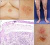
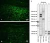
A 76-year-old woman presented with a 3-year history of blister formation but no history of malignancy or autoimmune disease. Physical examination revealed annular erythematous plaques with grouped peripheral vesicobullae with intensive itch on the trunk and legs (Fig. 1A, B). Oral mucosa was not affected. A skin biopsy specimen revealed intraepidermal blister containing neutrophils, eosinophils and lymphocytes (Fig. 1C). In the dermis, infiltration of lymphocytes and eosinophils was seen. Direct immunofluorescence (IF) study showed IgG and complement 3 (C3) depositions on keratinocyte cell surfaces (Fig. 2A, B). Indirect IF of normal human skin also revealed IgG anti-keratinocyte cell surface antibodies.
These findings suspected the diagnosis of pemphigus. However, repeated enzyme-linked immunosorbent assay (ELISA) for both anti-Dsg1 and 3 antibodies showed negative results. Immunoblotting with normal human epidermal extracts revealed a doublet of a-form (110-kDa) and b-form (100-kDa) Dscs (Fig. 2C). Finally, recently established ELISAs using recombinantly expressed human Dsc1-Dsc37 proteins in mammalian cells detected anti-Dsc3 (OD 2.263, cut-off >0.120) antibodies, but no antibodies for Dsc1 (OD 0.166, cut-off >0.200) and 2 (OD 0.015, cut-off >0.070).
Based on these clinical, histopathological and immunological findings, the patient was diagnosed as PH exclusively with anti-Dsc3 antibodies. The skin lesions responded well with oral methylprednisolone (4~12 mg/day) and dapsone (50 mg/day), and the patient achieved complete remission 4 months after the initiation of the treatment.

DISCUSSION

Desmosomes in keratinocytes are the most important intercellular adhering junctions that provide structural strength to the epidermis. Dsg3 and Dsc3 are the predominant isoforms expressed in the basal epidermis, the site of blister formation in pemphigus vulgaris. Dsg1 and Dsc1 are expressed in an inverse pattern, predominantly in the superficial epidermis with little to no expression in the basal layers. Although the major autoantigens for pemphigus are Dsgs, several studies have reported that Dsc3 homo- and heterophilic binding is required to maintain keratinocyte cohesion and interference with Dsc3 multimerization may contribute to skin blistering in pemphigus58.
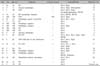
In our literature survey, we reviewed twenty-one cases of pemphigus with IgG anti-Dsc3 autoantibodies (Table 1). We found 1 case with autoantibodies exclusively against Dsc3 showing PV-like clinical and histopathological phenotypes, suggesting that Dsc3 and Dsg3 interact and have similar function in the lower epidermis3. In contrast, most other cases were those of atypical pemphigus, such as PH, paraneoplastic pemphigus and pemphigus vegetans, indicating that the pathogenic role of anti-Dsc3 antibodies is distinct from antibodies to either Dsg1 or Dsg3. These findings are consistent with the results of a recent study, which showed that sera from 30%~40% of patients with PH and pemphigus vegetans showed reactivity with Dsc1-Dsc3, and in contrast sera from only a few patients with PV and PF showed anti-Dsc antibodies at low titer7.
Among the 20 cases with description of oral involvement, nine cases had mucosal lesions, suggesting that anti-Dsc3 antibodies are responsible for oral mucosal lesions (Table 1)91011121314151617181920. This result is contradictory to the results in Dsc3-knock-out mice, which showed blisters on the skin but no mucosal lesions8. This discrepancy may be explained by the fact that high expression levels of Dsc2 compensated for the loss of Dsc3 in mucous membranes of mice.
Our patient had clinical and histopathologic features of PH, but ELISAs for both Dsg1 and Dsg3 showed negative results. In contrast, immunoblotting of normal epidermal extract showed strong reactivity with Dscs, and novel ELISAs for Dsc1-Dsc3 detected only anti-Dsc3 antibodies. Favorable responses to low-dose steroid and dapsone in this patient may indicate milder pathogenic activity of anti-Dsc3 than anti-Dsg3 antibodies, which was also suggested in a previous study3. Taken together, these results strongly indicate that this is a case of pemphigus herpetiformis and IgG anti-Dsc3 antibodies play an important role in pemphigus pathogenesis.

Figures and Tables

Fig. 1

Clinical and histopathologic findings. Clinical features. Annular erythematous plaques with grouped peripheral tense vesicobullae on the (A) trunk and (B) legs. (C) Histopathological features. Intraepidermal blister with infiltrates of neutrophils, eosinophils and lymphocytes (H&E, ×200).

ad-28-102-g001
Fig. 2

Immunologcal findings. DIF shows (A) immunoglobulin G (IgG) and (B) complement 3 (C3) deposition along the cell surface of keratinocytes. (C) Results of immunoblotting of normal human epidermal extracts. Pemphigus vulgaris (PV) control serum reacted with the 160-kDa desmoglein (Dsg) 1 and the 130-kDa Dsg3 (lane 1), paraneoplastic pemphigus (PNP) control serum reacted with the 210-kDa envoplakin and the 190-kDa periplakin (lane 2), bullous pemphigoid (BP) control serum reacted with the 230-kDa BP230 and the 180-kDa BP180 (lane 3), anti-desmocollin (Dsc) monoclonal antibody (mAb) (lane 4) and the patient serum (lane 5) reacted strongly with the 110-kDa a-form and the 100-kDa b-form Dsc3. DIF: direct immunofluorescence.

ad-28-102-g002
Table 1

Reported cases of autoimmune blistering disease in association with Dsc3-specific immunoglobulin G antibodies

ad-28-102-i001
Case Sex Age (yr) Diagnosis Mucosal lesions Autoantibodies Reference No.
1 M 42 PH - Dsc3, Dsg1 9
2 F 11 Atypical pemphigus + Dsc3, Dsg3, Desmoplakin 10
3 F 48 PNP + Dsc3, Dsc2, Dsg3,
Envoplakin/periplakin, BP180
11
4 M 63 BP+pemphigus vegetans - Dsc3, BP230, BP180 12
5 ND ND PNP ND Dsc3, Dsg3 13
6 M 53 Pemphigus+gastric carcinoma - Dsc3, Dsc1, Dsc2 14
7 F 55 PV + Dsc3 3
8 ND ND Pemphigus vegetans + Dsc3 4
9 ND ND Pemphigus vegetans - Dsc3, Dsg1 4
10 ND ND PH + Dsc3 4
11 ND ND PH - Dsc3 4
12 F 79 Atypical pemphigus - Dsc3 5
13 F 83 BP+PH + Dsc3, Dsc2, Dsc1, Dsg1, Dsg3 15
BP180
14 F 81 PNP+follicular B cell lymphoma + Dsc3, Dsc2, Dsg3 16
Periplakin
15 M 54 PV+SCC + Dsc3, Dsc 1, Dsc2,
Dsg1, Dsg3
17
16 F 84 PH - Dsc3, Dsg1 18
17 F 80s Pemphigus vegetans - Dsc3, Dsg1 19
18 F 70s Pemphigus vegetans - Dsc3 19
BP230, Periplakin
19 M 68 Herpetiform bullous dermatosis + Dsc3, Dsc1 20
LAD-1
20 M 57 PH - Dsc3 6
21 F 76 PH - Dsc3 Present case

Dsc: desmocollin, M: male, F: female, PH: pemphigus herpetiformis, Dsg: desmoglein, BP: bullous pemphigoid, PNP: paraneoplastic pemphigus, SCC: squamous cell carcinoma, PV: pemphigus vulgaris, LAD-1: linear IgA dermatosis antigen, ND: no description.

References

1. Matsuda-Hirose H, Ishikawa K, Goto M, Hatano Y, Fujiwara S. Selective elevation of antibodies to desmoglein 1 during the transition from mucocutaneous to cutaneous type pemphigus vulgaris. Ann Dermatol. 2013; 25:263–265.
crossref
2. Kasperkiewicz M, Kowalewski C, Jabłońska S. Pemphigus herpetiformis: from first description until now. J Am Acad Dermatol. 2014; 70:780–787.
3. Mao X, Nagler AR, Farber SA, Choi EJ, Jackson LH, Leiferman KM, et al. Autoimmunity to desmocollin 3 in pemphigus vulgaris. Am J Pathol. 2010; 177:2724–2730.
crossref
4. Rafei D, Müller R, Ishii N, Llamazares M, Hashimoto T, Hertl M, et al. IgG autoantibodies against desmocollin 3 in pemphigus sera induce loss of keratinocyte adhesion. Am J Pathol. 2011; 178:718–723.
crossref
5. Hatano Y, Hashimoto T, Fukuda S, Ishikawa K, Goto M, Kai Y, et al. Atypical pemphigus with exclusively anti-desmocollin 3-specific IgG antibodies. Eur J Dermatol. 2012; 22:560–562.
crossref
6. Nakamura Y, Takahata H, Teye K, Ishii N, Hashimoto T, Muto M. A case of pemphigus herpetiformis-like atypical pemphigus with IgG anti-desmocollin 3 antibodies. Br J Dermatol. 2014; 171:1588–1590.
crossref
7. Ishii N, Teye K, Fukuda S, Uehara R, Hachiya T, Koga H, et al. Anti-desmocollin autoantibodies in nonclassical pemphigus. Br J Dermatol. 2015; 173:59–68.
crossref
8. Chen J, Den Z, Koch PJ. Loss of desmocollin 3 in mice leads to epidermal blistering. J Cell Sci. 2008; 121:2844–2849.
crossref
9. Kozlowska A, Hashimoto T, Jarzabek-Chorzelska M, Amagai A, Nagata Y, Strasz Z, et al. Pemphigus herpetiformis with IgA and IgG antibodies to desmoglein 1 and IgG antibodies to desmocollin 3. J Am Acad Dermatol. 2003; 48:117–122.
crossref
10. Hisamatsu Y, Amagai M, Garrod DR, Kanzaki T, Hashimoto T. The detection of IgG and IgA autoantibodies to desmocollins 1-3 by enzyme-linked immunosorbent assays using baculovirus-expressed proteins, in atypical pemphigus but not in typical pemphigus. Br J Dermatol. 2004; 151:73–83.
crossref
11. Preisz K, Horváth A, Sárdy M, Somlai B, Hársing J, Amagai M, et al. Exacerbation of paraneoplastic pemphigus by cyclophosphamide treatment: detection of novel autoantigens and bronchial autoantibodies. Br J Dermatol. 2004; 150:1018–1024.
crossref
12. Bolling MC, Mekkes JR, Goldschmidt WF, van Noesel CJ, Jonkman MF, Pas HH. Acquired palmoplantar keratoderma and immunobullous disease associated with antibodies to desmocollin 3. Br J Dermatol. 2007; 157:168–173.
crossref
13. Müller R, Heber B, Hashimoto T, Messer G, Müllegger R, Niedermeier A, et al. Autoantibodies against desmocollins in European patients with pemphigus. Clin Exp Dermatol. 2009; 34:898–903.
crossref
14. Endo Y, Tsujioka K, Tanioka M, Minegaki Y, Ohyama B, Hashimoto T, et al. Bullous dermatosis associated with IgG antibodies specific for desmocollins. Eur J Dermatol. 2010; 20:620–625.
15. Ohata C, Koga H, Teye K, Ishii N, Hamada T, Dainichi T, et al. Concurrence of bullous pemphigoid and herpetiform pemphigus with IgG antibodies to desmogleins 1/3 and desmocollins 1-3. Br J Dermatol. 2013; 168:879–881.
crossref
16. Gallo E, García-Martín P, Fraga J, Teye K, Koga H, Hashimoto T, et al. Paraneoplastic pemphigus with eosinophilic spongiosis and autoantibodies against desmocollins 2 and 3. Clin Exp Dermatol. 2014; 39:323–326.
crossref
17. Kim J, Teye K, Koga H, Yeoh SC, Wakefield D, Hashimoto T, et al. Successful single-cycle rituximab treatment in a patient with pemphigus vulgaris and squamous cell carcinoma of the tongue and IgG antibodies to desmocollins. J Am Acad Dermatol. 2013; 69:e26–e27.
crossref
18. Matsukura S, Takahashi K, Hirokado M, Ikezawa Y, Nakamura K, Fukuda S, et al. Recalcitrant pemphigus herpetiformis with high titer of immunoglobulin G antibody to desmoglein 1 and positive IgG antibody to desmocollin 3, elevating thymus and activation-regulated chemokine. Int J Dermatol. 2014; 53:1023–1026.
crossref
19. Saruta H, Ishii N, Teye K, Ono F, Ohyama B, Koga H, et al. Two cases of pemphigus vegetans with IgG anti-desmocollin 3 antibodies. JAMA Dermatol. 2013; 149:1209–1213.
crossref
20. Ueda A, Ishii N, Teye K, Dainichi T, Ohyama B, Hamada T, et al. Unique herpetiform bullous dermatosis with IgG antibodies to desmocollins 1/3 and LAD-1. Br J Dermatol. 2013; 169:719–721.
crossref
TOOLS
Similar articles