

 PDF
PDF ePub
ePub Citation
 Print
Print


Dear Editor:
Multiple eruptive dermatofibromas (MEDF) are very rare and usually have been associated with autoimmune diseases, immunosuppressant therapy, hematologic malignancy, and other conditions. Zaccaria et al.1 noted that the incidence of MEDF is higher in patients with underlying diseases than in healthy persons.
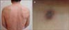
A 64-year-old man visited our department because of multiple lesions of the skin that had developed rapidly during the previous 3 months. Physical examination revealed firm, brownish, dome-shaped papules (>30) that were 5~10 mm in diameter, with lateral dimpled signs, on the trunk and legs (Fig. 1). He did not have any specific medical or family history. Excision biopsy of a papule on the right thigh showed a relatively well-demarcated dermal nodule composed of tightly interdigitating fascicles of spindle-shaped fibrohistiocytic cells (Fig. 2A, B). Immunohistochemical analysis revealed that many of the tumor cells were positive for factor XIIIa (Fig. 2C) and negative for CD34. Laboratory tests were conducted to rule out connective tissue disease and immunosuppressive state. Complete blood cell count, metabolic and hepatitis panels, erythrocyte sedimentation rate, analysis of T-cell subset, C3/C4, immunoglobulins, and thyroid-stimulating hormone showed normal results. Anti-human immunodeficiency virus antibody, antinuclear antibody, and double-stranded DNA antibody were all negative.
One or several dermatofibromas (DF; <5 lesions) are common; however, multiple DF (>15) are extremely rare. In 1970, Baraf and Shapiro2 first described multiple DF. Ammirati et al.3 proposed that MEDF should be defined as the presence of 5~8 DF appearing within a period of 4 months. However, this criterion is arbitrarily chosen, and therefore, it might not be entirely valid for all cases. It is important to note the dynamic changes in some lesions within a short period, in contrast to the static state usually observed in DF4. Indeed, some cases reported as MEDF did not meet the criteria or did not indicate the period of development. In our review of the literature published between 1973 and 2012, 21 of 72 subjects were otherwise healthy, whereas 51 (70%) had an underlying disease. Forty-one cases (80%) among the patients with underlying disease were related to various immunodeficiency states. The etiology of DF is unclear. It was proposed that DF represents an abortive immunoreactive process mediated by dermal dendritic cells, developing as a response to a putative pathogen that the suppressed immune system cannot clear5. Accordingly, the development of MEDF could be considered a manifestation of immunosuppression; however, in our case, no abnormal specific immune defect or a medical history of using immunosuppressive agents could be found.
Herein, we report a rare case of MEDF in a healthy adult, along with a literature review.

 XML Download