

 PDF
PDF ePub
ePub Citation
 Print
Print


INTRODUCTION
Transdermal drug delivery systems (TDDS) have impressive benefits, including the avoidance of poor drug absorption or enzymatic degradation in the gastrointestinal tract or liver, as well as avoidance of the pain that accompanies intravenous or intramuscular injection. However, transdermal delivery is severely limited by the inability of the large majority of drugs to cross the skin at therapeutic rates due to the excellently impervious nature of skin1,2. Thus, many approaches have been attempted to perturb the skin barrier and enhance the transdermal delivery of drugs. The major approaches for enhancing transdermal delivery are physical enhancers, such as ultrasound, iontophoresis, electroporation, magnetophoresis, and microneedles; vesicles; particulate systems, such as liposomes, niosomes, transfersomes, microemulsions, and solid lipid nanoparticles; and chemical enhancers, including sulphoxides, azones, glycols, alkanols, and terpenes3-8.
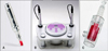
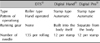
Since the initial application of microneedles to TDDS was reported in 19989, a variety of microneedle sizes, structures, and materials have been used in many types of drug delivery. The advantage of the microneedle is that it can perforate the skin directly and facilitate the passage of not only small molecules but also large molecules through the stratum corneum (the primary barrier) and reach the dermis without stimulation the dermal nerves. Also, there are many types, such as poke and patch; shapes, including solid and hollow; and materials, such as silicon, metal, and polymer, that can be used to fabricate microneedles. Therefore, it is possible to modify the design to match the purpose10-12. Digital Hand® and Digital Pro® (Bomtech Electronics Co., Ltd., Seoul, Korea) represent a novel type of microneedle (Fig. 1). They are automatized stamp type microneedles, which make them easy to handle, and the stamp mechanism reduces the risk of abrasion.
In the present study, two newly developed microneedle devices, Digital Hand® and Digital Pro®, were studied with respect to the safety of the skin in comparison with the commonly used DTS® (Disk type-microneedle Therapy System; DTS lab., Seoul, Korea).
MATERIALS AND METHODS
Subjects
Male skh-hairless-1 (albino) mice (5 weeks of age) were obtained from Hoshino Experimental Animal Center (Yashio, Japan), and housed in cages kept at 23℃ with a 12 hour light/dark cycle in pathogen-free conditions.
This study was approved by the Institutional Animal Care and Use Committee.
Equipment
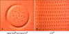
Digital Hand® and Digital Pro® are stamp type, motorized microneedle devices (Fig. 1). Therefore, when a doctor places them in contact with the patient's skin, the microneedle is automatically inserted into the skin. Digital Hand® and Digital Pro® operate by the mechanism, but the running gear of Digital Hand® is built into the body itself, and the running gear of Digital Pro® is separate from the body. The insertion power of Digital Pro® is stronger than Digital Hand®. We identified the number of needlings per unit area (2×2 cm) using paper clay. There were 12 pores per stamp for Digital Hand® and Digital Pro® versus 135 pores per rolling of the DTS® (Fig. 2, Table 1). General use of DTS® in animal experiments involves rolling the device five times; therefore, in our experiment we used 56 stamps of the new digital microneedle devices per unit area.
Pretreatment preparation
Topical anesthetic cream (2.5% lidocaine-2.5% prilocaine; AstraZeneca Korea, Seoul, Korea) was applied to the skin of hairless mice 30 minutes prior to the procedure. The anesthetic was washed off with mild soap and water immediately before the procedure.
Comparison of the new digital microneedle devices and DTS® using dermatoscopic inspection, transepidermal water loss (TEWL), and biopsy
We used the new digital microneedle devices and DTS® on the skin of hairless mice on day 0. Five-week-old albino hairless mice (n=12) were handled in one of the following ways: no application (control group, n=3); rolling with DTS® 1.5 mm five times in the horizontal, vertical, and diagonal directions (group A, n=3); using Digital Hand® 1.5 mm 56 times (group B, n=3); or using Digital Pro® 1.5 mm 56 times (group C, n=3). Then, visual and dermatoscopic inspections (Dermlite II Pro HR & Nikon COOLPIX995; Nikon Corporation, Tokyo, Japan), measurements of TEWL, and biopsies were performed on days 0, 1, and 7. TEWL was measured with a Tewameter TM 210 (Courage & Khazaka, Cologne, Germany).
Dermatoscopic inspection, TEWL, and biopsy for Digital Hand® using different needle sizes (0.25, 0.5, 1.0, 1.5, and 2.0 mm)
We applied the Digital Hand® with different needle sizes to the skin of hairless mice on day 0. Five-week-old albino hairless mice (n=15) were handled with either no application (control group, n=3) or using 56 repetitions of the Digital Hand® with the following needle sizes: 0.25 mm (group A, n=3), 0.5 mm (group B, n=3), 1.0 mm (group C, n=3), 1.5 mm (group D, n=3), or 2.0 mm (group E, n=3). Then, visual and dermatoscopic inspections (Dermlite II Pro HR & Nikon COOLPIX995), measurements of TEWL, and biopsies were performed on days 0, 1, and 7.
Dermatoscopic inspection, TEWL, and biopsy for Digital Pro® using different needle sizes (0.25, 0.5, 1.0, 1.5 and 2.0 mm)
We applied the Digital Pro® with different needle sizes to the skin of hairless mice on day 0. Five-week-old albino hairless mice (n=18) were handled with either no application (control group, n=3) or using with Digital Pro® with one of the following needle sizes: 0.25 mm (group A, n=3), 0.5 mm (group B, n=3), 1.0 mm (group C, n=3), 1.5 mm (group D, n=3), or 2.0 mm (group E, n=3) for 56 times. Then, visual and dermatoscopic inspections (Dermlite II Pro HR & Nikon COOLPIX995), measurements of TEWL, and biopsies were performed on days 0, 1, and 7.
RESULTS
Comparison between new digital microneedle devices and DTS®
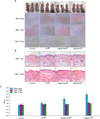
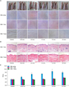
In regard to dermatoscopic inspection, we observed mild erythema and needle marks in groups A, B, and C on day 0. The erythema and needle marks were not present on days 1 and 7 (Fig. 3A). On histopathological examination, group B (Digital Hand®) and C (Digital Pro®) showed no significant abnormalities in comparison with group A (DTS®). There was no significant inflammatory cell infiltration, deszquamation of the stratum corneum, or disruption of the basal layer. Intact superficial and deep vascular plexuses in the dermis were observed in all experimental groups (Fig. 3B). The increase of TEWL immediately after treatment with DTS® and Digital Hand® were similar, but after treatment with Digital Pro®, TEWL markedly increased. However, TEWL had normalized in all experimental groups on days 1 and 7 (Fig. 3C). The increased TEWL is an indirect indicator of drugs being delivered through the skin. Therefore, DTS®, Digital Hand®, and Digital Pro® worked well for delivering drugs and skin disruptions caused by DTS®, Digital Hand®, and Digital Pro® were not severe.
Dermatoscopic inspection, TEWL, and biopsy for Digital Hand® using different needle sizes
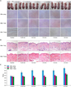
Dermatoscopy revealed mild erythema and needle marks in all experimental groups on day 0. The erythema and needle marks were not present on days 1 and 7 (Fig. 4A). There were no significant epidermal or dermal abnormalities on histopathological examination (Fig. 4B). The TEWL increased immediately after treatment with all sizes of the Digital Hand® and normalized in all experimental groups on days 1 and 7 (Fig. 4C).
Dermatoscopic inspection, TEWL, and biopsy for Digital Pro® using different needle sizes
We observed mild erythema and needle marks in all experimental groups on day 0. The erythema and needle marks were not present on day 1 and 7 (Fig. 5A). There were no significant epidermal or dermal abnormalities on histopathological examination (Fig. 5B). The TEWL increased immediately after treatment with all sizes of the Digital Pro® and normalized in all experimental groups on days 1 and 7 (Fig. 5C).
DISCUSSION
Transdermal drug delivery offers compelling opportunities to address the low bioavailability of many oral drugs. Different chemical and physical methods, such as chemical enhancers, ultrasound, electric energy, pressure driven flow, and lasers, have been used in attempts to disrupt the mass transfer resistance barrier of the stratum corneum to allow for the delivery of larger molecular weight and hydrophilic compounds. However, these methods have had limited success.
Microneedles are microscopic needles, a few hundred microns in size, and they are used to painlessly deliver large molecular compounds into the subcutaneous tissue at rates that allow the drug to reach therapeutic levels by enhancing skin permeability for transdermal delivery. Microneedles assembled on transdermal patches have been proposed as a hybrid between hypodermic needles and transdermal patches, in order to overcome the individual limitations of both injections and patches10. Unlike transdermal patches, microneedles have been successfully used to deliver a variety of large and hydrophilic compounds into the skin, including proteins and DNA. In vitro skin permeability enhancement of two to four orders of magnitude was observed for small molecules like calcein and large compounds like proteins and nanoparticles13,14. Prausnitz et al.15 studied the delivery of DNA plasmids into skin using microneedles. Lin et al.16 used microneedles either alone or in combination with iontophoresis to deliver 20-mer phosphorothioated oligodeoxynucleotides across the skin of hairless guinea pigs. A related study further demonstrated that microneedles enhance the delivery of desmopressin and human growth hormone using a similar approach17. A variety of microneedles can be fabricated for these applications by adapting the tools of the microelectronics industry for inexpensive mass production9. Microneedle technology is able to overcome some of the barriers preventing the transdermal administration of drugs with low bioavailability. The microneedle approach could make delivery across the skin feasible for a much wider range of drugs than is possible at present and may confer some safety advantages, according to researchers10.
Several recent advances in microneedle design and formulation are worthy of note. Microneedles can be fabricated by a number of different methods to yield a variety of needle sizes, shapes, and materials. Most of the work has focused on making microscopic holes in the skin by inserting solid microneedles made of silicon or metal. Solid microneedles have been shown to increase transdermal delivery by 'poke with patch,' 'coat and poke,' and 'dip and scrape' methods, and hollow microneedles have been shown to microinject into skin. Therapeutic responses have been achieved in vivo following delivery of proteins, DNA, and vaccines. Proper needle design can assure insertion into the skin, which prevents needle fracture or patient pain. Many studies suggest that microneedles may provide a powerful new approach to transdermal drug delivery18.
Data from animal and human studies have been published and generally report that there are no significant adverse reactions to microneedles. No infections caused by microneedles have been reported19-21. In addition, skin irritation has been reported to be mild and transient, when it exists at all12,16,19,22,23, and bleeding is generally not associated with the use of microneedles12,22,24. A variety of microneedle designs have been reported to be painless in human subjects. Additional studies are needed to fully assess their safety. A recent, randomized, double-blind study about the effect of microneedle design on pain in humans reported that microneedles are significantly less painful than a 26-gauge hypodermic needle over the range of dimensions investigated. Decreasing the length and number of microneedles reduces pain25.
The aim of this study was to obtain insight into the ability of digital microneedle devices to disrupt the barrier of the skin and to determine the safety of microneedle treatment in terms of skin irritation and histopathological change. We evaluated safety through dermatoscopic inspection, biopsy, and TEWL measurements. Measurements of TEWL are frequently used to evaluate the integrity of the skin barrier26. TEWL measurements may provide a rapid and non-invasive evaluation of skin barrier function before penetration experiments with Franz chambers27. We obtained insight into the ability of the digital microneedle devices to not disrupt the skin barrier. The safety of the digital microneedle devices is similar to the existing microneedle device (DTS®) in this study. However, the advantages of the digital microneedle devices are that the procedure can be performed with same and accurate power, and is more comfortable than the existing microneedle device (DTS®) because the digital microneedle devices are motorized microneedle devices and thus are automatically inserted into the skin. The digital microneedle devices work in the manner of a stamp, which avoids the minor abrasion often caused by microneedle rollers.
In conclusion, these results indicate that these novel digital microneedle devices can puncture sufficiently and safely to allow drugs to pass by the stratum corneum. However, this study was performed in mice. Therefore, a study with humans should be performed later for identifying overall safety toward human skin.
 XML Download