

 PDF
PDF ePub
ePub Citation
 Print
Print


INTRODUCTION
Hobnail hemangioma (targetoid hemosiderotic hemangioma) is a benign vascular tumor that typically presents as a small, single lesion on the skin of the trunk or limb of a young or middle-aged adult1,2. The lesion has a characteristic 'targetoid' appearance, in which a violaceous papule is surrounded by an ecchymotic or brown ring that can expand or subsequently disappear, with persistence of the central papule. In the Korean literature, there are 10 case reports. We report two additional cases of hobnail hemangioma revealed by typical clinical and histological appearance. Remarkably, one of them was a congenital lesion, never described before, and the other was a multiple hobnail hemangioma, which is also an atypical presentation.
CASE REPORT
Case 1
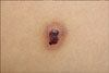
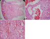
A 7-year-old boy presented with a dark brown papule with ecchymotic halo on the left upper back. According to the patient's mother, it had been there as a black macule since birth and recently the size had increased. The patient had no subjective symptoms and denied any history of trauma. On examination, the skin lesion was a 4 mm-sized, well-demarcated dark brown, irregularly shaped papule surrounded by a thin, pale area and a peripheral ecchymotic ring (Fig. 1). Histologic examination revealed dilated vascular channels with hobnail endothelial cells protruding into the lumen and occasional intraluminalpapillaryprojections in the upper dermis. Deeper in the dermis, the vascular channels were thinner and seeme d to dissect the collag en bundles. There were numerous extravasated erythrocytes (Fig. 2). Immunohistochemistry with monoclonal antibodies revealed strongly positive reaction with CD31 and focal positive reaction with CD34 and D2-40 (Fig. 3). The lesion disappeared completely following punch biopsy, and no recurrence was observed.
Case 2
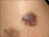
A 15-year-old boy presented with two dusky red to brown plaques on the medial and lateral aspect of the left knee that appeared 12 and 4 years ago, respectively. The patient had no subjective symptoms and denied any history of trauma. The patient said that these lesions had been indurated small papules but recently the size had increased and the color darkened progressively. Physical examination revealed two dusky erythematous plaques about 2 cm in size with surrounding ecchymotic macular rings (Fig. 4). A 4-mm skin biopsy was performed from the superolateral lesion to rule out verrucous hemangioma and Kaposi's sarcoma. Histologic examination was very similar to that of the first case, and additionally hemosiderin deposition was widely present in the dermis (Fig. 5). HHV-8 staining was negative. The lesions improved substantially following intermittent triamcinolone intralesional injections and pulsed dye laser treatment.
DISCUSSION
Hobnail hemangioma was first reported by Santa Cruz and Aronberg in 19883. A typical clinical appearance is a small solitary lesion consisting of a 2~3 mm-sized brown to violaceous papule surrounded by a thin, pale area and a peripheral ecchymotic ring. The characteristic targetoid appearance is due to peripheral hemorrhage and subsequent deposition of hemosiderin. However, these features are only present in a small percentage of cases and, most often, the clinical appearance is that of a red-blue or brown papule.
The etiology of hobnail haemangioma is unknown, but traumas to a pre-existing hemangioma and the influences of sex steroid hormones have been proposed4-6. Interestingly, some lesions in females change during the menstrual cyce or pregnancy.
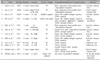
Published reports of hobnail hemangioma reveal an equal gender incidence, lwith an age range of presentation of 5~67 years. It is identified more frequently in younger persons3. There are 10 case reports in the Korean literature7-16 (Table 1). In those cases, skin lesions developed at 3~32-years-of-age. Nine patients had a solitary lesion, but one patient had two papules on the upper back and arm8. Five lesions arose in the lower extremities, four on the trunk and two on the upper extremities. Our 'case 1' is the youngest patient reported in Korea so far and is the first patient that has had the skin lesion since birth. 'Case 2' is the second case with two lesions after the first one reported in the Korean literature8.
Histologically, characteristic features are irregularly dilated vesHsels lined by hobnail endothelial cells in the superficial dermis and collagen-dissecting, rather narrow vessels in deeper dermis. The hobnail endothelial cell has scanty cytoplasm and rounded nuclei that protrude into theumina. Focally, intraluminal papillary projections can be seen in the superficial blood vessels. The vascular channels in the deeper dermis become much less conspicuous and eventually disappear completely. The whole architecture is wedge l-shaped, with a prominent superficial component. In the later stages, extensive stromal hemosiderin deposits are commonly seen.
The clinical differential diagnoses include melanocytic nevus, dermatofibroma, hemangioma and insect bite reaction. The microscopic differential diagnoses are the patch stage of Kaposi's sarcoma, retiform hemangioendothelioma, solitary angiokeratoma, progressive lymphangioma and eosinophilic hemangioma. It is especially important to distinguish hobnail hemangioma from the patch stage of Kaposi's sarcoma. Factors favoring Kaposi's sarcoma are the presence of plasma cells, spindle-shaped cells and apoptotic endothelial cells17.
The tumor origin is controversial. Santonja and Torrelo18 suggested a vascular origin for hobnail hemangioma since it had positive reaction with Factor VIII-related antigen, CD31 and CD34. However, Franke et al.19 proposed that this tumor has a lymphatic origin because it revealed positive reaction with D2-40 and CD31, and a negative reaction with CD34. In our first case, immunohistochemistry revealed a mild focal positive reaction with CD34 and D2-40 and strong positive reaction with CD31. These results imply that the origin is vascular endothelial cells, not lymphatic endothelial cells.
To our knowledge, all reported cases are acquired type. Here, we report two cases of hobnail hemangioma that are peculiar in that one is congenital and the other is multiple.


 XML Download